Dichloroacetate and Quercetin Prevent Cell Proliferation, Induce Cell Death and Slow Tumor Growth in a Mouse Model of HPV-Positive Head and Neck Cancer
- PMID: 38672607
- PMCID: PMC11048222
- DOI: 10.3390/cancers16081525
Dichloroacetate and Quercetin Prevent Cell Proliferation, Induce Cell Death and Slow Tumor Growth in a Mouse Model of HPV-Positive Head and Neck Cancer
Abstract
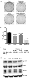
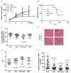
Elevated glucose uptake and production of lactate are common features of cancer cells. Among many tumor-promoting effects, lactate inhibits immune responses and is positively correlated with radioresistance. Dichloroacetate (DCA) is an inhibitor of pyruvate dehydrogenase kinase that decreases lactate production. Quercetin is a flavonoid compound found in fruits and vegetables that inhibits glucose uptake and lactate export. We investigated the potential role and mechanisms of DCA, quercetin, and their combination, in the treatment of HPV-positive head and neck squamous cell carcinoma, an antigenic cancer subtype in need of efficacious adjuvant therapies. C57Bl/6-derived mouse oropharyngeal epithelial cells, a previously developed mouse model that was retrovirally transduced with HPV type-16 E6/E7 and activated Ras, were used to assess these compounds. Both DCA and quercetin inhibited colony formation and reduced cell viability, which were associated with mTOR inhibition and increased apoptosis through enhanced ROS production. DCA and quercetin reduced tumor growth and enhanced survival in immune-competent mice, correlating with decreased proliferation as well as decreased acidification of the tumor microenvironment and reduction of Foxp (+) Treg lymphocytes. Collectively, these data support the possible clinical application of DCA and quercetin as adjuvant therapies for head and neck cancer patients.
Keywords: Dichloroacetate (DCA); head and neck oral cancer; human papilloma virus; lactate; quercetin.
Conflict of interest statement
The authors declare no conflicts of interest. The funders had no role in the design of the study; in the collection, analyses, or interpretation of data; in the writing of the manuscript; or in the decision to publish the results.
Figures


Similar articles
-
Improved clearance during treatment of HPV-positive head and neck cancer through mTOR inhibition.Neoplasia. 2013 Jun;15(6):620-30. doi: 10.1593/neo.13432. Neoplasia. 2013. PMID: 23730210 Free PMC article.
-
Phase II study of dichloroacetate, an inhibitor of pyruvate dehydrogenase, in combination with chemoradiotherapy for unresected, locally advanced head and neck squamous cell carcinoma.Invest New Drugs. 2022 Jun;40(3):622-633. doi: 10.1007/s10637-022-01235-5. Epub 2022 Mar 21. Invest New Drugs. 2022. PMID: 35312941 Free PMC article. Clinical Trial.
-
Activation of mitochondrial oxidation by PDK2 inhibition reverses cisplatin resistance in head and neck cancer.Cancer Lett. 2016 Feb 1;371(1):20-9. doi: 10.1016/j.canlet.2015.11.023. Epub 2015 Nov 23. Cancer Lett. 2016. PMID: 26607904
-
[Basics of tumor development and importance of human papilloma virus (HPV) for head and neck cancer].Laryngorhinootologie. 2012 Mar;91 Suppl 1:S1-26. doi: 10.1055/s-0031-1297241. Epub 2012 Mar 28. Laryngorhinootologie. 2012. PMID: 22456913 Review. German.
-
Combination antiangiogenic therapy and radiation in head and neck cancers.Oral Oncol. 2014 Jan;50(1):19-26. doi: 10.1016/j.oraloncology.2013.10.003. Epub 2013 Oct 23. Oral Oncol. 2014. PMID: 24269532 Review.
References
-
- Sadri G., Mahjub H. Tobacco smoking and oral cancer: A meta-analysis. J. Res. Health Sci. 2007;7:18–23. - PubMed
-
- Lubin J.H., Purdue M., Kelsey K., Zhang Z.F., Winn D., Wei Q., Talamini R., Szeszenia-Dabrowska N., Sturgis E.M., Smith E., et al. Total exposure and exposure rate effects for alcohol and smoking and risk of head and neck cancer: A pooled analysis of case-control studies. Am. J. Epidemiol. 2009;170:937–947. doi: 10.1093/aje/kwp222. - DOI - PMC - PubMed
-
- Wang J.T., Palme C.E., Morgan G.J., Gebski V., Wang A.Y., Veness M.J. Predictors of outcome in patients with metastatic cutaneous head and neck squamous cell carcinoma involving cervical lymph nodes: Improved survival with the addition of adjuvant radiotherapy. Head Neck. 2012;34:1524–1528. doi: 10.1002/hed.21965. - DOI - PubMed
Grants and funding
LinkOut - more resources
Full Text Sources
Miscellaneous

