[Preprint]. 2024 Apr 18:2024.04.17.589973.
doi: 10.1101/2024.04.17.589973.

De novo design of protein minibinder agonists of TLR3

Affiliations

De novo design of protein minibinder agonists of TLR3

Chloe S Adams et al. bioRxiv. .

Abstract

Toll-like Receptor 3 (TLR3) is a pattern recognition receptor that initiates antiviral immune responses upon binding double-stranded RNA (dsRNA). Several nucleic acid-based TLR3 agonists have been explored clinically as vaccine adjuvants in cancer and infectious disease, but present substantial manufacturing and formulation challenges. Here, we use computational protein design to create novel miniproteins that bind to human TLR3 with nanomolar affinities. Cryo-EM structures of two minibinders in complex with TLR3 reveal that they bind the target as designed, although one partially unfolds due to steric competition with a nearby N-linked glycan. Multimeric forms of both minibinders induce NF-κB signaling in TLR3-expressing cell lines, demonstrating that they may have therapeutically relevant biological activity. Our work provides a foundation for the development of specific, stable, and easy-to-formulate protein-based agonists of TLRs and other pattern recognition receptors.


Conflict of interest statement

Declaration of Interests A provisional patent application has been filed by the University of Washington on the TLR3 minibinders described here, listing C.S.A., B.C., and N.P.K. as co-inventors. The King lab has received unrelated sponsored research agreements from Pfizer and GSK. The other authors declare no competing interests.

Figures

Extended Data Fig. 1:
Extended Data Fig. 1:. Experimental characterization of hits obtained by yeast display.
From left to right, design model, SEC, and BLI for each minibinder. KD values are listed; N.D. indicates poor fit or no binding. Bottom, reducing SDS-PAGE of minibinders 1-11.
Extended Data Fig. 2:
Extended Data Fig. 2:. Affinity maturation of initial hits using yeast surface display.
a, Yeast displaying the SSM libraries for each design were pooled and incubated with fluorescently labeled hTLR3. Double positive cells were collected for sequencing and additional rounds of sorting for a total of 3 sorts. b, Yeast displaying the combinatorial libraries for each design were pooled and incubated with fluorescently labeled hTLR3. Double positive cells were collected for sequencing and additional rounds of sorting for a total of 5 sorts.
Extended Data Fig. 3:
Extended Data Fig. 3:. Biochemical and biophysical characterization of minibinder 7 variants.
Left, SEC, BLI, and Right, reducing SDS-PAGE of affinity-matured minibinders. KD values are listed for each minibinder.
Extended Data Fig. 4:
Extended Data Fig. 4:. Biochemical and biophysical characterization of minibinder 8 variants.
Left, SEC, BLI, and Right, reducing SDS-PAGE of affinity-matured minibinders. KD values are listed for each minibinder.
Extended Data Fig. 5:
Extended Data Fig. 5:. Cryo-EM analysis of TLR3/minibinder 7.7 complex.
a, Representative cryo-EM micrograph (left) and its Fourier transform (right). b, Data processing workflow of cryo-EM analysis and representative 2D class averages of the TLR3/minibinder 7.7 complex. c, Gold-standard Fourier shell correlation (FSC) between two independently refined half-maps in cryoSPARC (resolution cutoff at FSC = 0.143). d, FSC curves for cross-validation: model versus summed map (black), model versus half-map A (used in test refinement, green), and model versus half-map B (not used in test refinement, red). e, Final cryo-EM map colored by local resolution. f, Euler angle distribution of all particles used in the final 3D reconstructions. The height and color (from blue to red) of the cylinder bars are proportional to the number of particles in those views.
Extended Data Fig. 6:
Extended Data Fig. 6:. Cryo-EM analysis of TLR3/minibinder 8.6 complex.
a, Representative cryo-EM micrograph (left) and its Fourier transform (right). b, Data processing workflow of cryo-EM analysis and representative 2D class averages of the TLR3/minibinder 8.6 complex. c, Gold-standard Fourier shell correlation (FSC) between two independently refined half-maps in cryoSPARC (resolution cutoff at FSC = 0.143). d, FSC curves for cross-validation: model versus summed map (black), model versus half-map A (used in test refinement, green), and model versus half-map B (not used in test refinement, red). e, Final cryo-EM map colored by local resolution. f, Euler angle distribution of all particles used in the final 3D reconstructions. The height and color (from blue to red) of the cylinder bars are proportional to the number of particles in those views.
Extended Data Fig. 7:
Extended Data Fig. 7:. Helices and hydrophobic residues in minibinders.
a, Cryo-EM density for the third helix of minibinder 7.7 under high threshold (left, threshold = 0.2 σ) and low threshold (right, threshold = 0.08 σ). The C-terminal end of the second helix is marked with a red dot, and the potential density for the third helix is marked as α3. b,c, Hydrophobic residues in helices of minibinder 7.7 (b) and minibinder 8.6 (c). Interacting residues in the hydrophobic core are shown as yellow (minibinder 7.7) and purple (minibinder 8.6) sticks. Residues involved in hydrophobic interactions between each minibinder and TLR3 are colored in gray.
Extended Data Fig. 8:
Extended Data Fig. 8:. Interactions between minibinders and TLR3.
a,b, Residues making key interactions between TLR3 and minibinders 7.7 (a) and 8.6 (b) are provided, as well as the nature of each interaction.
Extended Data Fig. 9:
Extended Data Fig. 9:. Sequence alignments of minibinder 7 and 8 derivatives and TLR3.
a, Amino acid sequence alignments of left, minibinder 7, and 7.1–12, and right 7 KO or 8, 8.1–6, and 8 KO. Residues mutated from the parental designs 7 or 8 are colored red. α-helices are noted above the alignment. Red squares and gray triangles indicate residues involved in electrostatic and hydrophobic interactions in the structures of minibinders 7.7 or 8.6 and TLR3, respectively. Yellow and purple circles indicate helix-helix interacting residues in minibinders 7.7 and 8.6, respectively. b, Amino acid sequence alignment of human (H. sapiens, UniProt: O15455) and mouse (M. musculus, Uniprot: Q99MB1) TLR3. The LRR consensus sequence of TLR3 (xLxxLxxLxLxxNxLxxLxxxxFx) is provided under the LRR motif alignment, and residues conserved in both sequences are colored red. Domains (LRRNT, LRR1-LRR23 motif, and LRRCT) and secondary structure (arrows for β stands and helices for α-helices) elements are noted above the alignment. Yellow and purple squares indicate TLR3 residues interacting with minibinders 7.7 and 8.6, respectively. Disulfide bonds are also indicated by orange lines. The sequence alignment was created using T-Coffee (http://tcoffee.crg.cat).
Extended Data Fig. 10:
Extended Data Fig. 10:. Characterization of multimers and gating strategy.
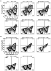
a, SEC of multimers. b,c, BLI of minibinder and knockout monomers (b) and multimers (c). d, Gating strategy for measuring GFP expression in HEK293-TLR3hi cells.
Fig. 1:
Fig. 1:. Computational design of TLR3 minibinders.
a, Left, TLR3 is natively dimerized and activated by dsRNA (PDB ID: 7WV5). Right, two hydrophobic patches on the TLR3 monomer were targeted for minibinder design (PDB ID: 1ZIW). b, Polyvaline scaffolds were used in the RifDock pipeline to design de novo miniprotein binders. Several structural metrics were used as filters to select 23,789 designs for experimental screening. c, Binders were identified using yeast surface display. During Sort 5, binding was clearly observed at 150 nM receptor, but approached levels of background signal at 100 nM.
Fig. 2:
Fig. 2:. Biochemical characterization and affinity maturation of lead TLR3 minibinders.
a, Design models of minibinders 7 (yellow) and 8 (purple) in complex with TLR3 (gray). The details of the predicted interface are shown at right. b, Size exclusion chromatograms of each minibinder on a Superdex 75 10/300 GL. c, Affinity determination for each minibinder by BLI. The concentrations of hTLR3 used are listed. The black lines represent experimental data and the colored lines represent fits. KD values are given. d, Site saturation mutagenesis heat maps of interface residues. The originally designed amino acid at each position is provided at the bottom and in the white square. Red indicates affinity improvement and blue indicates affinity reduction. e, Biolayer interferometry of affinity-matured minibinders 7.1 and 8.6. KD values are given. f, CD of affinity-matured constructs at various temperatures. Solid line, 25°C; dashed line, 95°C; dotted line, 95°C followed by 25°C.
Fig. 3:
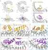
Fig. 3:. Structural characterization of TLR3 minibinders.
a, Two different views of the cryo-EM structures of human TLR3 in complex with minibinder 7.7 (left) or minibinder 8.6 (right). TLR3, glycans, minibinder 7.7, and minibinder 8.6 are in gray, brown, yellow, and purple, respectively. b, Comparison of experimental and AlphaFold2-predicted structures of the minibinders in complex with TLR3. c-h, Close-up views of key molecular interactions in TLR3/minibinder 7.7 (c-e) or TLR3/minibinder 8.6 (f-h). Each box is a close-up view of the same colored box in a. Residues involved in the TLR3/minibinder interaction are displayed as sticks and labeled.
Fig. 4:
Fig. 4:. Multimerization of minibinders leads to NF-κB activation.
a, Minibinder tetramers were generated by fusing four tandem repeats of the minibinder together using 16-residue (GlySer) linkers. b, Antiparallel 8.6 dimers were generated by fusing minibinder 8.6 to the C terminus of an antiparallel coiled-coil derived from myosin 10. Two views of a model of the 8.6 dimer in complex with hTLR3 are shown. c, TLR3 is expressed on the cell surface of TLR3hi cells, which express an NF-κB-linked GFP reporter. d, Histograms showing GFP signal in stimulated TLR3hi cells. The identities and concentrations of the stimuli are provided on the right and left sides of each panel, respectively. e, Mean fluorescence intensity (MFI) values from assay duplicates. Concentrations are provided in d.

Similar articles

References

    1. Taro Kawai S. A. Pathogen recognition with Toll-like receptors. Curr. Opin. Immunol. 17, 338–344 (2005). - PubMed
    1. Medzhitov R., Preston-Hurlburt P. & Janeway C. A. A human homologue of the Drosophila Toll protein signals activation of adaptive immunity. Nature 388, 394–397 (1997). - PubMed
    1. Akira S., Takeda K. & Kaisho T. Toll-like receptors: critical proteins linking innate and acquired immunity. Nat. Immunol. 2, 675–680 (2001). - PubMed
    1. Kim H. M. et al. Crystal Structure of the TLR4-MD-2 Complex with Bound Endotoxin Antagonist Eritoran. Cell 130, 906–917 (2007). - PubMed
    1. Bell J. K. et al. The molecular structure of the Toll-like receptor 3 ligand-binding domain. Proc. Natl. Acad. Sci. U. S. A. 102, 10976–10980 (2005). - PMC - PubMed

Publication types