. 2024 Feb 21;5(2):101827.
doi: 10.1016/j.xcrp.2024.101827. Epub 2024 Feb 12.

Neutron diffraction from a microgravity-grown crystal reveals the active site hydrogens of the internal aldimine form of tryptophan synthase

Affiliations

Neutron diffraction from a microgravity-grown crystal reveals the active site hydrogens of the internal aldimine form of tryptophan synthase

Victoria N Drago et al. Cell Rep Phys Sci. .

Abstract

Pyridoxal 5'-phosphate (PLP), the biologically active form of vitamin B6, is an essential cofactor in many biosynthetic pathways. The emergence of PLP-dependent enzymes as drug targets and biocatalysts, such as tryptophan synthase (TS), has underlined the demand to understand PLP-dependent catalysis and reaction specificity. The ability of neutron diffraction to resolve the positions of hydrogen atoms makes it an ideal technique to understand how the electrostatic environment and selective protonation of PLP regulates PLP-dependent activities. Facilitated by microgravity crystallization of TS with the Toledo Crystallization Box, we report the 2.1 Å joint X-ray/neutron (XN) structure of TS with PLP in the internal aldimine form. Positions of hydrogens were directly determined in both the α- and β-active sites, including PLP cofactor. The joint XN structure thus provides insight into the selective protonation of the internal aldimine and the electrostatic environment of TS necessary to understand the overall catalytic mechanism.


Conflict of interest statement

DECLARATION OF INTERESTS The authors declare no competing interests.

Figures

Figure 1.
Figure 1.. Biochemistry of PLP
(A) Pyridoxal 5’-phosphate (PLP) forms an internal aldimine with the ε-amine of an active site lysine (Lys87 in TS) through a Schiff base linkage, and the amino acid substrate forms a Michaelis complex. (B) Electronic overlap in the external aldimine promotes the deprotonation of Cα by the ε-amine of the released active site lysine. Indicated are the reaction specificities of PLP-dependent enzymes including transamination, decarboxylation, racemization, β- and γ-elimination (TS is β-elimination), replacement, and retro-aldol cleavage. (C) The natural substrate for TS is indole-3-glycerol phosphate (IGP).
Figure 2.
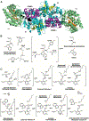
Figure 2.. Salmonella typhimurium TS is an abba linearly arranged heterotetramer
In the α-site reaction, indole-3-glycerol phosphate (IGP) is cleaved into indole and glyceraldehyde-3- phosphate. In the β-site reaction, indole is coupled to PLP-activated serine to form tryptophan. (A) Ribbon model of the αββα heterotetramer. In the α-domain (green), the core β-strands (orange) have a distinctive TIM barrel-like fold with IGP (green circle) bound in the active site (PDB: 1A5B). The b-domain (cyan) has two sub-domains. The large sub-domain forms contacts with the α-domain, the binding pocket for a monovalent cation (gray sphere), and the loops connecting the C termini of the core parallel b-strands (yellow) to the intervening a helices form the PLP binding site. A 25 Å hydrophobic, intramolecular tunnel connects the α- and β-active sites. The smaller, more flexible “COMM” β-subdomain (purple) forms the top of the channel and participates in interdomain communication. (B) The TS α-active site catalyzes the cleavage of IGP into glyceraldehyde 3-phosphate and indole. When protonated, Glu49 forms hydrogen bonds with water molecules near Tyr173 (left). Shown is the assumed intermediate (center) when IGP binds, in which Glu49 delivers a proton to indole C3 (red) and aligns to deprotonate the 3′ hydroxy of IGP (blue). Asp60 is positioned to form a short hydrogen bond to the protonated indole N1. The ensuing retro-aldol cleavage involves adding a proton to indole C3 (red) and yielding glyceraldehyde-3-phosphate and indole. The presence of glyceraldehyde-3-phosphate in the active site and release of indole initiates conformational changes, primarily in loop αL6, allowing indole to travel through the hydrophobic channel, which is coordinated to the activation of serine in the β-site. (C) In the β-reaction, L-Ser is first shown in the Michaelis complex near the PLP-Lys87 internal aldimine. Transimination through an intermediate gem-diamine forms the Ser-PLP external aldimine and a neutral Lys87. As the Ser-PLP external aldimine shifts into a more stable conformation, the neutral ε-amine of Lys87 is repositioned near the Ser-PLP Cα proton. Electronic overlap within the external aldimine coordinates the Cα for deprotonation by Lys87, acting as a general base, and the rearrangement of the transient carbanionic/quinonoid intermediate causes β-elimination of water (HOH). In the lower panel, from right to left, formation of the metastable α-aminoacrylate intermediate coincides with the arrival of indole transported through the intramolecular channel. Indole, positioned as a Michaelis complex, promotes coupling to the Cβ of the aminoacrylate through a second carbanionic intermediate. Lys87 then reprotonates the Cα position and the L-Trp external aldimine is produced. L-Trp is released through a gem-diamine intermediate regenerating the internal aldimine.
Figure 3.
Figure 3.. Observed dual positions of a-active site residue Glu49
Glu49a is the active position oriented toward Tyr175 hydrogen bonded to water W1 in which Glu49 can deliver a proton to the indole C3 and abstract a proton from IGP 3′-OH. Glu49b is in a hydrogen bonding network with two water molecules (W2 and W3) reinforced by residues Tyr173 and Tyr4 and is the suspected reservoir for the proton delivered to indole C3. Not shown in this view, W2 is linked to Ser125, and W3 has a short 2.5 Å (D–O 1.7 Å) H-bond to the phenolic oxygen of Tyr4. The 2| FO|-|FC| neutron scattering length density is shown in wheat and the electron density in blue mesh. (A) The room temperature X-ray model shows both Glu49a (60%) and b (40%) conformers. (B) The room temperature neutron model shows only Glu49a, and deuteron-donor H-bonding distances are shown in parentheses. (C) The cryo X-ray model shows Glu49b becoming the dominant conformer. (D) The low-pH, room temperature X-ray model shows Glu49a becoming dominant, suggesting Glu49 is protonated.
Figure 4.
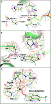
Figure 4.. β-Active site perspectives from a TS neutron structure
The 2|FO|-|FC| neutron scattering length density map is depicted in wheat mesh contoured at 1 σ, and the omit |FO|-|FC| neutron scattering length density is shown in purple mesh contoured at 2.2 σ. Hydrogen bonding distances between heavy atoms are shown with the deuteron-donor distances in parentheses. (A) The Schiff base nitrogen, NSB, is protonated and hydrogen bonded to the phenolic oxygen,O3′. The 2|FO|-|FC| electron density is shown in blue mesh. (B) Ser377 prevents protonation of pyridine nitrogen, N1, and is stabilized by an additional hydrogen bond with Ser351. His86 is neutral and monoprotonated on the ε-nitrogen, positioned above the cofactor. (C) The glycine-rich phosphate binding loop is composed of Gly232, Gly233, Gly234, Ser235, Asn236, and two crystallographic waters, as well as His86 and Thr109 (not shown for clarity).
Figure 5.
Figure 5.. Potential energy profile for proton transfer between PLP-NSB and PLP-O3′
The calculated energy barrier height for the intramolecular proton transfer from the Schiff base to the phenolic oxygen is reported relative to the NSB-protonated (reactant) structure. The energies for the reactant and product are denoted with blue circles, while the calculated transition state energy (‡) is signified with a red circle.

Similar articles

Cited by

References

    1. Percudani R, and Peracchi A (2003). A genomic overview of pyridoxal-phosphate-dependent enzymes. EMBO Rep. 4, 850–854. 10.1038/sj.embor.embor914. - DOI - PMC - PubMed
    1. Jansonius JN (1998). Structure, evolution and action of vitamin B6-dependent enzymes. Curr. Opin. Struct. Biol. 8, 759–769. 10.1016/s0959-440x(98)80096-1. - DOI - PubMed
    1. Eliot AC, and Kirsch JF (2004). Pyridoxal phosphate enzymes: mechanistic, structural, and evolutionary considerations. Annu. Rev. Biochem. 73, 383–415. 10.1146/annurev.biochem.73.011303.074021. - DOI - PubMed
    1. Amadasi A, Bertoldi M, Contestabile R, Bettati S, Cellini B, di Salvo ML, Borri-Voltattorni C, Bossa F, and Mozzarelli A (2007). Pyridoxal 5’-phosphate enzymes as targets for therapeutic agents. Curr. Med. Chem. 14, 1291–1324. 10.2174/092986707780597899. - DOI - PubMed
    1. Graber R, Kasper P, Malashkevich VN, Strop P, Gehring H, Jansonius JN, and Christen P (1999). Conversion of aspartate aminotransferase into an L-aspartate beta-decarboxylase by a triple active-site mutation. J. Biol. Chem. 274, 31203–31208. 10.1074/jbc.274.44.31203. - DOI - PubMed