. 2024 Apr 19;20(4):e1012175.
doi: 10.1371/journal.ppat.1012175. eCollection 2024 Apr.

Sensitive detection of pathological seeds of α-synuclein, tau and prion protein on solid surfaces

Affiliations

Sensitive detection of pathological seeds of α-synuclein, tau and prion protein on solid surfaces

Christina D Orrú et al. PLoS Pathog. .

Abstract

Prions or prion-like aggregates such as those composed of PrP, α-synuclein, and tau are key features of proteinopathies such as prion, Parkinson's and Alzheimer's diseases, respectively. Their presence on solid surfaces may be biohazardous under some circumstances. PrP prions bound to solids are detectable by ultrasensitive real-time quaking-induced conversion (RT-QuIC) assays if the solids can be immersed in assay wells or the prions transferred to pads. Here we show that prion-like seeds can remain detectable on steel wires for at least a year, or even after enzymatic cleaning and sterilization. We also show that contamination of larger objects with pathological seeds of α-synuclein, tau, and PrP can be detected by simply assaying a sampling medium that has been transiently applied to the surface. Human α-synuclein seeds in dementia with Lewy bodies brain tissue were detected by α-synuclein RT-QuIC after drying of tissue dilutions with concentrations as low as 10-6 onto stainless steel. Tau RT-QuIC detected tau seeding activity on steel exposed to Alzheimer's disease brain tissue diluted as much as a billion fold. Prion RT-QuIC assays detected seeding activity on plates exposed to brain dilutions as extreme as 10-5-10-8 from prion-affected humans, sheep, cattle and cervids. Sampling medium collected from surgical instruments used in necropsies of sporadic Creutzfeldt-Jakob disease-infected transgenic mice was positive down to 10-6 dilution. Sensitivity for prion detection was not sacrificed by omitting the recombinant PrP substrate from the sampling medium during its application to a surface and subsequent storage as long as the substrate was added prior to performing the assay reaction. Our findings demonstrate practical prototypic surface RT-QuIC protocols for the highly sensitive detection of pathologic seeds of α-synuclein, tau, and PrP on solid objects.


Conflict of interest statement

BC, CDO, BG and AH are inventors on patent applications pertaining to RT-QuIC technology. The other authors have declared that no competing interests exist. Mention of trade names or commercial products in this article is solely for the purpose of providing specific information and does not imply recommendation or endorsement by the US Government.

Figures

Fig 1
Fig 1. RT-QuIC detection of 263K prion seeding activity on stainless steel wires after 1 year.
Stainless-steel wires were coated with 10−3 brain equivalents of 263K or uninfected brain tissue, dried, and inserted in individual wells for RT-QuIC analysis (one wire per well). One set of wires was tested after drying for 24 h (263K, orange; uninfected, black; n = 3); another set (263K only) was tested after 1 y of storage at room temperature (blue; n = 3). ThT fluorescence for each reaction is plotted vs. time.
Fig 2
Fig 2. sfRT-QuIC experimental design schematic.
Panel A. Spotting of surfaces with serially diluted brain homogenate (BH, yellow liquid; BE: brain equivalents) followed by sfRT-QuIC testing of sampling medium (SM; blue liquid). Panel B. Spotting of surfaces with 10% BH followed by serial dilutions of the SM for sfRT-QuIC. Panel C. sfRT-QuIC of surgical instruments contaminated with CJD-infected humanized mouse brain tissue: sampling procedure and serial dilutions of the SM for sfRT-QuIC. Created with BioRender.com.
Fig 3
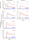
Fig 3. sfRT-QuIC of stainless steel- or acrylic-bound 263K, CWD, or uninfected brain homogenates.
Panel A-B. sfRT-QuIC of SM collected from a single spot from 263K (red, full line) or uninfected (blue, full line) BH (10−4 dilution) dried onto stainless steel (A) or acrylic (B) surfaces or in solution (A; red and blue dashed lines). Panel C-D. sfRT-QuIC of a 2 mL SM collection from 8 263K (red) or uninfected (blue) spottings placed in a random pattern within a test circle on stainless steel (C) or acrylic (D). Each collection was tested in quadruplicate. Panel E-F. sfRT-QuIC of SM collected as in C,D from spottings of CWD (red) or uninfected (blue) BH (10−4 dilutions) from either deer (E) or elk (F). The recombinant PrP substrates used are described in Methods.
Fig 4
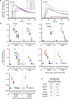
Fig 4. sfRT-QuIC detection of α-synuclein and tau seeding activity from brain homogenates.
Panel A. Designated dilutions of DLB or corticobasal degeneration (CBD, a non-synucleinopathy control) tested by solution or sfRT-QuIC. Curves are averages of 4 replicate reactions. Y-axis indicates mean ThT fluorescence from replicate wells and x-axis assay duration (h). Panel B 1/time to fluorescence positivity threshold for each DLB and CBD reaction versus the dilution (log10 tissue equivalents). The dotted horizontal line represents the inverse of the time of the final measurement. The dashed vertical line separates solution vs. surface data. The mean (heavy horizontal lines) and standard deviations (vertical) are displayed for each dilution tested. Panel C. Designated dilutions of sAD or cerebrovascular disease (CVD; a low tau seed control) BH tested by solution and sfRT-QuIC. Curves as in A. Panel D. 1/time to threshold for each reaction seeded with brain homogenate at the designated tissue dilutions from sAD, Tau KO (a tau-negative control), or CVD (a low, but not completely negative, tau seed control) with labeling as in B.
Fig 5
Fig 5. sfRT-QuIC detection of seeding activity from different strains of prion-infected brain homogenates.
Panel A. Designated dilutions of sCJD (MM1 type) and sAD (prion negative control) BHs tested by solution (left) or sfRT-QuIC (right). Curves are show mean ThT fluorescence from quadruplicate wells versus assay duration. Panel B. 1/time to threshold comparisons of solution and sfRT-QuIC data from dilutions of sCJD (MM1 subtype; extracted from data shown in A), genetic Creutzfeldt-Jakob disease (gCJD; E200K), Gerstmann-Sträussler-Scheinker disease (GSS; P102L), ovine classical scrapie and CWD and uninfected controls (Ctrl) BHs. X-axis indicates the dilution (log10 tissue equivalents). Panel C. Table of Log SD50/mg original tissue values estimated using Spearman-Karber analysis of the data shown in Panel B as described in Methods. Comparable results were seen in analogous experiments performed with sCJD, ovine classical scrapie and CWD, except for the inclusion of 0.1% SDS in the brain homogenate diluent (data available on request).
Fig 6
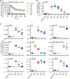
Fig 6. sfRT-QuIC of stainless-steel bound brain homogenate derived human, cervid, ovine, and bovine prions.
Panel A. sfRT-QuIC of diluted SM collections from sCJD (MM1 type) or Alzheimer’s disease (as a prion-negative control) spots on a stainless-steel plate: average ThT fluorescence of 4 replicate wells versus reaction time. Panel B. sfRT-QuIC results from panel A represented as 1/time to threshold for each reaction seeded with the designated dilutions (log10 tissue equivalents; x-axis) as described initially in legend of Fig 4B and 4D. Panel C. Analogous data from plates contaminated with 10% BH from humans with variant Creutzfeldt-Jakob disease (vCJD), fatal familial insomnia (FFI), genetic Creutzfeldt-Jakob disease (gCJD; E200K), and Gerstmann-Sträussler-Scheinker disease (GSS; P102L); cervids with CWD; sheep with scrapie; cows with classical (C-), high- (H-) and low- (L-) BSE; and species-matched uninfected controls.
Fig 7
Fig 7. sfRT-QuIC detection of prions bound to stainless-steel surgical instruments after use on sCJD-infected Tg66 humanized mice.
sfRT-QuIC data from serially diluted SM collected from forceps and scissors contaminated with brain tissue during routine surgery represented as 1/time to threshold for each reaction versus SM dilution as otherwise marked as in Fig 4B and 4D.
Fig 8
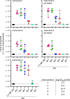
Fig 8. Effect of sterilization treatment of wires contaminated with sAD, DLB, and Parkinson’s disease (PD)-associated seeds.
BH (10% w/v)-treated stainless steel wires tested by α-syn RT-QuICR for DLB, PD, as well as a CBD non-synucleinopathy control (Panel A) or K12 Tau RT-QuIC for sAD, as well as tau KO and CVD non-tauopathy controls (Panel B). Data points show 1/time to threshold values for untreated (triangles) and treated (circles) wires. Other markings as in Fig 4B and 4D. Asterisk (*) denotes a statistically significant difference (p<0.05) as analyzed by One-Way ANOVA; ns means non-significant.

Similar articles

Cited by

References

    1. Scheres SHW, Ryskeldi-Falcon B, Goedert M. Molecular pathology of neurodegenerative diseases by cryo-EM of amyloids. Nature. 2023;621(7980):701–10. doi: 10.1038/s41586-023-06437-2 - DOI - PubMed
    1. Collinge J. Mammalian prions and their wider relevance in neurodegenerative diseases. Nature. 2016;539(7628):217–26. doi: 10.1038/nature20415 - DOI - PubMed
    1. Caughey B, Kraus A. Transmissibility versus Pathogenicity of Self-Propagating Protein Aggregates. Viruses. 2019;11(11). doi: 10.3390/v11111044 - DOI - PMC - PubMed
    1. Kraus A, Groveman BR, Caughey B. Prions and the potential transmissibility of protein misfolding diseases. Ann Rev Microbiol. 2013;67:543–64. doi: 10.1146/annurev-micro-092412-155735 - DOI - PMC - PubMed
    1. Soto C, Pritzkow S. Protein misfolding, aggregation, and conformational strains in neurodegenerative diseases. Nat Neurosci. 2018;21(10):1332–40. doi: 10.1038/s41593-018-0235-9 - DOI - PMC - PubMed

Publication types