. 2024 Apr 16;43(1):115.
doi: 10.1186/s13046-024-03041-8.

CCAAT enhancer binding protein delta activates vesicle associated membrane protein 3 transcription to enhance chemoresistance and extracellular PD-L1 expression in triple-negative breast cancer

Affiliations

CCAAT enhancer binding protein delta activates vesicle associated membrane protein 3 transcription to enhance chemoresistance and extracellular PD-L1 expression in triple-negative breast cancer

Yan Zhao et al. J Exp Clin Cancer Res. .

Abstract

Background: Chemoresistance and immunosuppression are two major obstacles in the current anti-cancer treatments. This study investigates the involvements of a CCAAT enhancer binding protein delta (CEBPD)/vesicle associated membrane protein 3 (VAMP3) axis in paclitaxel (PTX) resistance and immune evasion in triple-negative breast cancer (TNBC).

Methods: PTX resistance-related genes were screened by bioinformatics. CEBPD and VAMP3 expression in clinical TNBC samples was examined by immunohistochemistry. Three PTX-resistant TNBC cell lines (MDA-MB-231/PTX, MDA-MB-468/PTX and MDA-MB-453/PTX) were generated, and their drug resistance was analyzed. Autophagy of cells was analyzed by immunofluorescence staining. Interaction between CEBPD and VAMP3 promoter was identified by immunoprecipitation and luciferase assays. The extracellular expression of programmed cell death-ligand 1 (PD-L1) in TNBC cells was detected. Extracellular vesicles (EVs) from TNBC cells were isolated to examine their effects on CD8+ T cell exhaustion.

Results: CEBPD and VAMP3 were upregulated in chemo-resistant tissue samples and in PTX-resistant TNBC cells. The CEBPD downregulation enhanced PTX sensitivity of cells. However, further upregulation of VAMP3 in cells restored PTX resistance, which was likely due to the activation of autophagy, as the autophagy antagonist chloroquine enhanced PTX sensitivity of cells. CEBPD was found to bind to the VAMP3 promoter to activate its transcription. The CEBPD/VAMP3 axis also increased the PD-L1 expression in the conditioned medium of TNBC cells. The TNBC cell-derived EVs increased the exhaustion of co-cultured CD8+ T cells.

Conclusion: This study provides novel evidence that CEBPD plays a key role in enhancing PTX resistance in TNBC cells across various subtypes through VAMP3-mediated autophagy activation. Additionally, the CEBPD/VAMP3 axis also increases extracellular PD-L1 level, delivered by cancer cell-derived EVs, to suppress CD8+ T cell-mediated anti-tumor immune response. These significant observations may provide new insights into the treatment of TNBC, suggesting CEBPD and VAMP3 as promising targets to overcome treatment resistance.

Keywords: Autophagy; CEBPD; Immune evasion; Paclitaxel resistance; Triple-negative breast cancer; VAMP3.


Conflict of interest statement

The authors declare that they have no competing interests.

Figures
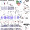
Fig. 1
Fig. 1
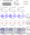
CEBPD is highly expressed in PTX-resistant TNBC cells. A volcano plots for DEGs identified using GSE28784 and GSE90564 datasets; B intersections between the two sets of DEGs and human transcription factors; C correlations between CEBPD and HEY1 (HESR-1) and OS of breast cancer analyzed by Kaplan–Meier Plotter analysis; D expression of CEBPD in biopsy tissues of SD/PD (n = 18) and CR/PR (n = 23) patients measured by IHC assay; E viability of parental (MDA-MB-231, MDA-MB-468, and MDA-MB-453) and PTX-resistant (MDA-MB-231/PTX, MDA-MB-468/PTX, and MDA-MB-453/PTX) cells and the PTX IC50 examined by CCK-8 assay (n = 3). Parental and PTX-resistant TNBC cells were treated with a fixed dose of 15 nM for 48 h for subsequent use. F colony formation ability of parental and PTX-resistant TNBC cells after PTX treatment (n = 3); G apoptosis of parental and PTX-resistant TNBC cells after PTX treatment determined by flow cytometry (n = 3); H migration and invasion of parental and PTX-resistant TNBC cells after PTX treatment determined by flow cytometry (n = 3); I CEBPD expression in parental and PTX-resistant TNBC cells measured by WB analysis (n = 3). Differences were analyzed by the t-test (D) or one-way ANOVA with Tukey's post-hoc test (F-I). IC50 of PTX in cells was analyzed by nonlinear fitting analysis (E). *p < 0.05
Fig. 2
Fig. 2
Knockdown of CEBPD enhances PTX sensitivity of TNBC cells. MDA-MB-231/PTX, MDA-MB-468/PTX, MDA-MB-453/PTX cells were administered lentivirus-carried sh-CEBPD or sh-NC. A CEBPD protein level in cells determined by WB analysis (n = 3); B viability of cells and the IC50 of PTX examined by CCK-8 assay (n = 3). Stably transfected cells were treated with 15 nM PTX for 48 h. C colony formation ability of cells analyzed by colony formation assay (n = 3); D apoptosis of cells determined by flow cytometry (n = 3); E migration and invasion of cells determined by Transwell assay (n = 3). Differences were analyzed by two-way ANOVA with Tukey's post-hoc test (A, C-E). IC50 of PTX to cells was analyzed by nonlinear fitting analysis (B). *p < 0.05
Fig. 3
Fig. 3
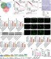
Knockdown of CEBPD suppresses VAMP3 transcription and cell autophagy. A intersections of top 1,000 candidate CEBPD targets with DEGs screened from GSE28784 and GSE90564 datasets; B correlations between CEBPD and HEY1 and OS of breast cancer analyzed by Kaplan–Meier Plotter analysis; C expression of VAMP3 in biopsy tissues of SD/PD (n = 18) and CR/PR (n = 23) patients measured by IHC assay. Parental or PTX-resistant TNBC cells were administered lentivirus-carried sh-CEBPD or sh-NC. D mRNA and protein levels of VAMP3 in cells detected by qPCR and WB analysis (n = 3); E autophagy activity in cells analyzed by immunofluorescence staining of LC3 (n = 3); F putative binding relationship and binding sites between CEBPD and VAMP3 promoter; G binding of CEBPD with VAMP3 promoter validated by ChIP-qPCR assay (n = 3); H regulatory role of CEBPD in VAMP3 transcription in 293 T cells with sh-NC or sh-CEBPD transfection determined by luciferase assay (H) (n = 3). Differences were analyzed by the t-test (C), one-way ANOVA followed by Tukey's multiple comparisons test (D-E) or by the two-way ANOVA followed by Sidak's post-hoc test (G-H). *p < 0.05
Fig. 4
Fig. 4
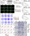
VAMP3 overexpression induces autophagy and reduces PTX sensitivity of TNBC cells. MDA-MB-231/PTX, MDA-MB-468/PTX, and MDA-MB-453/PTX cells stably transfected with sh-CEBPD were further administered oe-VAMP3. A mRNA and protein levels of VAMP3 in cells determined by qPCR and WB assays (n = 3). These cells were further treated with the autophagy antagonist CQ. B autophagy activity in cells analyzed by immunofluorescence staining of LC3; C viability of cells and the IC50 of PTX examined by CCK-8 assay (n = 3). These cells were additionally treated with 15 nM PTX for 48 h, D colony formation ability of cells analyzed by colony formation assay (n = 3); E apoptosis of cells determined by flow cytometry (n = 3); F migration and invasion of cells determined by Transwell assay (n = 3). Differences were analyzed by two-way ANOVA, followed by Tukey's (B, D-F) or Sidak's (A) post-hoc tests. IC50 of PTX to cells was analyzed by nonlinear fitting analysis (C). *p < 0.05
Fig. 5
Fig. 5
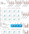
CEBPD/VAMP3 increases extracellular PD-L1 level in PTX-resistant TNBC cells. A expression of PD-L1 in biopsy tissues of SD/PD (n = 18) and CR/PR (n = 23) patients measured by IHC assay; B concentration of PD-L1 in the culture supernatant of sh-CEBPD- or sh-CEBPD + oe-VAMP3-treated PTX-resistant TNBC cells detected by ELISA kit (n = 3); C morphology of the isolated EVs under transmission electron microscopy; D particle size distribution and concentration of the isolated EVs determined by nanoparticle tracking analysis (n = 3). Differences were analyzed by the t-test (A) or two-way ANOVA with Tukey's post-hoc test (B, D). *p < 0.05
Fig. 6
Fig. 6
CEBPD/VAMP3 affects the cytotoxicity of CD8+ T cells to the PTX-resistant TNBC cells. EVs derived from differentially treated (sh-NC, sh-CEBPD, sh-CEBPD + oe-NC, and sh-CEBPD + oe-VAMP3) PTX-resistant cells were collected, which were co-cultured with activated CD8+ T cells for 48 h. A, concentrations of IFN-γ, Granzyme B, and Perforin in the supernatant of co-culture system examined using ELISA kits (n = 3); B, proportion of exhausted CD8+ T cells (Tim3+CD8+) analyzed by flow cytometry (n = 3). Differentially treated MDA-MB-231/PTX cells were injected into C57BL/6 J mice. When the tumor volume reached 100 mm3, PTX treatment was applied at 20 mg/kg via intraperitoneal injection twice a week. C, growth rate of xenograft tumors in mice (n = 5); D, weight of xenograft tumors on day 28 (n = 5); E, proportion of exhausted CD8+ T cells (Tim3+CD8.+) in the tumor-infiltrating lymphocytes analyzed by flow cytometry (n = 5). Differences were analyzed by the one-way (D-E) or two-way (A-C) ANOVA with Tukey's post-hoc test (B, D). *p < 0.05
Fig. 7
Fig. 7
Graphical abstract. In PTX-resistant TNBC cells, elevated CEBPD expression promotes transcription and expression of VAMP3, which increases formation of autophagosomes to aggravate chemoresistance. Moreover, VAMP3 increases the secretion of PD-L1 to induce immunosuppression

Similar articles

Cited by

References

    1. Sung H, Ferlay J, Siegel RL, Laversanne M, Soerjomataram I, Jemal A, et al. Global Cancer Statistics 2020: GLOBOCAN Estimates of Incidence and Mortality Worldwide for 36 Cancers in 185 Countries. CA Cancer J Clin. 2021;71(3):209–249. doi: 10.3322/caac.21660. - DOI - PubMed
    1. Yin L, Duan JJ, Bian XW, Yu SC. Triple-negative breast cancer molecular subtyping and treatment progress. Breast Cancer Res. 2020;22(1):61. doi: 10.1186/s13058-020-01296-5. - DOI - PMC - PubMed
    1. Tang Y, Tian W, Zheng S, Zou Y, Xie J, Zhang J, et al. Dissection of FOXO1-Induced LYPLAL1-DT Impeding Triple-Negative Breast Cancer Progression via Mediating hnRNPK/beta-Catenin Complex. Research (Wash D C) 2023;6:0289. - PMC - PubMed
    1. Li S, Bao C, Huang L, Wei JF. Current Therapeutic Strategies for Metastatic Triple-Negative Breast Cancer: From Pharmacists' Perspective. J Clin Med. 2022;11(20):6021. doi: 10.3390/jcm11206021. - DOI - PMC - PubMed
    1. Qi Y, Fu X, Xiong Z, Zhang H, Hill SM, Rowan BG, et al. Methylseleninic acid enhances paclitaxel efficacy for the treatment of triple-negative breast cancer. PLoS ONE. 2012;7(2):e31539. doi: 10.1371/journal.pone.0031539. - DOI - PMC - PubMed

MeSH terms