

Imaging extrachromosomal DNA (ecDNA) in cancer
- PMID: 38625562
- PMCID: PMC7616135
- DOI: 10.1007/s00418-024-02280-2
Imaging extrachromosomal DNA (ecDNA) in cancer
Abstract
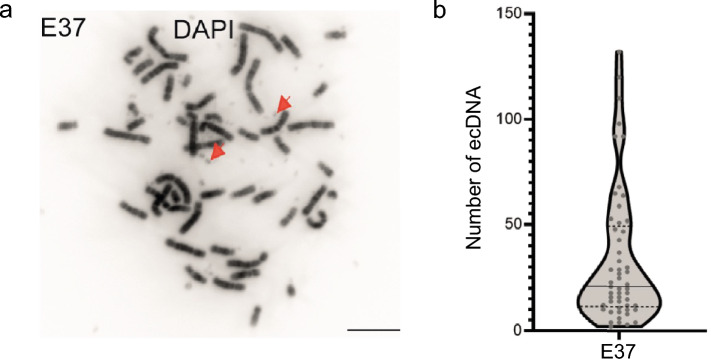
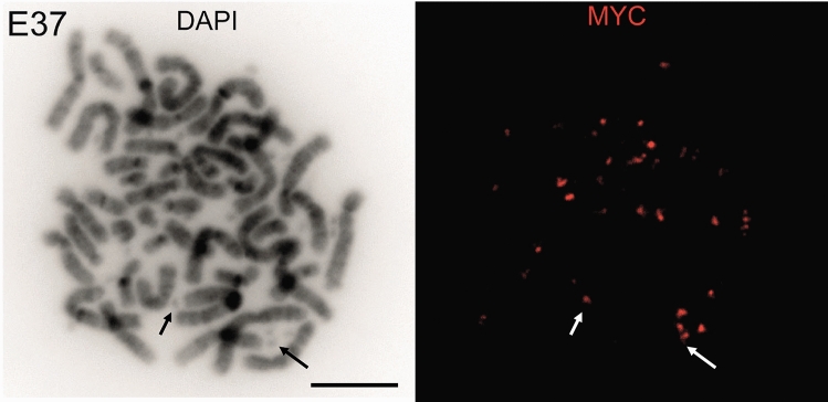
Extrachromosomal DNA (ecDNA) are circular regions of DNA that are found in many cancers. They are an important means of oncogene amplification, and correlate with treatment resistance and poor prognosis. Consequently, there is great interest in exploring and targeting ecDNA vulnerabilities as potential new therapeutic targets for cancer treatment. However, the biological significance of ecDNA and their associated regulatory control remains unclear. Light microscopy has been a central tool in the identification and characterisation of ecDNA. In this review we describe the different cellular models available to study ecDNA, and the imaging tools used to characterise ecDNA and their regulation. The insights gained from quantitative imaging are discussed in comparison with genome sequencing and computational approaches. We suggest that there is a crucial need for ongoing innovation using imaging if we are to achieve a full understanding of the dynamic regulation and organisation of ecDNA and their role in tumourigenesis.
Keywords: Double-minute; Fluorescence in situ hybridisation; Homogeneously staining regions; Oncogene; Transcription hubs.
© 2024. The Author(s).
Figures
Similar articles
-
Extrachromosomal DNA in Cancer.Annu Rev Genomics Hum Genet. 2022 Aug 31;23:29-52. doi: 10.1146/annurev-genom-120821-100535. Epub 2022 May 24. Annu Rev Genomics Hum Genet. 2022. PMID: 35609926 Free PMC article. Review.
-
Live-Cell Imaging Shows Uneven Segregation of Extrachromosomal DNA Elements and Transcriptionally Active Extrachromosomal DNA Hubs in Cancer.Cancer Discov. 2022 Feb;12(2):468-483. doi: 10.1158/2159-8290.CD-21-1376. Epub 2021 Nov 24. Cancer Discov. 2022. PMID: 34819316 Free PMC article.
-
Extrachromosomal DNA: An Emerging Hallmark in Human Cancer.Annu Rev Pathol. 2022 Jan 24;17:367-386. doi: 10.1146/annurev-pathmechdis-051821-114223. Epub 2021 Nov 9. Annu Rev Pathol. 2022. PMID: 34752712 Free PMC article. Review.
-
Guilt by association: EcDNA as a mobile transactivator in cancer.Trends Cancer. 2022 Sep;8(9):747-758. doi: 10.1016/j.trecan.2022.04.011. Epub 2022 Jun 23. Trends Cancer. 2022. PMID: 35753910 Free PMC article. Review.
-
Novel insights into extrachromosomal DNA: redefining the onco-drivers of tumor progression.J Exp Clin Cancer Res. 2020 Oct 12;39(1):215. doi: 10.1186/s13046-020-01726-4. J Exp Clin Cancer Res. 2020. PMID: 33046109 Free PMC article. Review.
Cited by
-
Seeing genomes.Histochem Cell Biol. 2024 Jul;162(1-2):1-2. doi: 10.1007/s00418-024-02301-0. Histochem Cell Biol. 2024. PMID: 38850309 No abstract available.
References
-
- Alitalo K, Schwab M, Lin CC, Varmus HE, Bishop JM. Homogeneously staining chromosomal regions contain amplified copies of an abundantly expressed cellular oncogene (c-MYC) in malignant neuroendocrine cells from a human colon carcinoma. Proc Natl Acad Sci U S A. 1983;80:1707–1711. doi: 10.1073/pnas.80.6.1707. - DOI - PMC - PubMed
-
- Balaban-Malenbaum G, Gilbert F. The proposed origin of double minutes from homogeneously staining region (HSR)-marker chromosomes in human neuroblastoma hybrid cell lines. Cancer Genet Cytogenet. 1980;2:339–348. doi: 10.1016/0165-4608(80)90065-5. - DOI
Publication types
MeSH terms
Substances
Grants and funding
LinkOut - more resources
Full Text Sources
