. 2024 May 7;92(5):e0006024.
doi: 10.1128/iai.00060-24. Epub 2024 Apr 15.

Flagellin-modulated inflammasome pathways characterize the human alveolar macrophage response to Burkholderia pseudomallei, a lung-tropic pathogen

Affiliations

Flagellin-modulated inflammasome pathways characterize the human alveolar macrophage response to Burkholderia pseudomallei, a lung-tropic pathogen

Lara Lovelace-Macon et al. Infect Immun. .

Abstract

Melioidosis is an emerging tropical infection caused by inhalation, inoculation, or ingestion of the flagellated, facultatively intracellular pathogen Burkholderia pseudomallei. The melioidosis case fatality rate is often high, and pneumonia, the most common presentation, doubles the risk of death. The alveolar macrophage is a sentinel pulmonary host defense cell, but the human alveolar macrophage in B. pseudomallei infection has never been studied. The objective of this study was to investigate the host-pathogen interaction of B. pseudomallei infection with the human alveolar macrophage and to determine the role of flagellin in modulating inflammasome-mediated pathways. We found that B. pseudomallei infects primary human alveolar macrophages but is gradually restricted in the setting of concurrent cell death. Electron microscopy revealed cytosolic bacteria undergoing division, indicating that B. pseudomallei likely escapes the alveolar macrophage phagosome and may replicate in the cytosol, where it triggers immune responses. In paired human blood monocytes, uptake and intracellular restriction of B. pseudomallei are similar to those observed in alveolar macrophages, but cell death is reduced. The alveolar macrophage cytokine response to B. pseudomallei is characterized by marked interleukin (IL)-18 secretion compared to monocytes. Both cytotoxicity and IL-18 secretion in alveolar macrophages are partially flagellin dependent. However, the proportion of IL-18 release that is driven by flagellin is greater in alveolar macrophages than in monocytes. These findings suggest differential flagellin-mediated inflammasome pathway activation in the human alveolar macrophage response to B. pseudomallei infection and expand our understanding of intracellular pathogen recognition by this unique innate immune lung cell.

Keywords: Burkholderia pseudomallei; alveolar macrophage; flagella.


Conflict of interest statement

The authors declare no conflict of interest.

Figures

Fig 1
Fig 1
Dose-dependent bacterial uptake, intracellular bacterial restriction, and progressive cytotoxicity in human alveolar macrophages infected with B. pseudomallei. Primary human alveolar macrophages were plated at a concentration of 2 × 10⁵ cells per well and infected with Bps for 30 minutes before extracellular bacteria were removed. Cells were incubated at 38°C under 5% CO2. At specified time points after infection, supernatants were removed, and cells were washed and lysed with Triton-x 100 0.1% before intracellular bacteria were quantified. (A) Alveolar macrophages were infected with Bps at MOIs ranging from 0.2 to 250.0. Uptake of bacteria was quantified 1 hour after infection. Each dot represents CFUs quantified in cells from an individual subject; the line demarcates the mean. (B) Alveolar macrophages were infected with Bps at an MOI of 5, and uptake (1 hour) and intracellular persistence (4 and 20 hours) of bacteria were quantified at serial time points after infection. Each dot represents CFUs quantified in cells from an individual subject; the line demarcates the mean. (C) The intrasubject fold change of log10-transformed bacterial CFU per milliliter over was determined relative to the 1-hour timepoint. (D) Alveolar macrophages were infected with Bps at an MOI of 5, and percent cytotoxicity, as determined by quantification of LDH release relative to uninfected cells, was determined for alveolar macrophages at serial time points after infection. Each dot represents cytotoxicity quantified in cells from an individual subject. Bars indicate mean values, and whiskers indicate standard deviations. **P < 0.01, ***P < 0.001. ns, not significant.
Fig 2
Fig 2
Electron microscopy of human alveolar macrophages infected with Burkholderia pseudomallei. (A) Viable human alveolar macrophage 20 hours after infection (N, nucleus; E, endolysomal vesicle; bar = 2 μm) with two bacteria in membrane-bound vacuoles (dashed box). (B) Higher magnification image of B. pseudomallei (arrowhead) from panel A in single membrane-bound cytoplasmic vacuoles (arrow, bar = 1 μm) with adjacent endolysomes containing heterogenous material and mitochondria (M). One of the bacteria is undergoing cellular division (* denotes a dividing bacterium). (C) Dead alveolar macrophage containing bacteria in single membrane-bound vacuoles (dashed box, bar = 2 μm). The plasma membrane is disrupted (arrow); the nuclear membrane (N, nucleus) is convoluted; the cytosol has lost most of its electron dense contents; endolyososomes (E) are dilated with heterogenous content; and mitochondria (M) are dilated with dilated christae and focal electron dense inclusions. (D) Higher magnification of one of the bacteria (arrowhead, bar = 200 μm) from panel C inside the cytosolic single membrane-bound vacuole (arrow)..
Fig 3
Fig 3
B. pseudomallei uptake and intracellular persistence in human blood monocytes are similar to alveolar macrophages, but monocytes are more resistant to cell death. Blood monocytes from the subjects in Fig. 1 were plated at a concentration of 2 × 10⁵ cells per well and infected with Bps at an MOI of 5 for 30 minutes before extracellular bacteria were removed. cells were incubated at 38°C under 5% CO2. At specified time points after infection, supernatants were removed, and cells were washed and lysed with Triton-x 100 0.1% before intracellular bacteria were quantified. (A) Uptake (1 hour) and intracellular persistence (4 and 20 hours) of bacteria were quantified at serial time points after infection of monocytes. Each dot represents CFUs quantified in cells from an individual subject; the line demarcates the mean. (B) The intraindividual fold change of log10-transformed bacterial CFU per milliliter in monocytes over time was determined relative to the 1-hour timepoint. (C) Log10-transformed bacterial counts in monocytes relative to alveolar macrophages from the same individuals are shown at 1, 4, and 20 hours after infection. (D) Percent cytotoxicity, as determined by quantification of LDH release relative to uninfected cells, was determined for monocytes at serial time points after infection. Each dot represents cytotoxicity quantified in cells from an individual subject. Bars indicate mean values and whiskers indicate standard deviations. (E) Cytotoxicity in monocytes relative to alveolar macrophages from the same individuals (from Fig. 1) are shown at 1, 4, and 20 hours after infection. Ratios significantly less than (or more than) one reflect less (or more) cytotoxicity in monocytes relative to AMs. *P < 0.05, **P < 0.01, ***P < 0.01. ns, not significant; TNF-α, tumor necrosis factor-alpha.
Fig 4
Fig 4
Human alveolar macrophages release more IL-18 than blood monocytes during B. pseudomallei infection. Paired alveolar macrophages (AM) and monocytes (Mono) from individual subjects were plated at a concentration of 2 × 10⁵ cells per well and infected with Bps at an MOI of 5 as described for Fig. 1 and 3. Cell supernatants were collected at 1, 4, and 20 hours post-infection, and a multiplex electrochemiluminescence assay was used to quantify (A) IL-1β, (B) IL-18, (C) TNF-α, and (D) G-CSF. *P < 0.05, ***P < 0.001.
Fig 5
Fig 5
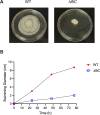
B. pseudomallei fliC exhibits reduced motility. A single colony of WT Bps orΔfliC mutant was plated on swim agar and incubated at 37°C for 77 hours. The diameter of bacterial growth was measured at 24, 48, and 77 hours and (A) photographed at 77 hours and (B) plotted over time.
Fig 6
Fig 6
Flagellin does not alter B. pseudomallei uptake or persistence in human alveolar macrophages or blood monocytes but drives alveolar macrophage cell death. Alveolar macrophages or blood monocytes were plated at a concentration of 2 × 10⁵ cells per well. Cells from the same individuals were infected with wild-type Bps at an MOI of 5 or with BpsΔfliC at an MOI of 5 as described in Fig. 1 and 3. (A and B) Uptake (1 hour) and intracellular persistence (4 and 20 hours) of bacteria were quantified at serial time points after infection of alveolar macrophages (A) and monocytes (B). (C and D) Cytotoxicity induced by ΔfliC relative to wild-type Bps infection, as determined by quantification of LDH release, was determined for (C) alveolar macrophages and (D) monocytes from the same individuals at serial time points after infection. Bars indicate mean values and whiskers indicate standard deviations. *P < 0.05. ns, not significant.
Fig 7
Fig 7
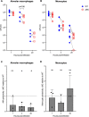
B. pseudomallei-induced inflammasome cytokine responses in human alveolar macrophages and blood monocytes are flagellin dependent. Alveolar macrophages (AM) or blood monocytes (Mono) were plated at a concentration of 2 × 10⁵ cells per well. Cells from the same individuals were infected with wild-type Bps at an MOI of 5 or with BpsΔfliC at an MOI of 5 as described in Fig. 4 and 6. (A) Cell supernatants were collected at 4 and 20 hours post-infection, and a multiplex electrochemiluminescence assay was used to quantify IL-1β, IL-18, TNF-α, and G-CSF. (B) The intraindividual proportion reduction in cytokine concentration attributable to absence of flagellin for each cell type. The center line denotes the median. The box extends from the 25th to the 75th percentile; whiskers extend from the smallest to the largest value. *P < 0.05, **P < 0.01, ***P < 0.001.

Similar articles

References

    1. Gee JE, Bower WA, Kunkel A, Petras J, Gettings J, Bye M, Firestone M, Elrod MG, Liu L, Blaney DD, et al. . 2022. Multistate outbreak of melioidosis associated with imported aromatherapy spray. N Engl J Med 386:861–868. doi:10.1056/NEJMoa2116130 - DOI - PMC - PubMed
    1. CDC . 2022. Melioidosis locally endemic in areas of the Mississippi Gulf coast after Burkholderia pseudomallei isolated in soil and water and linked to two cases – Mississippi, 2020 and 2022. Available from: https://emergency.cdc.gov/han/2022/han00470.asp. Retrieved 13 Jan 2023.
    1. Limmathurotsakul D, Golding N, Dance DAB, Messina JP, Pigott DM, Moyes CL, Rolim DB, Bertherat E, Day NPJ, Peacock SJ, Hay SI. 2016. Predicted global distribution of Burkholderia pseudomallei and burden of melioidosis. Nat Microbiol 1:15008. doi:10.1038/nmicrobiol.2015.8 - DOI - PubMed
    1. Chantratita N, Phunpang R, Yarasai A, Dulsuk A, Yimthin T, Onofrey LA, Coston TD, Thiansukhon E, Chaisuksant S, Tanwisaid K, Chuananont S, Morakot C, Sangsa N, Chayangsu S, Silakun W, Buasi N, Chetchotisakd P, Day NP, Lertmemongkolchai G, West TE. 2023. Characteristics and one year outcomes of melioidosis patients in northeastern Thailand: a prospective, multicenter cohort study. Lancet Reg Health Southeast Asia 9:100118. doi:10.1016/j.lansea.2022.100118 - DOI - PMC - PubMed
    1. Zaas AK, Schwartz DA. 2005. Innate immunity and the lung: defense at the interface between host and environment. Trends Cardiovasc Med 15:195–202. doi:10.1016/j.tcm.2005.07.001 - DOI - PubMed

Publication types

MeSH terms

LinkOut - more resources