. 2024 Apr 12;24(1):459.
doi: 10.1186/s12885-024-12177-x.

Partial recovery of peripheral blood monocyte subsets in head and neck squamous cell carcinoma patients upon radio(chemo)therapy is associated with decreased plasma CXCL11

Affiliations

Partial recovery of peripheral blood monocyte subsets in head and neck squamous cell carcinoma patients upon radio(chemo)therapy is associated with decreased plasma CXCL11

Christian Idel et al. BMC Cancer. .

Abstract

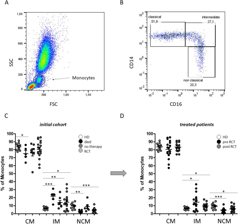
Background: Head and neck squamous cell carcinoma (HNSCC) represents a common and heterogeneous malignancy of the oral cavity, pharynx and larynx. Surgery and radio(chemo)therapy are the standard treatment options and also have great influence on the composition of the tumor microenvironment and immune cell functions. However, the impact of radio(chemo)therapy on the distribution and characteristics of circulating monocyte subsets in HNSCC are not fully understood.

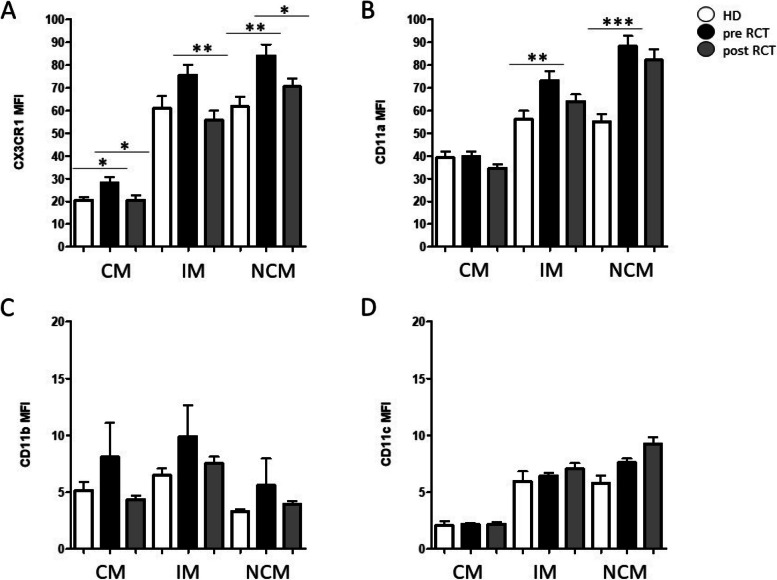
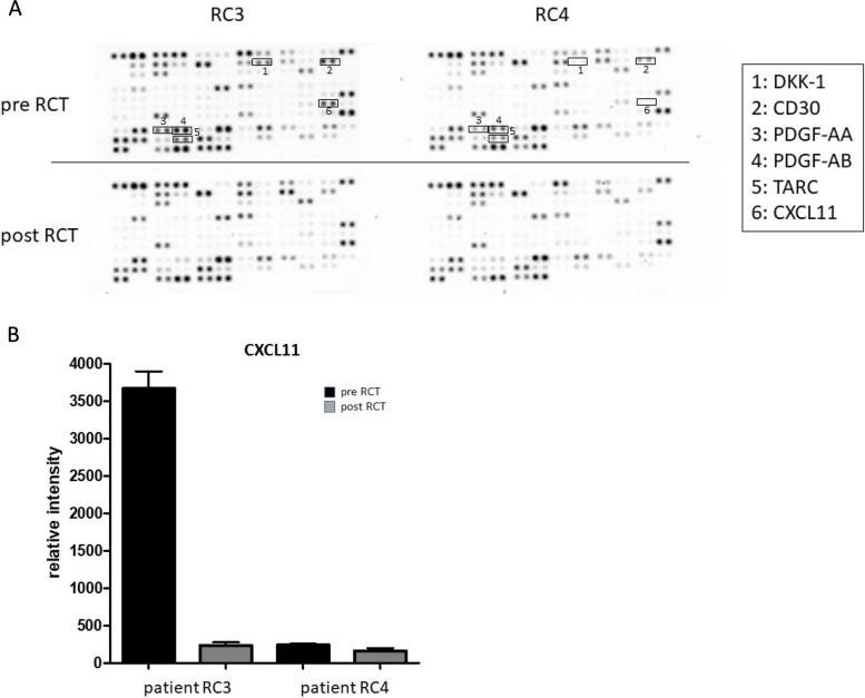
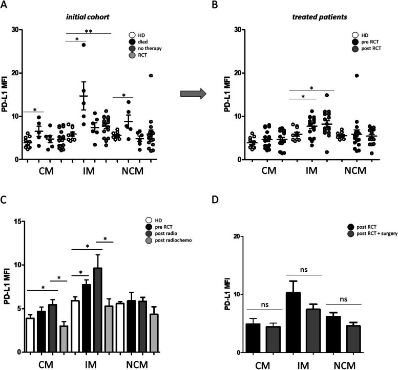
Methods: Expression patterns of adhesion molecules and chemokine receptors CD11a (integrin-α L; LFA-1), CD11b (integrin-α M; Mac-1), CD11c (integrin-α X), CX3CR1 (CX3CL1 receptor) and checkpoint molecule PD-L1 (programmed cell death ligand-1) were investigated upon radio(chemo)therapeutic treatment using flow cytometry. Furthermore, comprehensive analysis of plasma cytokines was performed before and after treatment using ELISA measurements.

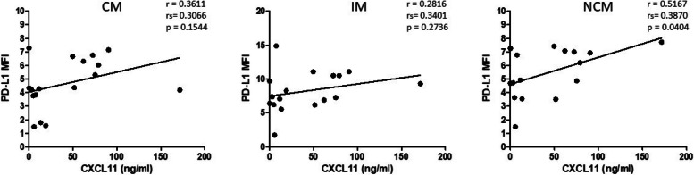
Results: Our data reveal a partial recovery of circulating monocytes in HNSCC patients upon radio(chemo)therapeutic treatment, with differential effects of the individual therapy regimen. PD-L1 expression on non-classical monocytes significantly correlates with the individual plasma levels of chemokine CXCL11 (C-X-C motif chemokine 11).

Conclusions: Further comprehensive investigations on larger patient cohorts are required to elucidate the meaningfulness of peripheral blood monocyte subsets and chemokine CXCL11 as potential bioliquid indicators in HNSCC with regard to therapy response and the individual immunological situation.

Keywords: Adhesion molecules; CXCL11; Head and Neck squamous cell carcinoma; Monocyte subsets; PD-L1; Radio(chemo)therapy.


Conflict of interest statement

The authors declare no competing interests.

Figures

Fig. 1
Fig. 1
Flow cytometric analysis of peripheral blood monocyte subsets. A Representative example gating scheme of monocyte subset analysis with regard to the forward scatter (FSC)/sideward scatter (SSC) characteristics and the B CD14/CD16 expression levels. C Percentages of circulating classical (CM), intermediate (IM) and non-classical monocytes (NCM) in the peripheral blood of HNSCC patients that unfortunately ‘died’ during the course of treatment, patients that received ‘no therapy’ and patients that were able to receive radio(chemo)therapeutic treatment (RCT). D Monocyte subset abundances of HNSCC patients before (pre RCT) and after (post RCT) radio(chemo)therapeutic treatment compared to healthy donors (HD). *: p < 0.05; **: p < 0.01; ***: p < 0.001
Fig. 2
Fig. 2
Adhesion molecules on peripheral blood monocyte subsets (CM: classical monocytes; IM: intermediate monocytes; NCM: non-classical monocytes) before and after radio(chemo)therapeutic treatment (RCT). Measurements revealed significant alterations of A CX3CR1 and B CD11a expression on intermediate and non classical monocytes prior to RCT treatment compared to healthy donors (HD), respectively. Measurements of adhesion molecules C CD11b and D CD11c revealed no significant differences. *: p < 0.05; **: p < 0.01; ***: p < 0.001. MFI: mean fluorescence intentsity
Fig. 3
Fig. 3
Cytokine screening upon radio(chemo)therapy of HNSCC patients. A Raw images of membrane based cytokine arrays of plasma samples of two HNSCC patients (RC3, RC4) before (pre RCT) and after (post RCT) radio(chemo)therapeutic treatment. Decreased expression patterns of certain cytokines (1: BDNF; 2: DKK1; 3: CD30; 4: PDGF-AA; 5: PDGF-AB; 6: TARC; 7: CXCL11) were identified in response to radio(chemo)therapeutic treatment. B Semiquantitative analysis was performed by measuring the density of the CXCL11 dots and revealed differential expression levels in the analyzed plasma samples of HNSCC patients RC3 and RC4 before and after radio(chemo)therapeutic treatment
Fig. 4
Fig. 4
Plasma CXCL11 in HNSCC patients upon radio(chemo)therapy (n = 17). A ELISA measurements revealed significantly increased CXCL11 levels in patients that unfortunately ‘died’ during the course of therapy (n = 5) and in patients that received ‘no therapy’ due to a bad health condition (n = 7) compared to healthy donors (n = 10). B CXCL11 measurements revealed no significant differences between the pre and post therapy situation or healthy donors. C Individual plasma CXCL11 pre and post therapy values (ng/ml) revealed decreases as well as increases upon radio(chemo)therapeutic treatment. CXCL11 levels of HNSCC patients that received only radiotherapy (RT) vs. HNSCC patients that received radiochemotherapy (RCT). Our data revealed that radiotherapy resulted in increased CXCL11 levels in some HNSCC patients, whereas radiochemotherapy resulted only in decreased CXCL11 levels. E Plasma CXCL11 levels of HPV positive vs. HPV negative HNSCC patients. Data revealed significantly increased CXCL11 levels in HPV + patients compared to healthy donors, but not in HPV negative individuals. F Plasma CXCL11 levels of HNSCC patients that received only RTC therapy vs. HNSCC patients that received RTC therapy + surgery. *: p < 0.05; **: p < 0.01. ns: not significant
Fig. 5
Fig. 5
Flow cytometric analysis of PD-L1 on peripheral blood monocyte subsets. A PD-L1 expression levels of circulating classical (CM), intermediate (IM) and non-classical monocytes (NCM) in the peripheral blood of HNSCC patients that unfortunately ‘died’ during the course of treatment, patients that received ‘no therapy’ and patients that were able to receive radio(chemo)therapeutic treatment (RCT) compared to healthy donors (HD). B PD-L1 expression levels on monocyte subsets of HNSCC patients before (pre RCT) and after (post RCT) radio(chemo)therapeutic treatment compared to healthy donors (HD). C PD-L1 expression levels on monocyte subsets of HNSCC patients before RCT and after radio-therapeutic treatment (post radio) and radiochemo-therapeutic treatment (post radiochemo) compared to healthy donors (HD). D To analyze the potential influence of surgical treatment, we compared the PDL-1 levels on the different monocyte subsets of HNSCC patients that received only RTC therapy vs. HNSCC patients that received RTC therapy + surgery. *: p < 0.05; **: p < 0.01; ***: p < 0.001. MFI: mean fluorescence intensity. ns: not significant
Fig. 6
Fig. 6
Correlation analysis between the PD-L1 expression of circulating classical (CM), intermediate (IM) and non-classical monocytes (NCM) and plasma levels of chemokine CXCL11 (ng/ml) of HNSCC patients after radio(chemo) therapeutic treatment. The correlation coefficient (r), spearman (rho) correlation coefficient (rs), and p values are given for each correlation. p < 0.05 was considered as significant. MFI: mean fluorescence intensity

Similar articles

References

    1. Pfister DG, Spencer S, Adelstein D, Adkins D, Anzai Y, Brizel DM, et al. Head and neck cancers, version 2.2020, NCCN Clinical Practice guidelines in Oncology. J Natl Compr Cancer Netw. 2020;18(7):873–898. doi: 10.6004/jnccn.2020.0031. - DOI - PubMed
    1. Zhang P, Li S, Zhang T, Cui F, Shi JH, Zhao F, et al. Characterization of molecular subtypes in head and neck squamous cell carcinoma with distinct prognosis and treatment responsiveness. Front Cell Dev Biology. 2021;9:711348. doi: 10.3389/fcell.2021.711348. - DOI - PMC - PubMed
    1. De Felice F, Cattaneo CG, Franco P. Radiotherapy and systemic therapies: focus on head and neck Cancer. Cancers. 2023;15(17):4232. doi: 10.3390/cancers15174232. - DOI - PMC - PubMed
    1. Lacas B, Carmel A, Landais C, Wong SJ, Licitra L, Tobias JS, et al. Meta-analysis of chemotherapy in head and neck cancer (MACH-NC): an update on 107 randomized trials and 19,805 patients, on behalf of MACH-NC Group. Radiotherapy Oncology: J Eur Soc Therapeutic Radiol Oncol. 2021;156:281–293. doi: 10.1016/j.radonc.2021.01.013. - DOI - PMC - PubMed
    1. Pardoll DM. The blockade of immune checkpoints in cancer immunotherapy. Nat Rev Cancer. 2012;12(4):252–264. doi: 10.1038/nrc3239. - DOI - PMC - PubMed