. 2024 Jun;31(6):711-721.
doi: 10.1038/s41418-024-01289-3. Epub 2024 Apr 6.

A novel inhibitory BAK antibody enables assessment of non-activated BAK in cancer cells

Affiliations

A novel inhibitory BAK antibody enables assessment of non-activated BAK in cancer cells

Hema Preethi Subas Satish et al. Cell Death Differ. 2024 Jun.

Abstract

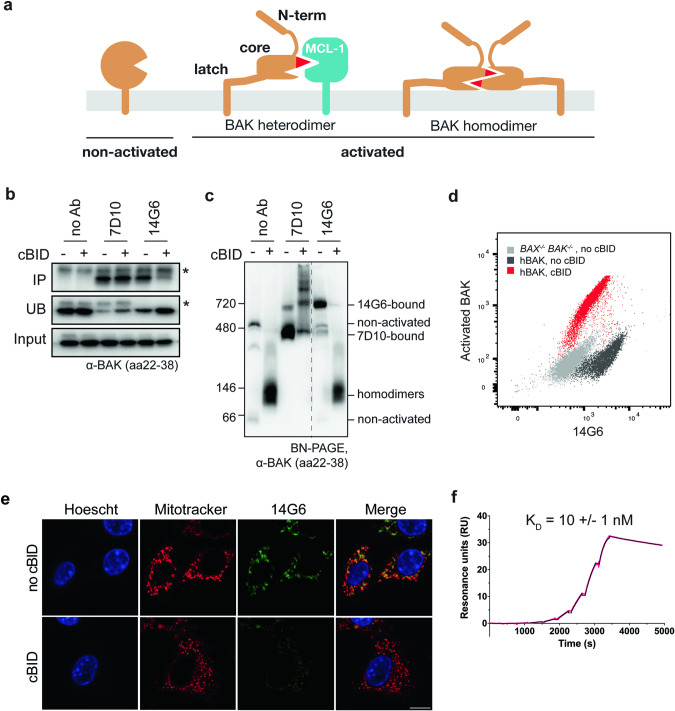
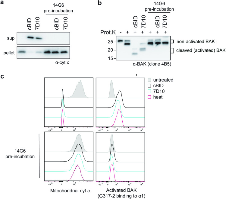
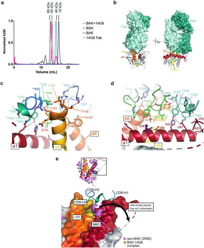
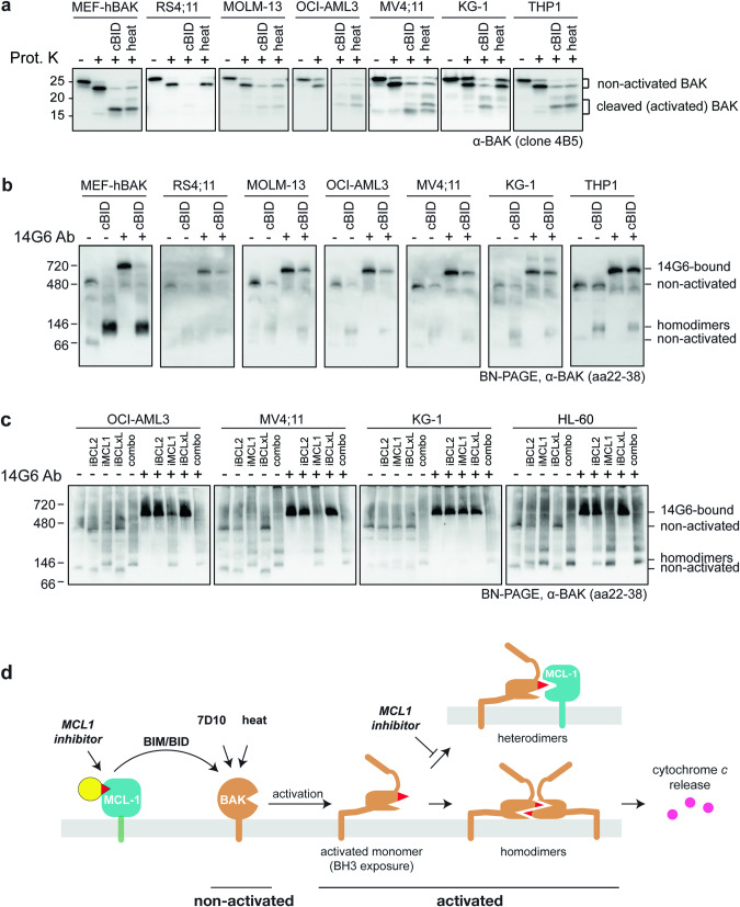
BAX and BAK are pro-apoptotic members of the BCL2 family that are required to permeabilize the mitochondrial outer membrane. The proteins can adopt a non-activated monomeric conformation, or an activated conformation in which the exposed BH3 domain facilitates binding either to a prosurvival protein or to another activated BAK or BAX protein to promote pore formation. Certain cancer cells are proposed to have high levels of activated BAK sequestered by MCL1 or BCLXL, thus priming these cells to undergo apoptosis in response to BH3 mimetic compounds that target MCL1 or BCLXL. Here we report the first antibody, 14G6, that is specific for the non-activated BAK conformer. A crystal structure of 14G6 Fab bound to BAK revealed a binding site encompassing both the α1 helix and α5-α6 hinge regions of BAK, two sites involved in the unfolding of BAK during its activation. In mitochondrial experiments, 14G6 inhibited BAK unfolding triggered by three diverse BAK activators, supporting crucial roles for both α1 dissociation and separation of the core (α2-α5) and latch (α6-α9) regions in BAK activation. 14G6 bound the majority of BAK in several leukaemia cell lines, and binding decreased following treatment with BH3 mimetics, indicating only minor levels of constitutively activated BAK in those cells. In summary, 14G6 provides a new means of assessing BAK status in response to anti-cancer treatments.


Conflict of interest statement

HPSS, SI, MXS, AWW, KCF, AZW, DL, JMB, RTU, PEC, MSM and RMK are or were employees of WEHI which receives royalties from AbbVie and Genentech from the sale of Venetoclax.

Figures

Fig. 1
Fig. 1. The 14G6 antibody binds specifically to non-activated BAK.
a Diagram of three BAK conformations present in cells. Non-activated BAK is anchored in the mitochondrial outer membrane via a C-terminal transmembrane domain (α9) [53], with the remaining 8 helices forming a globular protein with hydrophobic surface groove [44]. Upon activation, the N-terminus (α1) and latch (α6-α8) separate from the core (α2–α5). Activated BAK can be sequestered by prosurvival proteins (e.g. MCL1) or can pair up as homodimers capable of pore formation. b 14G6 immunoprecipitates BAK only prior to its activation by cBID. Membrane fractions from bak−/−bax−/− MEFs expressing hBAK were incubated with or without cBID (30 °C, 30 min). Samples were then solubilized in 1% digitonin and immunoprecipitated with no antibody or with 7D10 or 14G6. The immunoprecipitated (IP), unbound (UB) and input fractions were immunoblotted for BAK (clone aa23-38). Data are representative of two independent experiments. c 14G6 gel-shifts BAK only prior to its activation by cBID. Membrane fractions treated as in (b) were moved to ice and incubated with the indicated antibodies for 30 min, solubilized with 1% digitonin and run on blue-native PAGE (BN-PAGE) before immunoblotting for BAK (clone aa23–38). On BN-PAGE, non-activated BAK (lane 1) tends to run as a monomer (~66 kD) or in a complex with VDAC2 (~480 kD) [55]. Note that 7D10 gel-shifts BAK both before and after its activation, and when bound to BAK homodimers can generate a ladder indicating homodimers are in clusters. Note also that 14G6-bound BAK routinely runs at a significantly higher molecular weight (~720 kD) than 7D10 bound to non-activated BAK (~480 kD). Dashed line indicates deletion of lanes from the gel. Data are representative of two independent experiments. d 14G6 specificity for non-activated BAK on flow cytometry. Membrane fractions treated as in (b) were stained with 14G6 and with the G317-2 antibody to activated BAK prior to analysis by flow cytometry. (Also see Fig. S1a). Data are representative of two independent experiments. e 14G6 specificity for non-activated BAK on immunocytochemistry. Membrane fractions treated as in (b) were stained with 14G6-AF488, Mitotracker Deep Red and Hoescht prior to analysis by immunocytochemistry. Bar, 5 μm. Data are representative of two independent experiments. f 14G6 binds to BAK with a KD of 10 nM as determined by Surface Plasmon Resonance. Kinetic parameters were determined by capturing 14G6 on a Protein A chip and injecting a threefold dilution series of BAK in a single-cycle kinetic method (0, 0.45, 1.4, 4, 12, 37, 111, 333, 1000 nM). KD is the average of three independent experiments ± standard deviation. Sensorgram shown is representative of these experiments. Raw data are shown in pink, fitted curve in black.
Fig. 2
Fig. 2. 14G6 prevents BAK activation and cytochrome c release.
a 14G6 blocks cytochrome c release triggered by cBID or 7D10. bak−/−bax−/− MEFs expressing hBAK were permeabilized and incubated with or without 14G6 (on ice, 10 min) prior to incubation with or without cBID or 7D10 (30 °C, 30 min). Samples were then centrifuged and supernatant and pellet fractions immunoblotted for cytochrome c. Data are representative of two independent experiments. b 14G6 inhibits unfolding of BAK as shown by limited proteolysis. The pelleted mitochondrial fractions from (a) were treated with proteinase K and immunoblotted with antibody to the BAK BH3 domain (clone 4B5). After proteinase K, non-activated BAK runs as an ~23 kD fragment (lane 2), while cBID-activated BAK is seen as a ~16 kD fragment (lane 3) due to cleavage in the α1–α2 loop, and 7D10-activated BAK runs as a slightly larger fragment as 7D10 masks one of the cleavage sites [19]. Data is representative of two independent experiments. c 14G6 blocks BAK activation and cytochrome c release triggered by three distinct stimuli. bak−/−bax−/− MEFs expressing hBAK were permeabilized and incubated with or without 14G6 prior to incubation with or without cBID or 7D10 as indicated. Additional aliquots were also incubated at 43 °C (heat) to activate BAK. Cells were fixed and stained for cytochrome c or for activated BAK (clone G317-2) and analyzed by flow cytometry. Data are representative of at least two independent experiments.
Fig. 3
Fig. 3. Crystal structure of 14G6-bound human BAK reveals mechanism of inhibition.
a Size exclusion chromatography demonstrates that 14G6 Fab forms a 1:1 complex with BAK. BSA, bovine serum albumin, MW 66 kDa. b 14G6 binds BAK via interactions with the α5-α6 hinge (α5 and α6 shown in yellow and orange, respectively) and α1 (shown in red). The light and heavy chains of the Fab are shown in light and dark teal, respectively. c. Detailed interactions between 14G6 and BAK α5-α6 hinge. The light chain complementarity determining regions (CDRs) are shown in pale cyan (L1), light green (L2) and light blue (L3), and the heavy chain CDRs are shown in teal (H1), blue (H2), green (H3). d Detailed interactions between 14G6 and BAK α1. Colours as per (c). e 14G6 binding displaces the α1-α2 loop and inserts CDR-H3 L101 into the M60 pocket. apo-BAK (2IMS, pink) with closed α1-α2 loop overlaid with 14G6-bound BAK (grey, yellow, orange, red, chain D) showing open, unresolved loop.
Fig. 4
Fig. 4. BAK is not constitutively activated in blood cancer cell lines.
a The majority of BAK in untreated leukaemia cell lines is resistant to proteinase K. bak−/−bax−/− MEFs expressing hBAK and the indicated cancer cell lines were permeabilized and incubated with cBID (cBIDM97A) or incubated at 43 °C (heat) to activate BAK. Membrane fractions were incubated with proteinase K and blotted for BAK (as in Fig. 2b). Data are representative of at least two independent experiments. b The majority of BAK in untreated leukaemia cell lines is gel-shifted by 14G6. Membrane fractions from (a) were incubated with or without 14G6, run on BN-PAGE, and blotted for BAK (as in Fig. 1c). Data are representative of at least two independent experiments. c BAK activation by BH3 mimetic treatment demonstrated by loss of 14G6 binding. Four acute myeloid leukaemia cell lines were incubated with 1 µM iBCL2 (venetoclax), iMCL1 (S63845), iBCLxL (A-1331852), alone or in combination for 3 h. Membrane fractions were then incubated with or without 14G6, and analyzed as in (b). Cell lysates were also assessed for BAK and BAX cleavage by proteinase K (Fig. S6). Data are representative of at least two independent experiments. d Schematic of BAK activation by various stimuli demonstrating that in the cell types examined, the majority of BAK becomes activated only after apoptotic signalling. Here, MCL1 represents prosurvival proteins, as MCL1 inhibitor alone could trigger BAK activation in three cell lines (Figs. 4c and S6).

Similar articles

References

    1. Moldoveanu T, Czabotar PE. BAX, BAK, and BOK: a coming of age for the BCL-2 family effector proteins. Cold Spring Harb Perspect Biol. 2019;12:a036319. doi: 10.1101/cshperspect.a036319. - DOI - PMC - PubMed
    1. Westphal D, Dewson G, Czabotar PE, Kluck RM. Molecular biology of Bax and Bak activation and action. Biochim Biophys Acta. 2011;1813:521–31. doi: 10.1016/j.bbamcr.2010.12.019. - DOI - PubMed
    1. Dai H, Smith A, Meng XW, Schneider PA, Pang YP, Kaufmann SH. Transient binding of an activator BH3 domain to the Bak BH3-binding groove initiates Bak oligomerization. J Cell Biol. 2011;194:39–48. doi: 10.1083/jcb.201102027. - DOI - PMC - PubMed
    1. Czabotar PE, Westphal D, Dewson G, Ma S, Hockings C, Fairlie WD, et al. Bax crystal structures reveal How BH3 domains activate bax and nucleate its oligomerization to induce apoptosis. Cell. 2013;152:519–31. doi: 10.1016/j.cell.2012.12.031. - DOI - PubMed
    1. Brouwer JM, Westphal D, Dewson G, Robin AY, Uren RT, Bartolo R, et al. Bak core and latch domains separate during activation, and freed core domains form symmetric homodimers. Mol Cell. 2014;55:938–46. doi: 10.1016/j.molcel.2014.07.016. - DOI - PubMed

MeSH terms

Substances