Expression and significance of pigment epithelium-derived factor and vascular endothelial growth factor in colorectal adenoma and cancer
- PMID: 38577437
- PMCID: PMC10989378
- DOI: 10.4251/wjgo.v16.i3.670
Expression and significance of pigment epithelium-derived factor and vascular endothelial growth factor in colorectal adenoma and cancer
Abstract
Background: The incidence and mortality of colorectal cancer (CRC) are among the highest in the world, and its occurrence and development are closely related to tumor neovascularization. When the balance between pigment epithelium-derived factors (PEDF) that inhibit angiogenesis and vascular endothelial growth factors (VEGF) that stimulate angiogenesis is broken, angiogenesis is out of control, resulting in tumor development. Therefore, it is very necessary to find more therapeutic targets for CRC for early intervention and later treatment.
Aim: To investigate the expression and significance of PEDF, VEGF, and CD31-stained microvessel density values (CD31-MVD) in normal colorectal mucosa, adenoma, and CRC.
Methods: In this case-control study, we collected archived wax blocks of specimens from the Digestive Endoscopy Center and the General Surgery Department of Chengdu Second People's Hospital from April 2022 to October 2022. Fifty cases of specimen wax blocks were selected as normal intestinal mucosa confirmed by electronic colonoscopy and concurrent biopsy (normal control group), 50 cases of specimen wax blocks were selected as colorectal adenoma confirmed by electronic colonoscopy and pathological biopsy (adenoma group), and 50 cases of specimen wax blocks were selected as CRC confirmed by postoperative pathological biopsy after inpatient operation of general surgery (CRC group). An immunohistochemical staining experiment was carried out to detect PEDF and VEGF expression in three groups of specimens, analyze their differences, study the relationship between the two and clinicopathological factors in CRC group, record CD31-MVD in the three groups, and analyze the correlation of PEDF, VEGF, and CD31-MVD in the colorectal adenoma group and the CRC group. The F test or adjusted F test is used to analyze measurement data statistically. Kruskal-Wallis rank sum test was used between groups for ranked data. The chi-square test, adjusted chi-square test, or Fisher's exact test were used to compare the rates between groups. All differences between groups were compared using the Bonferroni method for multiple comparisons. Spearman correlation analysis was used to test the correlation of the data. The test level (α) was 0.05, and a two-sided P< 0.05 was considered statistically significant.
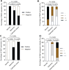
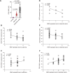
Results: The positive expression rate and expression intensity of PEDF were gradually decreased in the normal control group, adenoma group, and CRC group (100% vs 78% vs 50%, χ2 = 34.430, P < 0.001; ++~++ vs +~++ vs -~+, H = 94.059, P < 0.001), while VEGF increased gradually (0% vs 68% vs 96%, χ2 = 98.35, P < 0.001; - vs -~+ vs ++~+++, H = 107.734, P < 0.001). In the CRC group, the positive expression rate of PEDF decreased with the increase of differentiation degree, invasion depth, lymph node metastasis, distant metastasis, and TNM stage (χ2 = 20.513, 4.160, 5.128, 6.349, 5.128, P < 0.05); the high expression rate of VEGF was the opposite (χ2 = 10.317, 13.134, 17.643, 21.844, 17.643, P < 0.05). In the colorectal adenoma group, the expression intensity of PEDF correlated negatively with CD31-MVD (r = -0.601, P < 0.001), whereas VEGF was not significantly different (r = 0.258, P = 0.07). In the CRC group, the expression intensity of PEDF correlated negatively with the expression intensity of CD31-MVD and VEGF (r = -0.297, P < 0.05; r = -0.548, P < 0.05), while VEGF expression intensity was positively related to CD31-MVD (r = 0.421, P = 0.002).
Conclusion: It is possible that PEDF can be used as a new treatment and prevention target for CRC by upregulating the expression of PEDF while inhibiting the expression of VEGF.
Keywords: Colorectal adenoma; Colorectal cancer; Microvessel density; Pigment epithelium-derived factors; Targeted therapy; Vascular endothelial growth factor.
©The Author(s) 2024. Published by Baishideng Publishing Group Inc. All rights reserved.
Conflict of interest statement
Conflict-of-interest statement: The authors declare no competing interests.
Figures


Similar articles
-
Expression of Vascular Endothelial Growth Factor (VEGF) in Colorectal Adenoma and Carcinoma in a Tertiary Care Center.Cureus. 2022 Nov 11;14(11):e31393. doi: 10.7759/cureus.31393. eCollection 2022 Nov. Cureus. 2022. PMID: 36514651 Free PMC article.
-
Correlation between vascular endothelial growth factor-A expression and tumor location and invasion in patients with colorectal cancer.J Gastrointest Oncol. 2018 Dec;9(6):1099-1108. doi: 10.21037/jgo.2018.07.01. J Gastrointest Oncol. 2018. PMID: 30603129 Free PMC article.
-
Role of VEGF, CD105, and CD31 in the Prognosis of Colorectal Cancer Cases.J Gastrointest Cancer. 2019 Mar;50(1):23-34. doi: 10.1007/s12029-017-0014-y. J Gastrointest Cancer. 2019. PMID: 29110224
-
[Relationship of hypoxia-inducible factor 1 alpha (HIF-1alpha) gene expression with vascular endothelial growth factor (VEGF) and microvessel density (MVD) in human colorectal adenoma and adenocarcinoma].Ai Zheng. 2003 Nov;22(11):1170-4. Ai Zheng. 2003. PMID: 14613646 Chinese.
-
Pigment Epithelial-Derived Factor in Pancreatic and Liver Cancers-From Inflammation to Cancer.Biomedicines. 2024 Oct 4;12(10):2260. doi: 10.3390/biomedicines12102260. Biomedicines. 2024. PMID: 39457573 Free PMC article. Review.
References
-
- Sung H, Ferlay J, Siegel RL, Laversanne M, Soerjomataram I, Jemal A, Bray F. Global Cancer Statistics 2020: GLOBOCAN Estimates of Incidence and Mortality Worldwide for 36 Cancers in 185 Countries. CA Cancer J Clin. 2021;71:209–249. - PubMed
-
- Morgan E, Arnold M, Gini A, Lorenzoni V, Cabasag CJ, Laversanne M, Vignat J, Ferlay J, Murphy N, Bray F. Global burden of colorectal cancer in 2020 and 2040: incidence and mortality estimates from GLOBOCAN. Gut. 2023;72:338–344. - PubMed
-
- Leslie A, Carey FA, Pratt NR, Steele RJ. The colorectal adenoma-carcinoma sequence. Br J Surg. 2002;89:845–860. - PubMed
LinkOut - more resources
Full Text Sources
Miscellaneous

