Renal interstitial fibrotic assessment using non-Gaussian diffusion kurtosis imaging in a rat model of hyperuricemia
- PMID: 38570748
- PMCID: PMC10988851
- DOI: 10.1186/s12880-024-01259-8
Renal interstitial fibrotic assessment using non-Gaussian diffusion kurtosis imaging in a rat model of hyperuricemia
Abstract
Background: To investigate the feasibility of Diffusion Kurtosis Imaging (DKI) in assessing renal interstitial fibrosis induced by hyperuricemia.
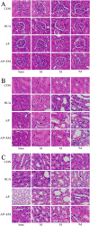
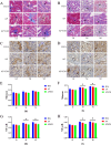
Methods: A hyperuricemia rat model was established, and the rats were randomly split into the hyperuricemia (HUA), allopurinol (AP), and AP + empagliflozin (AP + EM) groups (n = 19 per group). Also, the normal rats were selected as controls (CON, n = 19). DKI was performed before treatment (baseline) and on days 1, 3, 5, 7, and 9 days after treatment. The DKI indicators, including mean kurtosis (MK), fractional anisotropy (FA), and mean diffusivity (MD) of the cortex (CO), outer stripe of the outer medulla (OS), and inner stripe of the outer medulla (IS) were acquired. Additionally, hematoxylin and eosin (H&E) staining, Masson trichrome staining, and nuclear factor kappa B (NF-κB) immunostaining were used to reveal renal histopathological changes at baseline, 1, 5, and 9 days after treatment.
Results: The HUA, AP, and AP + EM group MKOS and MKIS values gradually increased during this study. The HUA group exhibited the highest MK value in outer medulla. Except for the CON group, all the groups showed a decreasing trend in the FA and MD values of outer medulla. The HUA group exhibited the lowest FA and MD values. The MKOS and MKIS values were positively correlated with Masson's trichrome staining results (r = 0.687, P < 0.001 and r = 0.604, P = 0.001, respectively). The MDOS and FAIS were negatively correlated with Masson's trichrome staining (r = -626, P < 0.0014 and r = -0.468, P = 0.01, respectively).
Conclusion: DKI may be a non-invasive method for monitoring renal interstitial fibrosis induced by hyperuricemia.
Keywords: Diffusion kurtosis imaging; Hyperuricemia; Magnetic resonance imaging; Renal interstitial fibrosis.
© 2024. The Author(s).
Conflict of interest statement
The authors declare no competing interests.
Figures


Similar articles
-
Based on functional and histopathological correlations: is diffusion kurtosis imaging valuable for noninvasive assessment of renal damage in early-stage of chronic kidney disease?Int Urol Nephrol. 2024 Jan;56(1):263-273. doi: 10.1007/s11255-023-03632-y. Epub 2023 Jun 16. Int Urol Nephrol. 2024. PMID: 37326823
-
Renal functional and interstitial fibrotic assessment with non-Gaussian diffusion kurtosis imaging.Insights Imaging. 2022 Apr 8;13(1):70. doi: 10.1186/s13244-022-01215-6. Insights Imaging. 2022. PMID: 35394225 Free PMC article.
-
Evaluating the renal mild tubulointerstitial damage and renal function in IgAN patients: a comparative study based on diffusion kurtosis imaging and diffusion tensor imaging.Abdom Radiol (NY). 2023 Apr;48(4):1350-1362. doi: 10.1007/s00261-023-03822-3. Epub 2023 Feb 7. Abdom Radiol (NY). 2023. PMID: 36749369
-
Assessment of renal fibrosis in a rat model of unilateral ureteral obstruction with diffusion kurtosis imaging: Comparison with α-SMA expression and 18F-FDG PET.Magn Reson Imaging. 2020 Feb;66:176-184. doi: 10.1016/j.mri.2019.08.035. Epub 2019 Sep 1. Magn Reson Imaging. 2020. PMID: 31484043
-
Diffusional kurtosis imaging of kidneys in patients with hyperuricemia: initial study.Acta Radiol. 2020 Jun;61(6):839-847. doi: 10.1177/0284185119878362. Epub 2019 Oct 14. Acta Radiol. 2020. PMID: 31610679
References
-
- Ejaz AA, Nakagawa T, Kanbay M, Kuwabara M, Kumar A, Garcia Arroyo FE, Roncal-Jimenez C, Sasai F, Kang DH, Jensen T, et al. Hyperuricemia in kidney disease: a major risk factor for cardiovascular events, vascular calcification, and renal damage. Semin Nephrol. 2020;40(6):574–585. doi: 10.1016/j.semnephrol.2020.12.004. - DOI - PMC - PubMed
MeSH terms
Grants and funding
- no. 82202284/National Natural Science Foundation of China
- no. A2022337/Medical Scientific Research Foundation of Guangdong Province
- no. 20221110/Administration of Traditional Chinese Medicine of Guangdong Province
- no. 20232025/Administration of Traditional Chinese Medicine of Guangdong Province
- no. 20221113/Administration of Traditional Chinese Medicine of Guangdong Province
LinkOut - more resources
Full Text Sources

