

Peste des petits ruminants virus infection induces endoplasmic reticulum stress and apoptosis via IRE1-XBP1 and IRE1-JNK signaling pathways
- PMID: 38568823
- PMCID: PMC10990917
- DOI: 10.4142/jvs.23236
Peste des petits ruminants virus infection induces endoplasmic reticulum stress and apoptosis via IRE1-XBP1 and IRE1-JNK signaling pathways
Abstract
Background: Peste des petits ruminants (PPR) is a contagious and fatal disease of sheep and goats. PPR virus (PPRV) infection induces endoplasmic reticulum (ER) stress-mediated unfolded protein response (UPR). The activation of UPR signaling pathways and their impact on apoptosis and virus replication remains controversial.
Objectives: To investigate the role of PPRV-induced ER stress and the IRE1-XBP1 and IRE1-JNK pathways and their impact on apoptosis and virus replication.
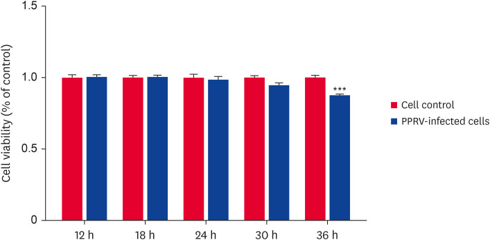
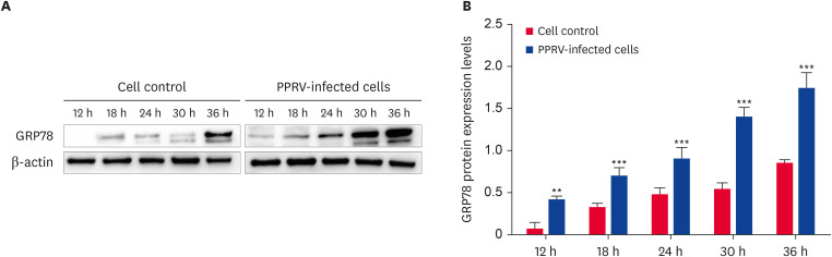
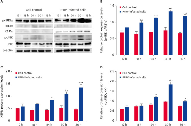
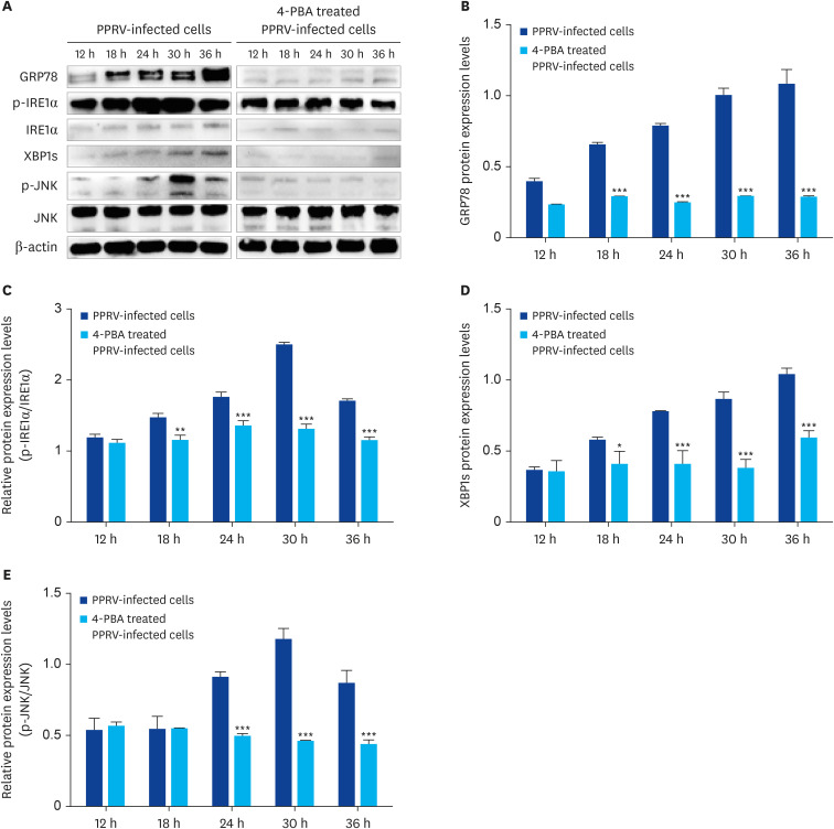
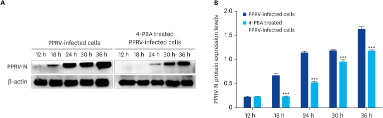
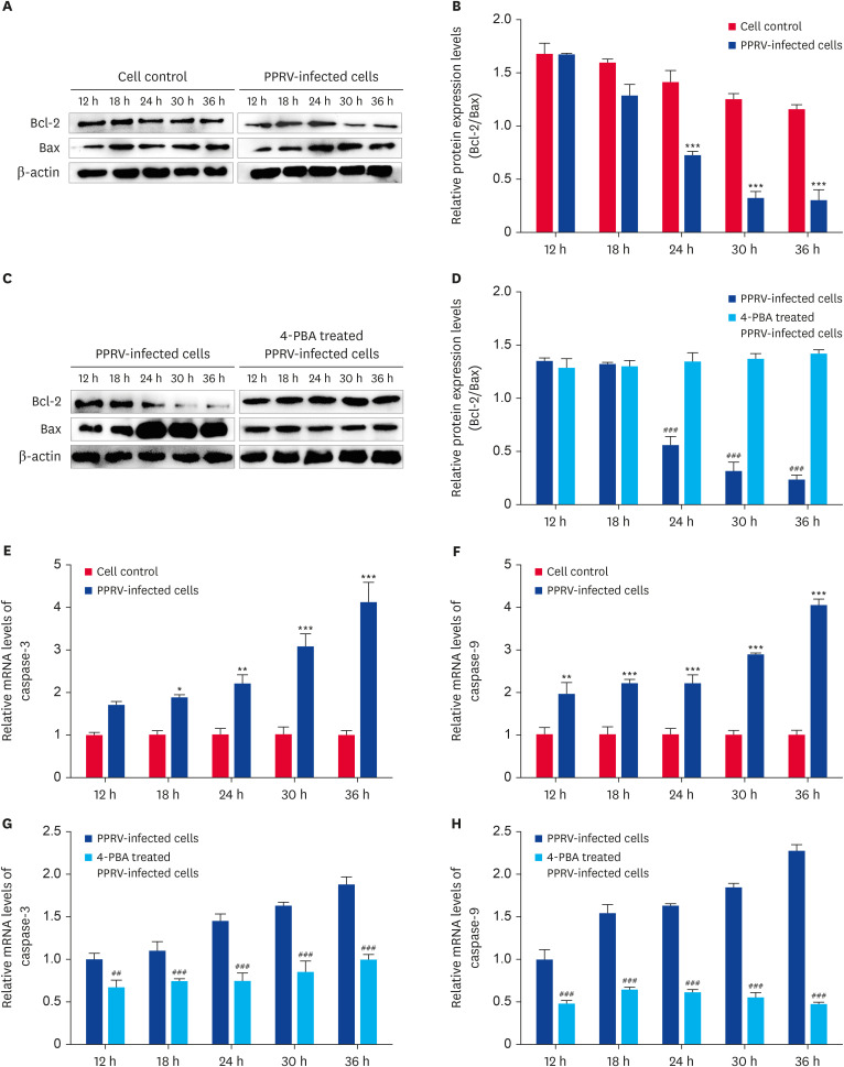
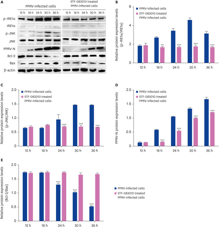
Methods: The cell viability and virus replication were assessed by 3-(4,5-Dimethyl-2-thiazolyl)-2,5-diphenyl-2H-tetrazolium bromide assay, immunofluorescence assay, and Western blot. The expression of ER stress biomarker GRP78, IRE1, and its downstream molecules, PPRV-N protein, and apoptosis-related proteins was detected by Western blot and quantitative reverse transcription-polymerase chain reaction, respectively. 4-Phenylbutyric acid (4-PBA) and STF-083010 were respectively used to inhibit ER stress and IRE1 signaling pathway.
Results: The expression of GRP78, IRE1α, p-IRE1α, XBP1s, JNK, p-JNK, caspase-3, caspase-9, Bax and PPRV-N were significantly up-regulated in PPRV-infected cells, the expression of Bcl-2 was significantly down-regulated. Due to 4-PBA treatment, the expression of GRP78, p-IRE1α, XBP1s, p-JNK, caspase-3, caspase-9, Bax, and PPRV-N were significantly down-regulated, the expression of Bcl-2 was significantly up-regulated. Moreover, in PPRV-infected cells, the expression of p-IRE1α, p-JNK, Bax, and PPRV-N was significantly decreased, and the expression of Bcl-2 was increased in the presence of STF-083010.
Conclusions: PPRV infection induces ER stress and IRE1 activation, resulting in apoptosis and enhancement of virus replication through IRE1-XBP1s and IRE1-JNK pathways.
Keywords: IRE1-JNK pathway; IRE1-XBP1 pathway; Peste des petits ruminants virus; apoptosis; endoplasmic reticulum stress.
© 2024 The Korean Society of Veterinary Science.
Conflict of interest statement
The authors declare no conflicts of interest.
Figures

Similar articles
-
Inhibition of caspase-1-dependent apoptosis suppresses peste des petits ruminants virus replication.J Vet Sci. 2023 Sep;24(5):e55. doi: 10.4142/jvs.22288. Epub 2023 Jul 7. J Vet Sci. 2023. PMID: 37638708 Free PMC article.
-
Peste des petits ruminants virus induces ERS-mediated autophagy to promote virus replication.Vet Microbiol. 2022 Jul;270:109451. doi: 10.1016/j.vetmic.2022.109451. Epub 2022 May 14. Vet Microbiol. 2022. PMID: 35594636
-
Plasminogen activator urokinase interacts with the fusion protein and antagonizes the growth of Peste des petits ruminants virus.J Virol. 2024 Apr 16;98(4):e0014624. doi: 10.1128/jvi.00146-24. Epub 2024 Mar 5. J Virol. 2024. PMID: 38440983 Free PMC article.
-
Peste Des Petits Ruminants in Atypical Hosts and Wildlife: Systematic Review and Meta-Analysis of the Prevalence between 2001 and 2021.Arch Razi Inst. 2021 Dec 30;76(6):1589-1606. doi: 10.22092/ari.2021.356900.1939. eCollection 2021 Dec. Arch Razi Inst. 2021. PMID: 35546985 Free PMC article. Review.
-
The role of the ER stress sensor IRE1 in cardiovascular diseases.Mol Cell Biochem. 2024 May 8. doi: 10.1007/s11010-024-05014-z. Online ahead of print. Mol Cell Biochem. 2024. PMID: 38717685 Review.
References
MeSH terms
Substances
Grants and funding
LinkOut - more resources
Full Text Sources
Research Materials
Miscellaneous
