. 2024 Apr 10;146(14):9779-9789.
doi: 10.1021/jacs.3c14380. Epub 2024 Apr 1.

Targeted Protein O-GlcNAcylation Using Bifunctional Small Molecules

Affiliations

Targeted Protein O-GlcNAcylation Using Bifunctional Small Molecules

Bowen Ma et al. J Am Chem Soc. .

Abstract

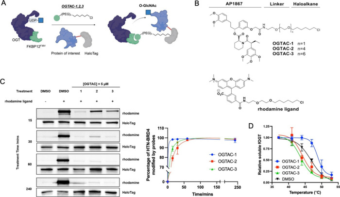
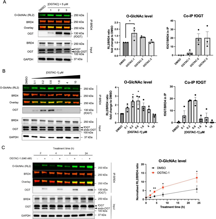
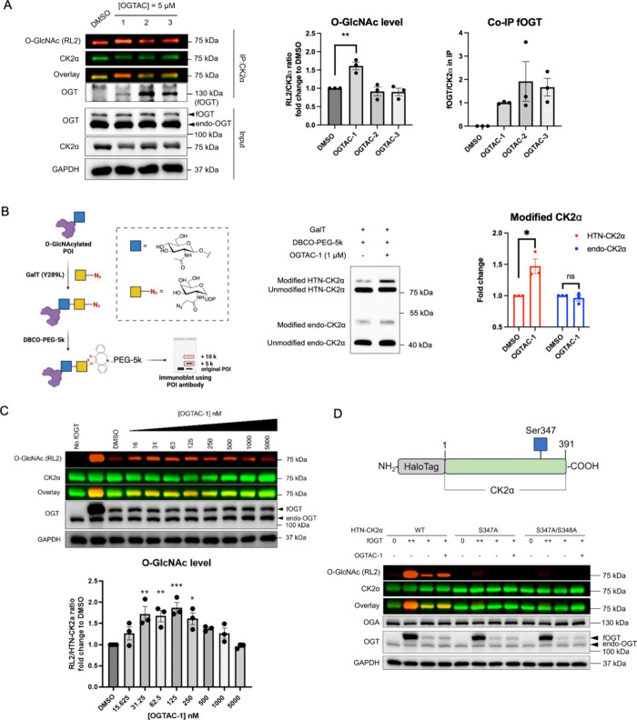
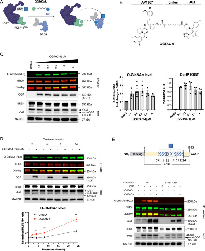
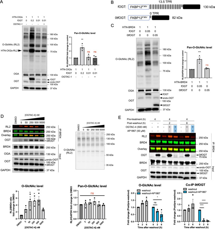
Protein O-linked β-N-acetylglucosamine modification (O-GlcNAcylation) plays a crucial role in regulating essential cellular processes. The disruption of the homeostasis of O-GlcNAcylation has been linked to various human diseases, including cancer, diabetes, and neurodegeneration. However, there are limited chemical tools for protein- and site-specific O-GlcNAc modification, rendering the precise study of the O-GlcNAcylation challenging. To address this, we have developed heterobifunctional small molecules, named O-GlcNAcylation TArgeting Chimeras (OGTACs), which enable protein-specific O-GlcNAcylation in living cells. OGTACs promote O-GlcNAcylation of proteins such as BRD4, CK2α, and EZH2 in cellulo by recruiting FKBP12F36V-fused O-GlcNAc transferase (OGT), with temporal, magnitude, and reversible control. Overall, the OGTACs represent a promising approach for inducing protein-specific O-GlcNAcylation, thus enabling functional dissection and offering new directions for O-GlcNAc-targeting therapeutic development.


Conflict of interest statement

The authors declare the following competing financial interest(s): B.W.M and B.W.-L.N. are inventors on a U.S. non-provisional patent application submitted by The Chinese University of Hong Kong that covers the described bifunctional molecules.

Figures

Figure 1
Figure 1
General concept of OGTAC technology and target engagement studies of OGTAC molecules. (A) Conceptual scheme of general OGTACs, which induce POI-specific O-GlcNAcylation by recruiting FKBP12F36V-OGT to HaloTag-fused POIs. (B) Chemical structure of OGTAC-1,2,3, and rhodamine ligand for the pulse-chase experiment. (C, D) Confirmation of OGTAC-1,2,3 target engagement in cells. HEK293T cells were transiently transfected with plasmids HTN-BRD4:fOGT at a 1:0.05 ratio. (C) Pulse-chase assay to verify the engagement of the OGTACs with HTN-BRD4. OGTAC-1,2,3 (5 μM) or vehicle was added to cells for increasing periods of time, followed by replacement with rhodamine ligand to label any remaining HTN-BRD4 proteins that were not engaged by OGTACs. Then, cells were lysed and subjected to SDS-PAGE for in-gel fluorescence using the rhodamine channel and immunoblotting using a HaloTag antibody to verify equal loading. (D) After 4 h treatment of 5 μM OGTAC-1,2,3, CETSA was conducted to verify their direct binding to fOGT. Data in (C, D) represent mean ± s.d. of n = 3 biologically independent replicates.
Figure 2
Figure 2
OGTAC-1 induces dose-dependent, rapid, targeted O-GlcNAcylation of HTN-BRD4 by recruiting fOGT in cells. (A) OGTAC-1 showed the best O-GlcNAcylation-inducing potency among three OGTACs at 5 μM. Endo-OGT, endogenous OGT. (B) Dose-dependent O-GlcNAcylation profile of HTN-BRD4 by the OGTAC-1 treatment. HEK293T cells expressing HTN-BRD4:fOGT = 1:0.05 were treated with increasing concentration of OGTAC-1 for 4 h. Then, the O-GlcNAcylation level of HTN-BRD4 was assessed by immunoblot after IP using BRD4 antibody. Co-IP of fOGT was also observed in an OGTAC-1 dose-dependent manner; (C) OGTAC-1 (640 nM) O-GlcNAcylation-inducing effects on HTN-BRD4 over the indicated time course. Notably, the marginal divergence between the red and black curves after 4 h suggests a diminished effect of OGTAC-1. For (A) and (B), the right panels show the quantifications of the immunoblot signal of RL2 relative to HTN-BRD4 and Co-IPed fOGT relative to IPed BRD4, as the mean ± s.e.m. of n = 3 biologically independent experiments. Statistical significance of (A) and (B) was calculated with ordinary one-way ANOVA; that of (C) was calculated with unpaired multiple t test. *p < 0.05; **p < 0.01.
Figure 3
Figure 3
OGTAC technology extended to HTN-CK2α. (A) IP-WB method showed OGTAC-induced HTN-CK2α O-GlcNAcylation. (B) Mass shift assay to validate the OGTAC-1-induced specific HTN-CK2α O-GlcNAcylation (data for other two repeats can be found in Figure S4); (C) OGTAC-1-induced dose-dependent O-GlcNAcylation of HTN-CK2α in cells. HEK293T cells were transfected with HTN-CK2α:fOGT = 1:0.01 for 24 h, followed by treatment with increasing concentration of OGTAC-1 for 8 h. Transfection with HTN-CK2α:fOGT = 1:0.2 was set as positive control; (D) OGTAC-1-induced physiologically relevant S347 O-GlcNAcylation in HTN-CK2α. “++” denotes the HTN-CK2α:fOGT = 1:0.2, while “+” denotes the HTN-CK2α:fOGT = 1:0.01. All quantifications are shown as mean ± s.e.m. of 3 independent biological repeats. Statistical significance for (B) was calculated with two-tailed Student’s t test, and remaining significance were calculated with ordinary one-way ANOVA comparing DMSO- to OGTAC-1-treated samples. ns, p ≥ 0.05; *p < 0.05; **p < 0.01; ***p < 0.001.
Figure 4
Figure 4
OGTAC-4 induces BRD4 O-GlcNAcylation by recruiting fOGT in cells. (A) Conceptual scheme of OGTAC-4, which induces BRD4-specific O-GlcNAcylation by recruiting FKBP12F36V-OGT to BRD4 through the JQ1 motif. (B) Chemical structure of OGTAC-4. (C) Dose-dependent O-GlcNAcylation profile of HTN-BRD4 by OGTAC-4 treatment. HEK293T cells expressing HTN-BRD4:fOGT = 1:0.05 were treated with an increasing concentration of OGTAC-4 for 4 h. Then, the O-GlcNAcylation level of HTN-BRD4 was assessed by immunoblot after IP using BRD4 antibody. Co-IP of fOGT was also observed in an OGTAC-4 dose-dependent manner; (D) OGTAC-4 (640 nM) O-GlcNAcylation-inducing effects on HTN-BRD4 over the indicated time course; (E) OGTAC-1 and OGTAC-4 induced HTN-BRD4 O-GlcNAcylation in the C-terminal region (S1367-F1224). “+” denotes the HTN-BRD4:fOGT = 1:0.05. The rectangles with blue stripes represent the truncated fragment used for validating O-GlcNAcylation sites. HAT, histone acetyltransferase catalytic domain; statistical significance was calculated with ordinary one-way ANOVA comparing DMSO- to OGTAC-4-treated samples. The quantifications of the immunoblot show the signal of RL2 relative to HTN-BRD4 and Co-IPed fOGT relative to IPed BRD4, as the mean ± s.e.m. of n = 3 biologically independent experiments. Statistical significance for (C) was calculated with ordinary one-way ANOVA; for (D), it was calculated with unpaired multiple t test. *p < 0.05; **p < 0.01; ***p < 0.001.
Figure 5
Figure 5
Specificity and reversibility of the OGTAC strategy. (A) Under the HTN-CK2α:fOGT = 1:0.01 cotransfection system, the cellular global O-GlcNAc level is not significantly changed with or without the treatment of the OGTAC-1 (125 nM). (B) Constructs of fOGT with 13.5 (rounded to 13) TPR and 0TPR-fOGT(0tfOGT); (C) 0tfOGT but not fOGT does not significantly change the cellular global O-GlcNAc level. (D) Dose-dependent O-GlcNAcylation profile of HTN-BRD4 by the OGTAC-4 treatment. HEK293T cells expressing HTN-BRD4:0tfOGT= 1:0.05 were treated with an increasing concentration of OGTAC-4 for 4 h. Then, the O-GlcNAcylation level of HTN-BRD4 was assessed by immunoblot after IP using the BRD4 antibody. All quantifications are shown as mean ± s.e.m. of n = 3 biologically independent experiments. (E) Reversible effect of OGTAC-4 on HTN-BRD4 can be achieved by addition of AP1867. HEK293T cells expressing HTN-BRD4:0tfOGT= 1:0.05 were pretreated with OGTAC-4 (500 nM) or DMSO for 4 h, and culturing media were replaced by fresh media with DMSO or AP1867 (50 μM) for the indicated time. The quantifications in right panel were conducted by the normalizing the fold-change (OGTAC-4/DMSO) for each postwashout time point to the value of 4 h pretreatment. All quantifications are shown as mean ± s.e.m. of 3 independent biological repeats. Statistical significance was calculated with ordinary one-way ANOVA comparing samples with different transfection conditions or OGTAC-treatment. ns, p ≥ 0.05; *p < 0.05; **p < 0.01; ***p < 0.001; ****p < 0.0001.

Similar articles

Cited by

References

    1. Slawson C.; Hart G. W. O-GlcNAc Signalling: Implications for Cancer Cell Biology. Nat. Rev. Cancer 2011, 11 (9), 678–684. 10.1038/nrc3114. - DOI - PMC - PubMed
    1. Yang X.; Qian K. Protein O-GlcNAcylation: Emerging Mechanisms and Functions. Nat. Rev. Mol. Cell Biol. 2017, 18 (7), 452–465. 10.1038/nrm.2017.22. - DOI - PMC - PubMed
    1. Eustice M.; Bond M. R.; Hanover J. A. O-GlcNAc Cycling and the Regulation of Nucleocytoplasmic Dynamics. Biochem. Soc. Trans. 2017, 45 (2), 427–436. 10.1042/BST20160171. - DOI - PubMed
    1. Hart G. W. Nutrient Regulation of Signaling and Transcription. J. Biol. Chem. 2019, 294 (7), 2211–2231. 10.1074/jbc.AW119.003226. - DOI - PMC - PubMed
    1. Ferrer C. M.; Sodi V. L.; Reginato M. J. O-GlcNAcylation in Cancer Biology: Linking Metabolism and Signaling. J. Mol. Biol. 2016, 428 (16), 3282–3294. 10.1016/j.jmb.2016.05.028. - DOI - PMC - PubMed

MeSH terms