. 2024 May 14;98(5):e0006024.
doi: 10.1128/jvi.00060-24. Epub 2024 Apr 1.

PDCD4 restricts PRRSV replication in an eIF4A-dependent manner and is antagonized by the viral nonstructural protein 9

Affiliations

PDCD4 restricts PRRSV replication in an eIF4A-dependent manner and is antagonized by the viral nonstructural protein 9

Ruiping Wei et al. J Virol. .

Abstract

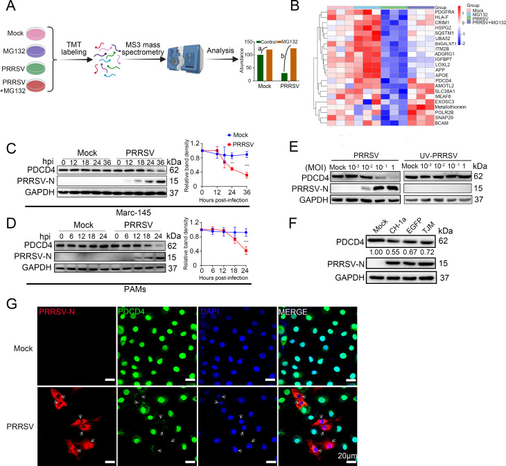
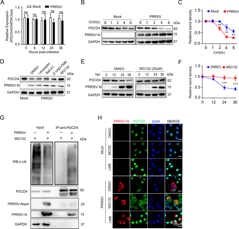
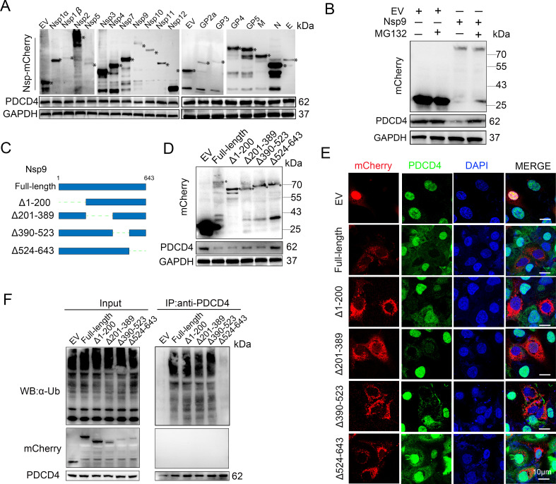
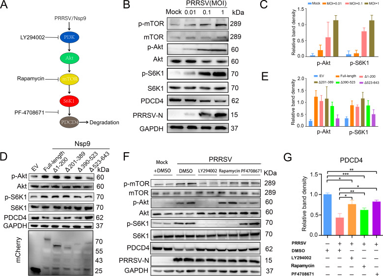
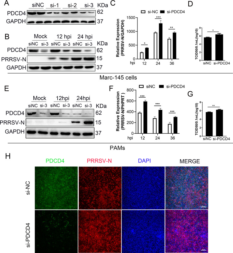
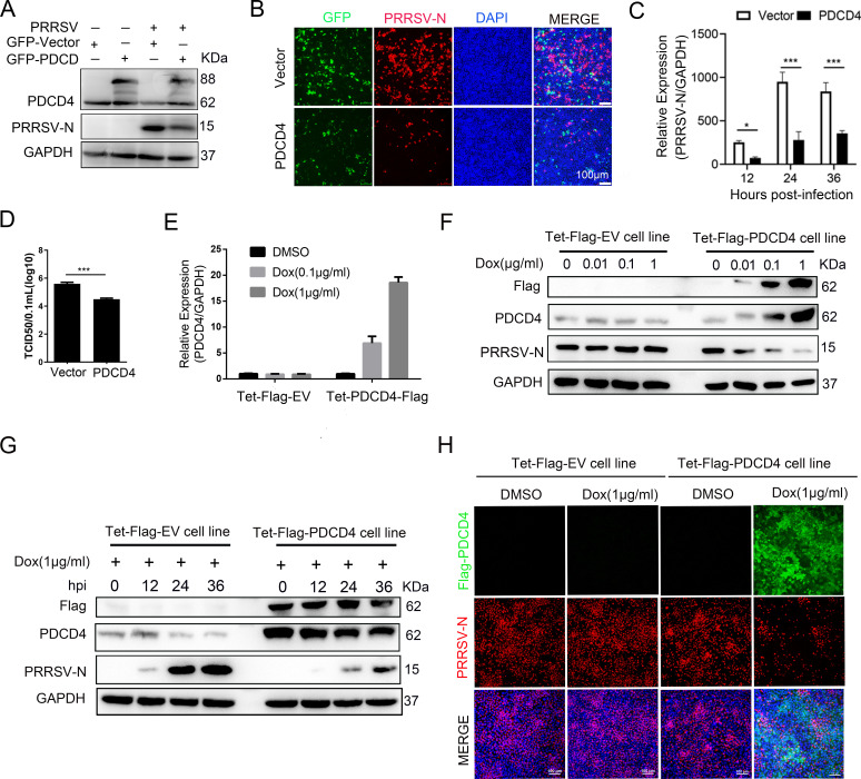
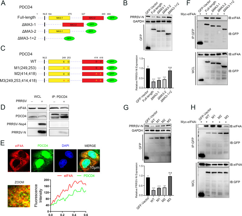
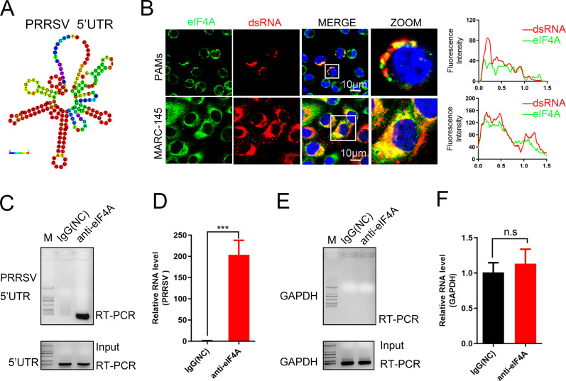
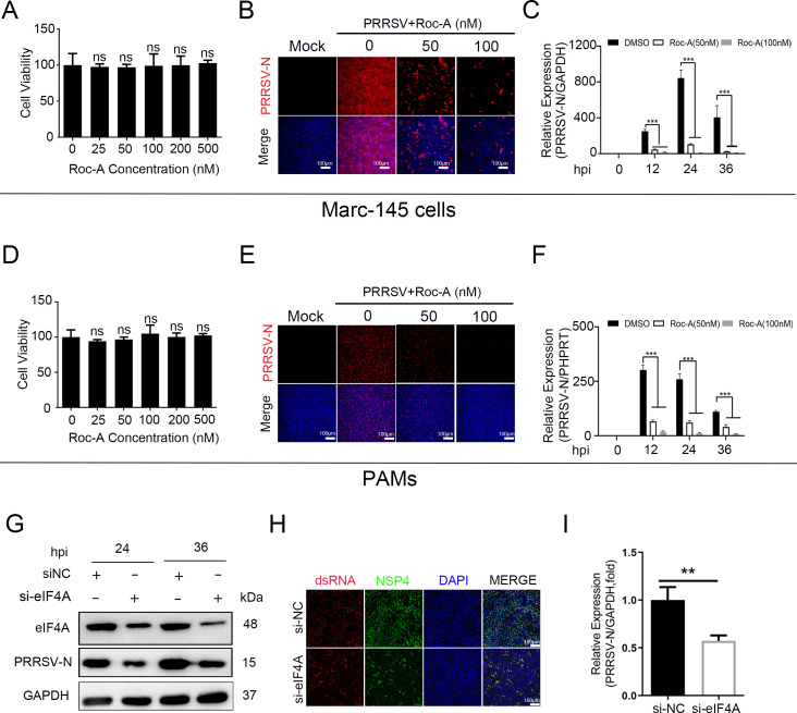
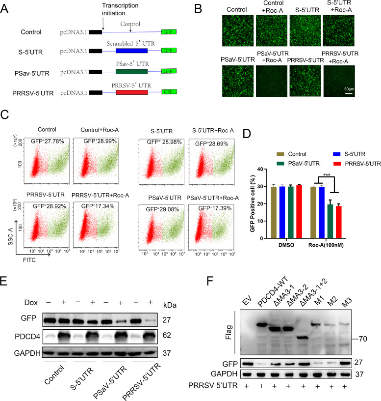
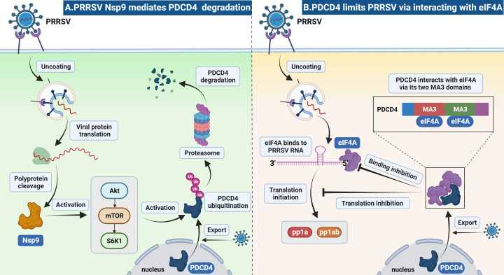
As obligate parasites, viruses have evolved multiple strategies to evade the host immune defense. Manipulation of the host proteasome system to degrade specific detrimental factors is a common viral countermeasure. To identify host proteins targeted for proteasomal degradation by porcine reproductive and respiratory syndrome virus (PRRSV), we conducted a quantitative proteomics screen of PRRSV-infected Marc-145 cells under the treatment with proteasome inhibitor MG132. The data revealed that the expression levels of programmed cell death 4 (PDCD4) were strongly downregulated by PRRSV and significantly rescued by MG132. Further investigation confirmed that PRRSV infection induced the translocation of PDCD4 from the nucleus to the cytoplasm, and the viral nonstructural protein 9 (Nsp9) promoted PDCD4 proteasomal degradation in the cytoplasm by activating the Akt-mTOR-S6K1 pathway. The C-terminal domain of Nsp9 was responsible for PDCD4 degradation. As for the role of PDCD4 during PRRSV infection, we demonstrated that PDCD4 knockdown favored viral replication, while its overexpression significantly attenuated replication, suggesting that PDCD4 acts as a restriction factor for PRRSV. Mechanistically, we discovered eukaryotic translation initiation factor 4A (eIF4A) was required for PRRSV. PDCD4 interacted with eIF4A through four sites (E249, D253, D414, and D418) within its two MA3 domains, disrupting eIF4A-mediated translation initiation in the 5'-untranslated region of PRRSV, thereby inhibiting PRRSV infection. Together, our study reveals the antiviral function of PDCD4 and the viral strategy to antagonize PDCD4. These results will contribute to our understanding of the immune evasion strategies employed by PRRSV and offer valuable insights for developing new antiviral targets.IMPORTANCEPorcine reproductive and respiratory syndrome virus (PRRSV) infection results in major economic losses in the global swine industry and is difficult to control effectively. Here, using a quantitative proteomics screen, we identified programmed cell death 4 (PDCD4) as a host protein targeted for proteasomal degradation by PRRSV. We demonstrated that PDCD4 restricts PRRSV replication by interacting with eukaryotic translation initiation factor 4A, which is required for translation initiation in the viral 5'-untranslated region. Additionally, four sites within two MA3 domains of PDCD4 are identified to be responsible for its antiviral function. Conversely, PRRSV nonstructural protein 9 promotes PDCD4 proteasomal degradation in the cytoplasm by activating the Akt-mTOR-S6K1 pathway, thus weakening the anti-PRRSV function. Our work unveils PDCD4 as a previously unrecognized host restriction factor for PRRSV and reveals that PRRSV develops countermeasures to overcome PDCD4. This will provide new insights into virus-host interactions and the development of new antiviral targets.

Keywords: Nsp9; PDCD4; PRRSV; eIF4A; host restriction factor; virus-host interactions.


Conflict of interest statement

The authors declare no conflict of interest.

Figures

Fig 1
Fig 1
PRRSV infection causes a decrease in PDCD4 protein. (A) Workflow schematic showing how the inhibitor-based proteomics screen was employed to identify host proteins targeted for proteasomal degradation by PRRSV. (B) Heatmap displaying 22 candidate proteins downregulated by PRRSV and partially rescued by MG132, as screened from total proteomics data. (C and D) Western blot analysis of PDCD4 protein levels in MARC-145 cells or porcine alveolar macrophages infected with PRRSV at a multiplicity of infection (MOI) of 0.1. GAPDH serves as a loading control, and the viral nucleocapsid protein N is used as an infection indicator. Protein abundance was quantified using Image J software. (E) Marc-145 cells were infected with PRRSV or UV-inactivated PRRSV at different MOIs for 24 hours, and the cells were harvested to detect the protein levels of PDCD4 by western blot. (F) Marc-145 cells were mock-infected or infected with different PRRSV strains, respectively, at an MOI of 0.1 for 24 hours, and the cells were harvested to detect the protein levels of PDCD4 by western blot. (G) Immunofluorescence of cells labeled with PDCD4 and PRRSV-N antibodies. Marc-145 cells were mock-infected or infected with PRRSV (MOI = 0.1) for 18 hours, and the cells were fixed, permeabilized, and stained with the antibodies against PDCD4 and PRRSV N. Images of cells were acquired by laser-scanning fluorescent confocal microscopy. Data presented as means ± SE of three independent experiments, and asterisks (*) indicate the statistical significance: **P < 0.01 and ***P < 0.001.
Fig 2
Fig 2
PRRSV targets PDCD4 for degradation via the proteasomal pathway. (A) Relative abundance of PDCD4 mRNA in PRRSV-infected Marc-145 cells at indicated time points post-infection. (B and C) PRRSV shortens the half-life of PDCD4. Marc-145 cells were infected with PRRSV at an MOI of 0.1. The cells were treated with cycloheximide (CHX) at 24 hpi and harvested at indicated time points for western blot analysis. The graph shows the levels of PDCD4 protein normalized against GAPDH. (D) MG132 treatment restores PDCD4 levels in PRRSV-infected cells. Marc-145 cells were infected with PRRSV at an MOI of 0.1, and at 24 hpi, the cells were treated with leupeptin (20 µM), Z-VAD-FMK (20 µM), Bafilomycin A1 (1 µM), or MG132 (20 µM) for 6 hours and then harvested for western blot analyses. (E and F) Marc-145 cells were infected with PRRSV at an MOI of 0.1. At the indicated time points, the cells were treated with DMSO or MG132 (20 µM) for an additional 6 hours before being harvested for western blot analyses. PDCD4 protein abundance was quantified by Image J software. (G) Ubiquitination of endogenous PDCD4. Marc-145 cells were infected with PRRSV at an MOI of 0.1 for 24 hours and treated with MG132 (20 µM) for an additional 6 hours. The cells were lysed for co-immunoprecipitation using an antibody against PDCD4. The immunoprecipitates were analyzed by western blot with antibodies against Ub, PDCD4, GAPDH, PRRSV N, and PRRSV Nsp4. (H) The effect of LMB and MG132 on PRRSV-induced PDCD4 degradation by using immunofluorescence analysis. Marc-145 cells were mock-infected or infected with PRRSV for 24 hours and then treated with LMB (10 µM) or MG132 (20 µM) for 6 hours. The cells were fixed and reacted with rabbit monoclonal antibody against PDCD4 (1:200) and mouse monoclonal antibody against PRRSV N (1:500). Data are presented as means ± SE of three independent experiments, and asterisks (*) indicate the statistical significance: ns, no significance; **P < 0.01; and ***P < 0.001.
Fig 3
Fig 3
The C-terminal domain of PRRSV Nsp9 mediates the degradation of PDCD4. (A) The effect of viral nonstructural and structural proteins on the reduction of PDCD4 protein. HEK-293T cells were transfected with different mCherry-tagged plasmids encoding non-structural and structural proteins for 24 hours. The cells were lysed for western blotting with antibodies against mCherry, PDCD4, and GAPDH. Asterisks mark the expressed mCherry-fusion proteins of viral nonstructural and structural proteins. EV, empty vector (pmCherry-N). (B) HEK-293FT cells grown on coverslips in 6-well plates were transfected with Nsp9-expressing plasmid. At 24 hours post-transfection, these cells were treated with DMSO or MG132 (20 µM) for 6 hours and then were collected for western blot analyses. (C) Schematic structure of full-length and truncated Nsp9 proteins. (D) Full-length Nsp9 or other Nsp9 fragments were transfected into HEK-293T cells. After transfection for 24 hours, the cells were harvested for western blot analyses. (E) Marc-145 cells were transfected with mCherry-tagged plasmids encoding full-length and truncated Nsp9 proteins for 24 hours followed by immunofluorescence analysis. The PDCD4 protein (green) was stained with specific antibodies, and nucleic acids were stained with 4,6-diamidino-2-phenylindole dihydrochloride (DAPI) (blue). (F) Marc-145 cells were transfected with plasmids expressing full-length and truncated Nsp9. At 24 hours post-transfection, proteasome inhibitor MG132 (20 µM) was added. At 30 hours post-transfection, cell lysates were harvested for co-immunoprecipitation analysis with antibodies against PDCD4. Western blot analysis was conducted with the whole-cell lysate or immunoprecipitated proteins using the antibodies against mCherry, PDCD4, and Ub.
Fig 4
Fig 4
The Akt-mTOR-S6K1 pathway is involved in the degradation of PDCD4 during PRRSV infection. (A) Model for inhibitor targets associated with the Akt-mTOR-S6K1 pathway. (B and C) PRRSV infection promotes the activation of the Akt-mTOR-S6K1 pathway. Marc-145 cells were infected with PRRSV at different MOIs for 24 hours. Western blot showed the detection of phosphorylated Akt, mTOR, and S6K1 using specific antibodies. Protein abundance was quantified by Image J software. (D and E) Marc-145 cells were transfected with plasmids expressing full-length and truncated Nsp9 for 24 hours followed by western blot analysis. (F and G) Marc-145 cells were infected with PRRSV (MOI = 0.1), and at 24 hpi, the cells were treated with DMSO or indicated inhibitors (dissolved in DMSO), during the last 6 hours before harvesting. Protein extracts were analyzed by immunoblotting with antibodies to the indicated proteins. The PDCD4 protein abundance was quantified by Image J software. Data are presented as means ± SE of three independent experiments, and asterisks (*) indicate the statistical significance: *P < 0.05 and **P < 0.01.
Fig 5
Fig 5
PDCD4 knockdown facilitates PRRSV infection. (A) Marc-145 cells were transfected with scrambled siRNA (siNC) or three siRNAs targeting PDCD4 for 24 hours, and protein expression of PDCD4 is shown using western blot analysis. (B–D) Marc-145 cells depleted for PDCD4 by siRNA were infected with PRRSV at an MOI of 0.1 for the indicated periods. PRRSV replication was monitored by western blot (B) and RT-qPCR (C). At 24 hpi, viral production in cells was measured and is shown as TCID50 (D). (E–G) The same as panels B–D, except that PAMs were used. PHPRT, the hypoxanthine phosphoribosyltransferase of Sus scrofa, was used as an endogenous reference in RT-qPCR. (H) The experiment was performed as in panels B–D, except that cells were collected and fixed for immunofluorescent staining of PDCD4 (green) and PRRSV-N (red). Data are presented as means ± SE of three independent experiments, and asterisks (*) indicate the statistical significance: *P < 0.05, **P < 0.01, and ***P < 0.001.
Fig 6
Fig 6
Overexpression of PDCD4 restrains PRRSV infection. (A–D) Marc-145 cells were transiently transfected with plasmids expressing GFP-PDCD4 or the empty vector for 24 hours, and the cells were infected with PRRSV at an MOI of 0.1. At 24 hpi, PRRSV replication was monitored by western blot (A), immunofluorescence staining (B), and RT-qPCR (C). Virus titers were quantified by TCID50 assay (D). (E–H) Marc-145 cells were stably transduced with control lentivirus (Tet-Flag-EV) or lentivirus containing tetracycline-inducible PDCD4-Flag gene (Tet-Flag-PDCD4). The Tet-Flag-EV or Tet-Flag-PDCD4 was induced with an indicated dose of doxycycline for 24 hours (E). The mRNA levels of PDCD4 were measured by RT-qPCR (F). Cells were infected with PRRSV at an MOI of 0.1. At 24 hpi, PDCD4 and viral N protein were measured by western blot. (G) Cells were infected with PRRSV at an MOI of 0.1 for the indicated periods. Western blot was conducted to analyze the expression of PDCD4 and viral N. (H) Cells were infected with PRRSV at an MOI of 0.1 for 24 hours, and the expression of PRRSV-N and PDCD4 was analyzed by immunofluorescence. Data are presented as means ± SE of three independent experiments, and asterisks (*) indicate the statistical significance: *P < 0.05 and ***P < 0.001.
Fig 7
Fig 7
PDCD4 inhibits PRRSV infection by binding to the RNA-helicase eIF4A via the two MA3 domains. (A and C) Schematic structure of full-length and mutant PDCD4 proteins. (B) Full-length PDCD4 or its mutants were transfected into Marc-145 cells. At 24 hours post-transfection, the cells were infected with PRRSV at an MOI of 0.1. At 24 hpi, the expression of GFP-fusion proteins and PRRSV-N was analyzed by western blot. The PRRSV-N protein abundance was quantified by Image J software. (D) PDCD4 was immunoprecipitated from mock- or PRRSV-infected Marc-145 cells, and the immunoprecipitated proteins were detected by western blot. (E) Marc-145 cells were plated onto 15-mm cover glass-bottomed petri dishes and then stained with antibodies directed against PDCD4 (green) and eIF4A (red). The fluorescence plot profiles were analyzed by Image J. (F) PDCD4 binds eIF4A via each MA3 domain. HEK-293T cells were co-transfected with plasmids encoding myc-eIF4A and full-length or mutants of GFP-PDCD4. Cell lysates were immunoprecipitated with an anti-GFP Ab, followed by immunoblotting. (G and H) The same as panels B and F, except that point-mutant plasmids were transfected. Data are presented as means ± SE of three independent experiments, and asterisks (*) indicate the statistical significance: ns, no significance; **P < 0.01; and ***P < 0.001.
Fig 8
Fig 8
eIF4A interacts with PRRSV RNA. (A) Secondary structure of PRRSV 5′ UTR predicted by RNAfold. (B) Immunofluorescence analysis of the co-localization between eIF4A (green) and dsRNA of viral origin (red). Marc-145 cells or PAMs infected with PRRSV at an MOI of 0.1 were subjected to immunofluorescence with antibodies against PDCD4 and dsRNA. The fluorescence plot profiles were analyzed by Image J. (C–F) Marc-145 cells were infected with PRRSV (MOI = 0.1, 24 hours). Cell lysates were immunoprecipitated with rabbit IgG or antibodies specific against eIF4A. The viral RNA was analyzed by RT-qPCR targeting the 5′ UTR sequence (C and D) or GAPDH (E and F), which is the designated negative control.
Fig 9
Fig 9
The eIF4A is required for PRRSV infection. (A) Marc-145 cells were treated with an indicated dose of Roc-A, at 24 hours post-treatment, and the cell viability was examined by using the CCK8 kit. (B and C) Roc-A inhibits PRRSV infection. Marc-145 cells were infected with PRRSV at an MOI of 0.1 after Roc-A treatment (indicated concentration). At 24 hpi, PRRSV replication was monitored by Immunofluorescence analysis (B) and RT-qPCR (C). (D–F) The experiment was performed as in panels A–C, except that PAMs were used. (H–J) The eIF4A knockdown suppresses PRRSV infection. At 24 hours post-transfection with indicated siRNA, Marc-145 cells were infected with PRRSV (MOI = 0.1, 24 hours). The mRNA or protein levels of PRRSV-N were analyzed by western blot (H), immunofluorescence analysis (I), and RT-qPCR (J). Data are presented as means ± SE of three independent experiments, and asterisks (*) indicate the statistical significance: ns, no significance; **P < 0.01; and ***P < 0.001.
Fig 10
Fig 10
PDCD4 disrupts eIF4A-dependent translation initiation in the 5′ UTR of PRRSV. (A) Schematic structure of mRNA expressed from plasmids containing the viral or scrambled 5′ UTR. (B) HEK-293T cells were transfected with GFP-tagged plasmids (above described). At 6 hours post-transfection, the medium was replaced with a medium containing DMSO or Roc-A (100 nM). At 24 hours post-transfection, cells were fixed and observed with a fluorescence microscope. (C and D) The experiment was performed as in panel B, except that flow cytometry was used to determine the percentage of GFP-positive cells. The GFP-positive cells were analyzed using GraphPad Software. Data are presented as means ± SE of three independent experiments, and asterisks (*) indicate the statistical significance: ns, no significance; **P < 0.01; and ***P < 0.001. (E) Tet-Flag-PDCD4 cells were treated with Dox for 24 hours to induce PDCD4 overexpression and then transfected with GFP-tagged plasmids as described in panel A. At 24 hours post-transfection, cell lysates were analyzed by immunoblotting with antibodies to the indicated proteins. (F) HEK-293T cells were co-transfected with plasmids encoding PRRSV 5′ UTR and full-length or mutants of Flag-PDCD4. Protein extracts were analyzed by immunoblotting with indicated antibodies.
Fig 11
Fig 11
PRRSV Nsp9 antagonizes the antiviral property of PDCD4. (A) PRRSV Nsp9 mediates PDCD4 degradation. Upon PRRSV entry into cells, the Nsp9 activates the Akt-mTOR-S6K1 signaling pathway, resulting in PDCD4 protein polyubiquitination and proteasomal degradation in the cytoplasm. (B) PDCD4 limits PRRSV via interacting with eIF4A. In response to PRRSV infection, PDCD4 undergoes translocation from the nucleus to the cytoplasm, where it interacts with eIF4A via its two MA3 domains. This interaction disrupts the binding of eIF4A to viral RNA, consequently impeding translation initiation in viral 5′ UTR.

Similar articles

References

    1. Cho JG, Dee SA. 2006. Porcine reproductive and respiratory syndrome virus. Theriogenology 66:655–662. doi:10.1016/j.theriogenology.2006.04.024 - DOI - PubMed
    1. Goyal SM. 1993. Porcine reproductive and respiratory syndrome. J Vet Diagn Invest 5:656–664. doi:10.1177/104063879300500435 - DOI - PubMed
    1. Neumann EJ, Kliebenstein JB, Johnson CD, Mabry JW, Bush EJ, Seitzinger AH, Green AL, Zimmerman JJ. 2005. Assessment of the economic impact of porcine reproductive and respiratory syndrome on swine production in the United States. J Am Vet Med Assoc 227:385–392. doi:10.2460/javma.2005.227.385 - DOI - PubMed
    1. Han J, Zhou L, Ge X, Guo X, Yang H. 2017. Pathogenesis and control of the Chinese highly pathogenic porcine reproductive and respiratory syndrome virus. Vet Microbiol 209:30–47. doi:10.1016/j.vetmic.2017.02.020 - DOI - PubMed
    1. Harding JCS, Ladinig A, Novakovic P, Detmer SE, Wilkinson JM, Yang T, Lunney JK, Plastow GS. 2017. Novel insights into host responses and reproductive pathophysiology of porcine reproductive and respiratory syndrome caused by PRRSV-2. Vet Microbiol 209:114–123. doi:10.1016/j.vetmic.2017.02.019 - DOI - PubMed

MeSH terms

LinkOut - more resources