. 2024 Apr 16;5(4):101489.
doi: 10.1016/j.xcrm.2024.101489. Epub 2024 Mar 29.

Multicellular ecotypes shape progression of lung adenocarcinoma from ground-glass opacity toward advanced stages

Affiliations

Multicellular ecotypes shape progression of lung adenocarcinoma from ground-glass opacity toward advanced stages

Yulan Deng et al. Cell Rep Med. .

Abstract

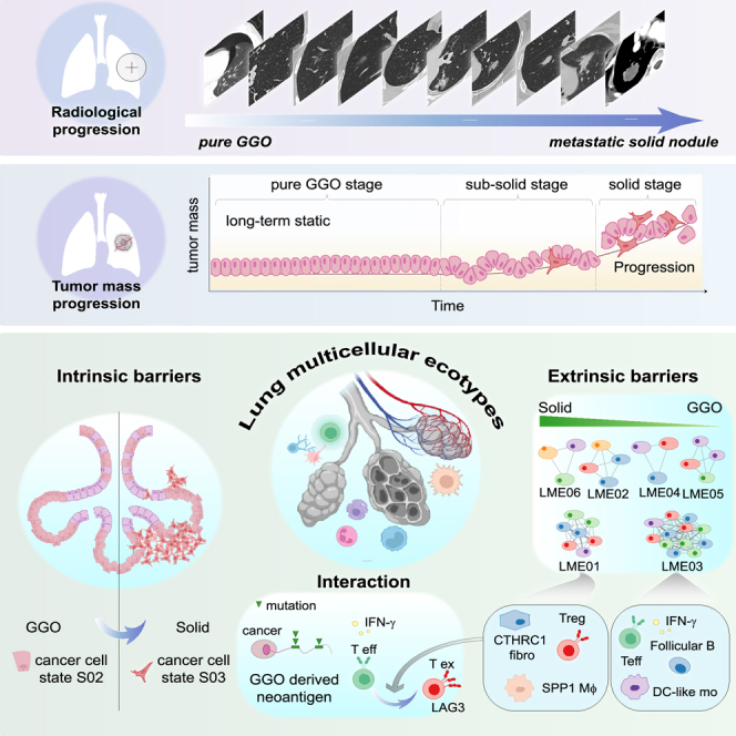
Lung adenocarcinoma is a type of cancer that exhibits a wide range of clinical radiological manifestations, from ground-glass opacity (GGO) to pure solid nodules, which vary greatly in terms of their biological characteristics. Our current understanding of this heterogeneity is limited. To address this gap, we analyze 58 lung adenocarcinoma patients via machine learning, single-cell RNA sequencing (scRNA-seq), and whole-exome sequencing, and we identify six lung multicellular ecotypes (LMEs) correlating with distinct radiological patterns and cancer cell states. Notably, GGO-associated neoantigens in early-stage cancers are recognized by CD8+ T cells, indicating an immune-active environment, while solid nodules feature an immune-suppressive LME with exhausted CD8+ T cells, driven by specific stromal cells such as CTHCR1+ fibroblasts. This study also highlights EGFR(L858R) neoantigens in GGO samples, suggesting potential CD8+ T cell activation. Our findings offer valuable insights into lung adenocarcinoma heterogeneity, suggesting avenues for targeted therapies in early-stage disease.


Conflict of interest statement

Declaration of interests The authors declare no competing interests.

Figures

None
Graphical abstract
Figure 1
Figure 1
Characterization of GGO and solid lung adenocarcinoma cell states and LMEs (A) Schematic depicting the analytical framework and stratifications of patients based on the radiological patterns. Yellow dashed lines highlight the malignant lesions. Scale bars, 1 cm (identical for all the CT images). (B) Overview of clinical characteristics of primary untreated individuals with lung adenocarcinoma. (C) The t-SNE (t-distributed stochastic neighbor embedding) by major cell partitions (left), tissue types (middle), or specimens from different patients (right). (D) Lung malignant ecotypes detected in untreated individuals with lung adenocarcinoma. Top: LME compositions are depicted as network diagrams. The width of each edge represents the Jaccard index across tumor samples. Bottom: cell state compositions in the corresponding LMEs. (E) Characteristics of LMEs in the discovery cohort. Top: proportions of major cell types (averaged and scaled). Bottom: mutation frequencies of driver genes. (F) The fraction of LMEs in different radiological clinical stages. Endo, endothelium; Epi, epithelium; Fib, fibroblast; NK, natural killer cell; DC, dendritic cell; PMN, polymorphonuclear cell (mainly neutrophil in our datasets); Mo/Mφ, monocyte and macrophage.
Figure 2
Figure 2
Identification of malignant cell states upon GGO progression toward solid nodules (A) The t-SNE by epithelial cells characterized based on CNV inferences. The middle panel represents single cells from normal lung tissues (n = 6). The right panel represents single cells from individuals with lung adenocarcinoma. (B) Heatmaps displaying marker genes among cancer cell states for scRNA-seq dataset, with genes as rows and tumor/adjacent normal tissue samples as columns. Heatmaps are organized by the most abundant cell state per sample. The expression of marker genes is scaled by its maximal value. (C) The cancer cell S03 is enriched in the late stage upon the progression of lung adenocarcinoma, with significance determined by Fisher’s exact test. (D and E) The fraction of Epi S02 and Epi S03 cell states in the SRP238929 cohort (D) and the GSE131907 cohort (E). (F) Kaplan-Meier plot showing differences in disease-free survival between patients with high/low expression of Epi S03 cell state signature in TCGA stage I–II cohort. (G) Kaplan-Meier plot showing differences in disease-free survival between patients with high/low expression of Epi S03 cell state signature in treatment-naive early-stage lung adenocarcinoma patient cohort (GSE31210). (H) The expression levels of epithelial cell-state-specific markers at single-cell level. (I) Multiplex immunofluorescence validation on formalin-fixed paraffin-embedded tissues from an independent cohort of patients for the specific cell state marker expressions in malignant cells. Left: CT scan. Middle: corresponding H&E staining and QuPath segmented immunofluorescence staining. Scale bar, 1 mm. Right: zoomed representative images of each cell state marker. Scale bar, 25 μm. (J) QuPath-based quantifications of expression levels of cell state markers. nLung, normal lung; tLung, early-stage primary tumors; tL/B, advanced-stage tumors; mLN, advanced tumors with lymph node metastases; mBrain, advanced tumors with cerebral metastases.
Figure 3
Figure 3
Early manifestations of microenvironmental cell states in GGO and subsolid nodules (A) Compositional changes in immune cell states in GGOs and solid nodular tumors relative to normal lung tissues. Cell states are connected by colored lines if they are within the same LMEs. Kruskal-Wallis false discovery rate (FDR) < 0.05 for solid nodules versus GGOs is marked with asterisks. (B) The UMAP (Uniform Manifold Approximation and Projection) representation for Mo/Mφ, B cells, CD4+ T cells, and CD8+ T cells. Right: specific cell-type marker expression is shown. (C) Heatmap showing expression of select memory, exhaustion, and progenitor exhausted lymphocyte-associated markers among CD8T and CD4T cell states from (A). (D) Multiplex immunofluorescence for the formation of tertiary lymphoid structure (TLS) upon the progression of lung adenocarcinoma. Matched H&E staining and QuPath-based cell segmentation are shown. Scale bar, 1 mm. Right: representative TLS-like structures are shown. Scale bar, 100 μm. (E) QuPath-based quantifications of cell count in tumor center and tumor periphery (n = 5). ns, not significant.
Figure 4
Figure 4
Immune and stromal ecotype involved in the progression from GGO to subsolid nodules of lung adenocarcinoma (A) Matrix representation of cell-to-cell interactions within LME01 determined by CellChat algorithm. Sender cell ligands are listed on the y axis and receiver cell receptors are on the x axis. Fibroblast-macrophage, mast cell-macrophage, and macrophage-CD4T cell interactions are highlighted. (B) Multiplex-immunofluorescence-based quantifications of the distances of B cells, CD4+ T cells, and CD8+ T cells to XCL1+ cells and CTHCR1+ cells upon the progression of lung adenocarcinoma. Bottom panel: exemplified images of mIF. Scale bar, 100 μm. (C) Monocle-based trajectory inference analysis of Mo/Mφ, colored by subclusters and pseudotime. (D) Marker gene expression during Mo/Mφ maturation along the pseudotemporal trajectory. (E) The mast-cell- and tumor-cell-derived annexin A1 (ANXA1) interacts with FPR2 receptor expressed by Mo/Mφ. (F) The expression levels of receptor and downstream signaling molecules at the single-cell level. (G) The expression levels of ANXA1-regulated target genes expressed in different cell states of Mo/Mφ. The targets were selected as significantly expressed genes varying among monocle branch points by function “BEAM.” (H–J) Kaplan-Meier plot showing that high LME01 score indicates a poorer clinical outcome of treatment-naive patients with lung adenocarcinoma in TCGA (H), GSE31210 (I), and GSE140343 (J). +, censored observations. Cox proportional hazards model was performed.
Figure 5
Figure 5
GGO-associated neoantigens detected by WES and HLA immunopeptidome (A) The number of neoantigens in different radiological stages of lung adenocarcinoma. (B and C) Pearson correlation analyses between copy-number variations and the number of neoantigens (B) and the diversity of single-cell dataset-derived T cell receptors (C). The fraction of genome altered (FGA) was defined as the fraction of the genome with copy-number alteration. (D) Schematic view of the neoantigen detection in two independent patient cohorts. (E) The patient-level number of clonal and subclonal expressed neoantigens derived from single-cell data is shown. (F) HLA immunopeptidome-detected neoantigens in validation cohort are shown. (G) Diversity of putative presentation of mutation-associated neoantigens in patients with EGFR(L858R) mutations predicted by netMHCpan algorithm. The red line represents neoantigens derived from EGFR(L858R) mutations. (H) Tandem mass spectrometry (MS/MS) spectrum of the HLA antibody immunopurified EGFR(L858R) peptides from lung adenocarcinoma patients with GGO radiological features.
Figure 6
Figure 6
GGO-associated EGFR(L858R) neoantigens can be recognized by CD8+ T cells (A) Machine-learning-based modeling to classify EGFR(L858R) neoantigen-recognizing CD8+ T cells. A receiver operating characteristic curve determines the best threshold for the model. (B) The model predicted most abundant neoantigen-recognizing T cells in 2-month treatment with antigens, which is consistent with an IFN-γ ELISPOT assay reported in the literature. (C) CD8+ T cell clones recognizing EGFR(L858R) neoantigens are more represented in GGO-associated samples. (D) Early-stage LME03 showed higher frequency of EGFR(L858R) neoantigen presentation compared with other EGFR wild-type samples. (E) The distribution of EGFR(L858R) neoantigen-recognizing CD8+ T cells in each cell state. (F) The expression of effector and exhaustion factors for neoantigen-recognizing T cells upon the progression of lung adenocarcinoma. (G) Schematic view of in vitro MHC tetramer assay on HLA-A11.01-matched CD8+ T cells. (H) Representative images of flow cytometry analysis of EGFR(L858R) neoantigen-associated MHC tetramer binding and intracellular IFN-γ concentrations. (I) Statistical analyses of CD8+ T cell reactivity to EGFR(L858R) neoantigens in four different HLA-A11.01-matched donors (n = 4).
Figure 7
Figure 7
LME04 ecotype-directed therapeutic indications (A) Matrix of cell-to-cell communications within LME04 predicted by both CellChat and NicheNet algorithms. The ligand-receptor interactions between lymphatic endothelial cells and CD8+ T or CD4+ T cells are highlighted. (B) Expression profiles of CD8+ T cell-associated target gene signatures regulated by lymphatic endothelial cells in different CD8+ T cell states. (C and D) Monocle-based trajectory inference analysis of CD8+ T cells, colored by cell states and pseudotime. (E) Exhaustion gene expressions kinetic curves from the root CD8T S03 through CD8T S04 to CD8T S01 (solid line) and CD8T S02. (F) Violin plots of average progenitor score, exhaustion score, tumor reactive score, and viral reactive score based on published corresponding gene signatures. Statistical testing by paired two-sided t test. (G–I) Boxplots of LME04 ecotype gene signatures in three independent immunotherapy treatment datasets. NR, non-responder; R, responder. Statistical testing by unpaired Wilcoxon two-sided t test. (J) Patient survival stratification testing LME04 ecotype signature in 27 advanced non-small cell lung carcinoma patients who were treated with anti-PD-1/PD-L1. +, censored observations.

Similar articles

References

    1. Gettinger S., Horn L., Jackman D., Spigel D., Antonia S., Hellmann M., Powderly J., Heist R., Sequist L.V., Smith D.C., et al. Five-Year Follow-Up of Nivolumab in Previously Treated Advanced Non-Small-Cell Lung Cancer: Results From the CA209-003 Study. J. Clin. Oncol. 2018;36:1675–1684. doi: 10.1200/JCO.2017.77.0412. - DOI - PubMed
    1. Fu F., Zhang Y., Wen Z., Zheng D., Gao Z., Han H., Deng L., Wang S., Liu Q., Li Y., et al. Distinct Prognostic Factors in Patients with Stage I Non-Small Cell Lung Cancer with Radiologic Part-Solid or Solid Lesions. J. Thorac. Oncol. 2019;14:2133–2142. doi: 10.1016/j.jtho.2019.08.002. - DOI - PubMed
    1. Gao J.W., Rizzo S., Ma L.-H., Qiu X.-Y., Warth A., Seki N., Hasegawa M., Zou J.-W., Li Q., Femia M., et al. Pulmonary ground-glass opacity: computed tomography features, histopathology and molecular pathology. Transl. Lung Cancer Res. 2017;6:68–75. doi: 10.21037/tlcr.2017.01.02. - DOI - PMC - PubMed
    1. Yu P.S.Y., Chan J.W.Y., Lau R.W.H., Ng C.S.H. Screening-detected pure ground-glass opacities: malignant potential beyond conventional belief? Transl. Lung Cancer Res. 2020;9:816–818. doi: 10.21037/tlcr.2020.03.19. - DOI - PMC - PubMed
    1. Nakata M., Saeki H., Takata I., Segawa Y., Mogami H., Mandai K., Eguchi K. Focal ground-glass opacity detected by low-dose helical CT. Chest. 2002;121:1464–1467. doi: 10.1378/chest.121.5.1464. - DOI - PubMed