. 2024 Jul:308:122545.
doi: 10.1016/j.biomaterials.2024.122545. Epub 2024 Mar 22.

Biomaterial-mediated intracellular control of macrophages for cell therapy in pro-inflammatory and pro-fibrotic conditions

Affiliations

Biomaterial-mediated intracellular control of macrophages for cell therapy in pro-inflammatory and pro-fibrotic conditions

Tina Tylek et al. Biomaterials. 2024 Jul.

Abstract

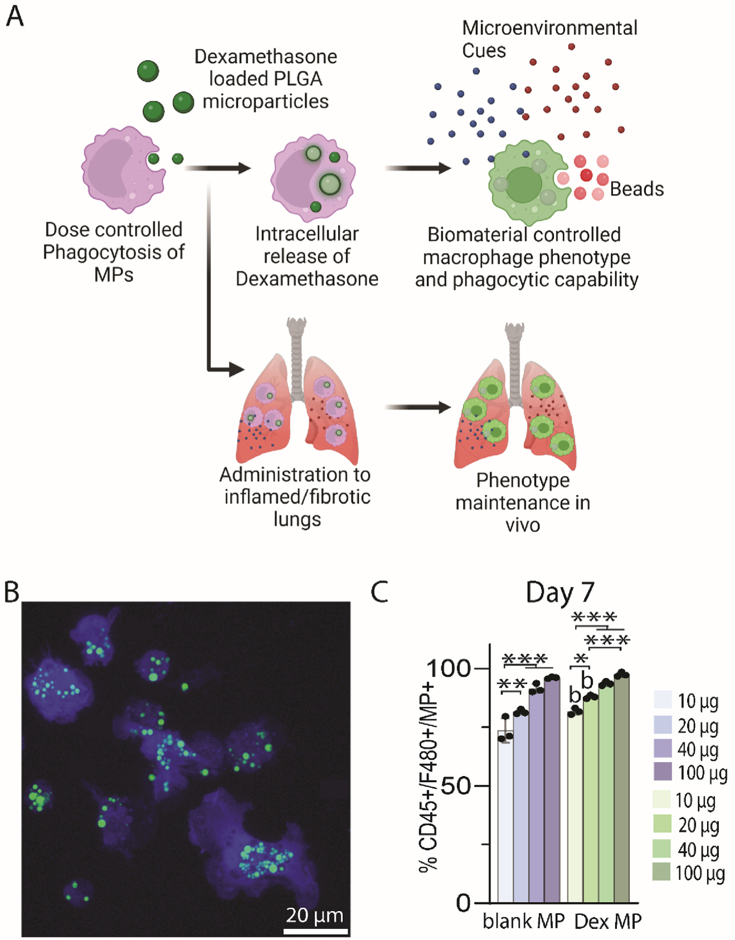
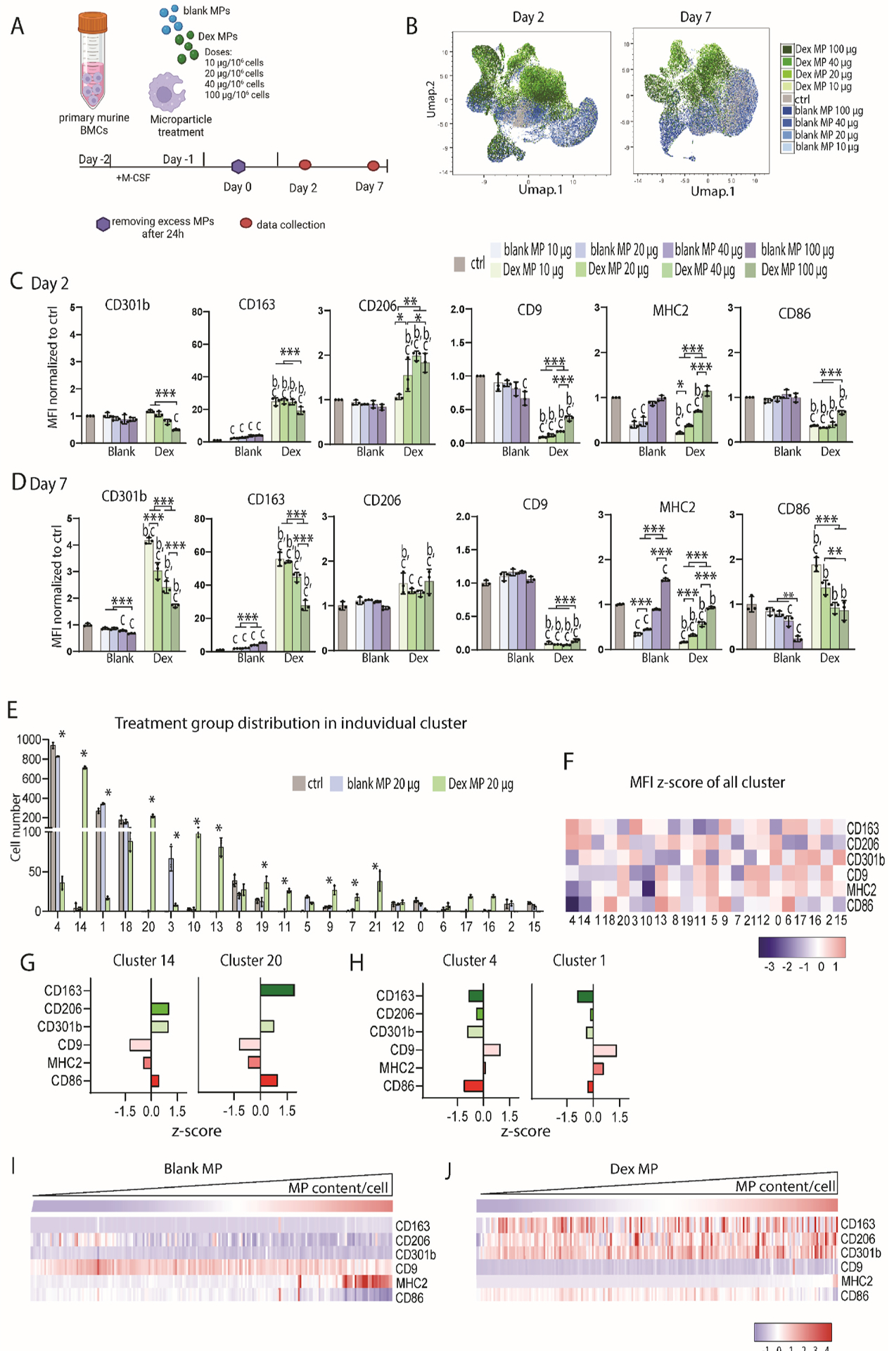
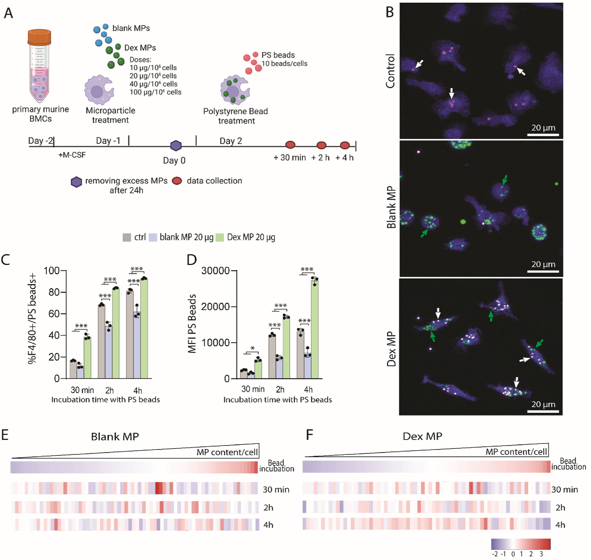
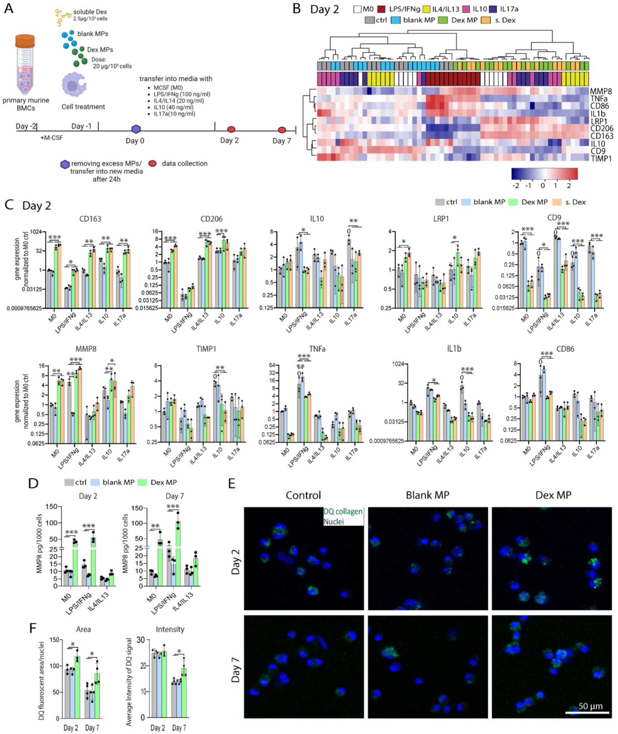
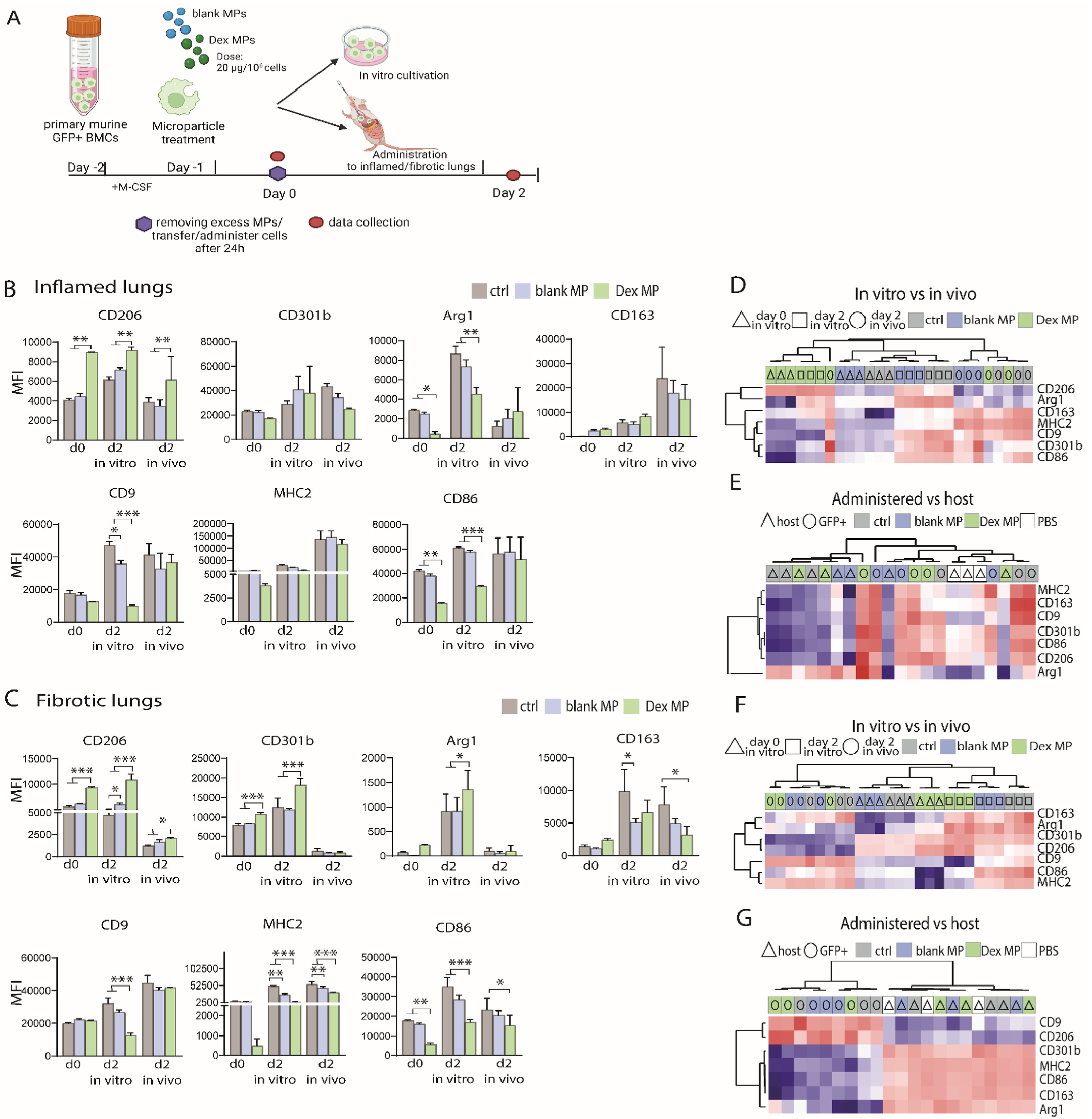
Macrophages are key modulators of all inflammatory diseases and essential for their resolution, making macrophage cell therapy a promising strategy for regenerative medicine. However, since macrophages change rapidly in response to microenvironmental cues, their phenotype must be controlled post-administration. We present a tunable biomaterial-based strategy to control macrophages intracellularly via small molecule-releasing microparticles. Poly(lactic-co-glycolic acid) microparticles encapsulating the anti-inflammatory and anti-fibrotic drug dexamethasone were administered to macrophages in vitro, with uptake rates controlled by different loading regimes. Microparticle dose and dexamethasone content directly affected macrophage phenotype and phagocytic capacity, independent of particle content per cell, leading to an overall pro-reparative, anti-inflammatory, anti-fibrotic phenotype with increased phagocytic and ECM degrading functionality. Intracellularly controlled macrophages partially maintained this phenotype in vivo in a murine pulmonary fibrosis model, with more prominent effects in a pro-fibrotic environment compared to pro-inflammatory. These results suggest that intracellular control using biomaterials has the potential to control macrophage phenotype post-administration, which is essential for successful macrophage cell therapy.

Keywords: Cell therapy; Immunomodulation; Macrophage polarization; Pulmonary fibrosis.


Conflict of interest statement

Declaration of competing interest The authors declare the following financial interests/personal relationships which may be considered as potential competing interests: Kara L. Spiller reports financial support was provided by National Heart Lung and Blood Institute. Kara L. Spiller reports financial support was provided by Coulter-Drexel Translational Research Partnership. Kara L. Spiller has patent pending to Drexel University. If there are other authors, they declare that they have no known competing financial interests or personal relationships that could have appeared to influence the work reported in this paper.

Figures

Fig. 1.
Fig. 1.
Biomaterial mediated intracellular control of macrophage phenotype. (A) Study overview. Dexamethasone-loaded microparticles are phagocytosed by macrophages. Over time, dexamethasone is intracellularly released and promotes an anti-inflammatory, anti-fibrotic phenotype in vitro and in vivo. (B) Representative image of macrophages (stained with CellTracker in blue) loaded with dexamethasone-loaded MPs (false-colored in green) 2 days after incubation with MPs for 24 h. Scale bar: 20 μm. (C) Percentage of macrophages that contain microparticles (MPs) (blank or dexamethasone-loaded) 7 days after administration of the MPs for 24 h. n = 3. Two-way ANOVA with Tukey post hoc, *p < 0.05,**p < 0.01, ***p < 0.001, bp < 0.05 compared to respective blank MP group.
Fig. 2.
Fig. 2.
Microparticle dosing affects macrophage phenotype. (A) Experimental set-up. Bone marrow-derived cells (BMCs) were cultivated for 24 h before blank or Dexamethasone encapsulated (Dex) MPs with varying doses (10 μg/106 cells, 20 μg/106 cells, 40 μg/106 cells, 100 μg/106 cells) were administered to the cells for 24 h. Then, MPs were removed, and cells were cultivated for up to 7 days following the removal of excess MPs. (B) Umap cluster analysis based on macrophage treatment groups 2 days and 7 days after incubation with MPs presenting differences between groups. (C,D) Protein expression analysis via flow cytometry 2 days (C) and 7 days (D) after incubation with MPs. One-way ANOVA with Tukey post hoc, *p < 0.05,**p < 0.01, ***p < 0.001, bp < 0.05 compared to respective blank MP group, cp < 0.05 compared to control. Statistics were calculated based on Log 2 of unnormalized data. (E–H) Multidimensional analysis of protein marker expression 7 days after incubation with blank and Dex MPs. Cluster analysis was performed via FlowSOM (Flowjo) after the cluster number was determined via Phenograph. (E) Distribution of treatment groups in each cluster. *indicates significances of p < 0.05 between treatment groups. Two-way ANOVA with Tukey post hoc. (F) Heatmap showing MFI values of each cluster and tested marker. (G,H) Bar graphs showing MFI Z-score for each marker in clusters with the greatest differences between treatment groups of Dex-MP dominant (G) and untreated and blank MP dominant cluster (H). (I,J) Phenograph single-cell cluster analysis based on MP content per cell (MFI of TRITC-labeled MPs) 7 days after incubation with blank MPs (I) and Dex MPs (J). n = 3. For simplification, only the 20 μg dose is shown, with other doses shown in Figs. S6–8.
Fig. 3.
Fig. 3.
Influence of MP dose and content per cell on phagocytic capacity of macrophages. (A) Experimental set-up. Bone marrow-derived cells (BMCs) were cultivated for 24 h before blank or dexamethasone encapsulated (Dex) MPs with varying doses (10 μg/106 cells, 20 μg/106 cells, 40 μg/106 cells, 100 μg/106 cells) were administered to the cells for 24 h followed by removal of excess MPs. After 2 days cells were incubated with fluorescent polystyrene (PS) beads (10 beads/cell) for up to 4 h and uptake was analyzed via flow cytometry and live cell imaging. (B) Representative images of nontreated control, blank MP treated and Dex MP-treated macrophages (blue with false-colored green MPs indicated by green arrows) after incubation with PS beads (false-colored in red indicated by white arrows) for 4 h. Scale bar: 20 μm. (C) Percentage of PS bead-positive cells after incubation for 30 min, 2 h and 4 h. (D) MFI values of PS beads in cells after incubation for 30 min, 2 h, and 4 h. (E,F) Phenograph single-cell cluster analysis based on MP content per cell (MFI of TRITC-labeled MPs) of blank MP (E) and Dex MP (F) treated cells 30 min, 2 h and 4 h after incubation with PS beads. Z-scores represented in the heatmap were calculated within bead incubation time point groups. Two-way ANOVA with Tukey post hoc, *p < 0.05,**p < 0.01, ***p < 0.001, bp < 0.05 compared to respective blank MP group, cp < 0.05 compared to control. n = 3. For simplification, only the 20 μg MP dose is shown, whereas other doses are shown in Fig. S9.
Fig. 4.
Fig. 4.
Microenvironmental stimuli challenge of macrophage phenotypes in vitro. (A) Experimental set-up. Bone marrow-derived cells (BMCs) were cultivated for 24 h before 20 μg/106 cells blank or dexamethasone encapsulated (Dex) MPs or soluble dexamethasone (s.Dex, 2.5 μg/106 cells) were administered to the cells for 24 h. Then, cells were washed, transferred into new media containing stimuli that were pro-inflammatory (LPS + IFNg), Th2/pro-fibrotic (IL4+IL13), immunosuppressive (IL10), or Th17/fibro-inflammatory (IL17a), and cultivated for up to 7 days. (B) Hierarchical clustering of samples and genes in the heatmap represents Z-score of relative gene expression 2 days after transfer to media supplemented with MCSF (M0) or additional stimuli. (C) Single marker presentation of relative gene expression of macrophages 2 days after transfer to environmental stimuli supplemented media. (D) MMP8 secretion analyzed by ELISA 2 and 7 days after transfer to media supplemented with MCSF (M0) or supplemented additionally with LPS/IFNg or IL4/IL13. n = 3. (E) Representative images of untreated control macrophages, blank MP-loaded and Dex-MP-loaded macrophages 2 and 7 days following incubation with MPs after 24-h cultivation on DQ-collagen I-containing collagen I coatings. The nuclei of macrophages were stained with Hoechst 33342 in blue. Green fluorescent areas indicate collagen I degradation. (F) Quantitative image analysis of the DQ collagen I signal for area and intensity after 24-h macrophage cultivation on DQ collagen-containing collagen coatings 2 and 7 days after MP incubation. For area calculations, total area per image was divided by the number of nuclei per image. For intensity calculations, the average intensity per DQ fluorescent event was calculated per image. For each sample, the average of 4 images are presented. Two-way ANOVA with Tukey post hoc, *p < 0.05,**p < 0.01, ***p < 0.001, 0p < 0.05 compared to M0 ctrl.
Fig. 5.
Fig. 5.
Phenotype analysis of macrophages administered to inflamed and fibrotic lungs after influenza infection. (A) Experimental set-up. Bone marrow-derived cells (BMCs) were cultivated for 24 h before blank or dexamethasone encapsulated (Dex) MPs at a dose of 20 μg/106 cells were administered to the cells for 24 h. Then, cells were washed and either incubated in vitro or administered to inflamed or fibrotic lungs of mice for 2 days. (B,C) Protein marker expression of GFP + administered macrophages was analyzed via flow cytometry 2 days after administration into inflamed (B) and fibrotic (E) lungs. (D,F) Hierarchical clustering based on MFI values of protein expression of in vitro cultivated vs. GFP + macrophages administered into inflamed (C) or fibrotic lungs (F). (E,G) Hierarchical clustering based on MFI values of administered GFP + macrophages vs. host macrophages under inflamed (D) or fibrotic (G) conditions. Two-way ANOVA with Tukey post hoc, n = 3. *p < 0.05,**p < 0.01, ***p < 0.001.

Similar articles

References

    1. Zhang L, Wang Y, Wu G, Xiong W, Gu W, Wang C-Y, Macrophages: friend or foe in idiopathic pulmonary fibrosis? Respir. Res 19 (1) (2018) 170. - PMC - PubMed
    1. Ardura JA, Rackov G, Izquierdo E, Alonso V, Gortazar AR, Escribese MM, Targeting macrophages: friends or foes in disease? Front. Pharmacol 10 (2019). - PMC - PubMed
    1. Watanabe S, Alexander M, Misharin AV, Budinger GRS, The role of macrophages in the resolution of inflammation, J. Clin. Invest 129 (7) (2019) 2619–2628. - PMC - PubMed
    1. Riario Sforza GG, Marinou A, Hypersensitivity pneumonitis: a complex lung disease, Clin. Mol. Allergy 15 (1) (2017) 6. - PMC - PubMed
    1. Huang WJ, Tang XX, Virus infection induced pulmonary fibrosis, J. Transl. Med 19 (1) (2021) 496. - PMC - PubMed

Publication types

MeSH terms

LinkOut - more resources