. 2024 Feb 24;17(3):293.
doi: 10.3390/ph17030293.

TRPV4 Activation during Guinea Pig Airway Smooth Muscle Contraction Promotes Ca2+ and Na+ Influx

Affiliations

TRPV4 Activation during Guinea Pig Airway Smooth Muscle Contraction Promotes Ca2+ and Na+ Influx

Luis M Montaño et al. Pharmaceuticals (Basel). .

Abstract

Airway smooth muscle (ASM) contraction is determined by the increase in intracellular Ca2+ concentration ([Ca2+]i) caused by its release from the sarcoplasmic reticulum (SR) or by extracellular Ca2+ influx. Major channels involved in Ca2+ influx in ASM cells are L-type voltage-dependent Ca2+ channels (L-VDCCs) and nonselective cation channels (NSCCs). Transient receptor potential vanilloid 4 (TRPV4) is an NSCC recently studied in ASM. Mechanical stimuli, such as contraction, can activate TRPV4. We investigated the possible activation of TRPV4 by histamine (His)- or carbachol (CCh)-induced contraction in guinea pig ASM. In single myocytes, the TRPV4 agonist (GSK101) evoked an increase in [Ca2+]i, characterized by a slow onset and a plateau phase. The TRPV4 antagonist (GSK219) decreased channel activity by 94%, whereas the Ca2+-free medium abolished the Ca2+ response induced by GSK101. Moreover, GSK101 caused Na+ influx in tracheal myocytes. GSK219 reduced the Ca2+ peak and the Ca2+ plateau triggered by His or CCh. TRPV4 blockade shifted the concentration-response curve relating to His and CCh to the right in tracheal rings and reduced the maximal contraction. Finally, the activation of TRPV4 in single myocytes increased the Ca2+ refilling of the SR. We conclude that contraction of ASM cells after stimulation with His or CCh promotes TRPV4 activation, the subsequent influx of Ca2+ and Na+, and the opening of L-VDCCs. The entry of Ca2+ into ASM cells via TRPV4 and L-VDCCs contributes to optimal smooth muscle contraction.

Keywords: TRPV4; airway smooth muscle; asthma; intracellular Ca2+ concentration; smooth muscle contraction.


Conflict of interest statement

The authors declare no conflicts of interest. The funders had no role in the design of the study; in the collection, analyses, or interpretation of data; in the writing of the manuscript; or in the decision to publish the results. Figure 12 of this manuscript was created with BioRender.com.

Figures

Figure 1
Figure 1
TRPV4 expression in guinea pig airway smooth muscle (ASM). Tracheal smooth muscle from two animals (ASM 1, ASM 2) was collected and dissected free from epithelium and connective tissue. Total protein was extracted, and electrophoresis (SDS-PAGE) was performed with 25 µg of total protein. A TRPV4-specific antibody (#ACC-034) was used, followed by an HRP-conjugated secondary antibody. The molecular weight (MW) is given according to the information in the commercial data sheets of the antibody. ASM 1 and ASM 2 show the blots illustrating one band around 100 kDa corresponding to TRPV4. Each blot represents a tissue from two different animals.
Figure 2
Figure 2
The increase in intracellular Ca2+ concentration ([Ca2+]i) in guinea pig tracheal myocytes induced by TRPV4 stimulation depends on the influx of Ca2+ from the extracellular medium. (A) Tracheal smooth muscle cells were constantly perfused with Krebs solution at 37 °C. The addition of 32 nM GSK101 (a selective TRPV4 agonist) increased the [Ca2+]i ~70 nanomoles, with a slow onset and plateau phase (1). Preincubation of cells with the selective TRPV4 antagonist (GSK219, 100 nM) reduced the increase in the [Ca2+]i caused by a second stimulation with GSK101 (32 nM) by ~94% (2). After the TRPV4 antagonist was washed out, a third stimulation with 32 nM GSK101 caused a Ca2+ response that was similar to the first stimulation (3). (B) The graph summarizes the percentage of Ca2+ response caused by stimulation of TRPV4 in the presence and after using the antagonist GSK219, 100 nM. ** p < 0.01 compared with the control group (the first stimulus with 32 nM GSK101). (C) Perfusion of TRPV4 agonist in a Ca2+-free medium did not increase [Ca2+]i, indicating that the Ca2+ response through the activation of this channel is mediated solely by the influx of extracellular Ca2+. (D) The bar graph shows the statistical analysis of the Ca2+ response (expressed as a percentage) after TRPV4 activation in a medium without or with Ca2+. Bars represent the mean plus the standard error of the mean (S.E.M.), n = 5. Repeated-measures analysis of variance followed by Dunnett’s Multiple Comparison Test was performed. ** p < 0.01. The data in the graph bars are expressed as the percentage of the first stimulus with GSK101 (corresponding to 100% of the total Ca2+ response).
Figure 3
Figure 3
Possible involvement of TRPV4 in activating L-type voltage-dependent Ca2+ channels in guinea pig tracheal myocytes. (A) When 32 nM GSK101 (a selective TRPV4 agonist) was added, the fluorescence increased slowly and reached a plateau phase (1). This indicates that when TRPV4 is opened, Na+ influx into the cytoplasm is promoted. However, when cells were preincubated with GSK219 (a selective TRPV4 antagonist, 100 nM), the increase in fluorescence caused by a second stimulation with GSK101 (32 nM) was reduced by 99% (2). After the washout of the TRPV4 antagonist, a third stimulus with 32 nM GSK101 elicited a response similar to the first stimulus (3). (B) The graph shows the increase in SBFI-AM fluorescence (expressed as a percentage) caused by TRPV4 stimulation in the presence and after the use of the antagonist GSK219 (100 nM). (C) Methoxy verapamil (D-600, 30 µM), a blocker of L-type voltage-gated Ca2+ channels (L-VDCCs), significantly decreased the Ca2+ increase provoked by stimulation with the selective TRPV4 agonist (GSK101, 32 nM). The original record shows that the addition of 32 nM GSK101 caused an increase in [Ca2+]i, characterized by a slow onset and a plateau phase. After a second stimulus with the TRPV4 agonist, a 5 min preincubation with D-600 led to a significant decrease in the Ca2+ response by approximately 20%. These results suggest that TRPV4 opening causes Na+ influx into the cytoplasm of tracheal myocytes, which leads to depolarization of the plasma membrane and opening of L-VDCCs. (D) The bar graph depicts the inhibition expressed as a percentage of Ca2+ increase by adding D-600. * p < 0.05, ** p < 0.01 (compared with the control group of 4–5 experiments). Repeated-measures analysis of variance followed by Dunnett’s Multiple Comparison Test was performed. The data in the graph bars are expressed as the percentage of the first stimulus with GSK101 (corresponding to 100% of the total Ca2+ or Na+ response).
Figure 4
Figure 4
Contraction induced by TRPV4 activation is mediated in part by the opening of L-VDCCs. (A) The original traces show smooth muscle contraction induced by a cumulative concentration–response curve to TRPV4 agonist (GSK101) after a third stimulus with 60 mM KCl. The black arrows illustrate the addition of the different tested concentrations of GSK101 (32, 56, 100, 170, 320, 560, 1000, 1700, and 3200 nM). (B) The graph shows the cumulative effect of the administration of the TRPV4 agonist GSK101 (32–3200 nM). The antagonist GSK219 (100 nM) almost abolished the contraction response, while 30 µM D-600 significantly reduced the contraction response to GSK101. * p < 0.05, ** p < 0.01. n = 6–11. A one-way analysis of variance followed by Dunnett’s Multiple Comparison Test was performed.
Figure 5
Figure 5
TRPV4 antagonist decreases carbachol (CCh) responsiveness in guinea pig tracheal smooth muscle. (A) Cumulative administration of CCh (10, 32, 100, and 320 nM, and 1 and 3.2 µM) caused concentration-dependent contraction of tracheal tissue (control group). Acute exposure of GSK219 at different concentrations (10, 100, and 1000 nM) shifted the concentration–response curve of CCh to the right and decreased the maximal contraction response. (B) Bar graphs show the analysis of effective concentration 50 (EC50) and maximum response of CCh. Symbols and bars illustrate mean ± S.E.M. Repeated measures analysis of variance followed by Dunnett’s multiple comparison test was performed. ** p < 0.01, n = 7.
Figure 6
Figure 6
Pharmacological blockade of TRPV4 decreased the response to Histamine (His) during guinea pig tracheal contraction. (A) Cumulative administration of His (0.1, 0.32, 1, 3.2, 10, and 32 μM) elicited concentration-dependent contraction of guinea pig tracheal rings (control group). Different concentrations of GSK219 (10, 100, and 1000 nM) shifted the concentration–response curve relating to His to the right. (B) Bar graphs illustrate the analysis of the effective concentration 50 (EC50) and maximum response of His. Notice that the reduction in EC50 relating to His caused by GSK219 in the nanomolar range was concentration–dependent. Symbols and bars depict mean ± S.E.M. Repeated measures analysis of variance followed by Dunnett’s multiple comparison test was performed. * p < 0.05, ** p < 0.01, n = 7.
Figure 7
Figure 7
TRPV4 is activated during the stimulation of airway smooth muscle cells with carbachol. (A) Smooth muscle cells from the trachea were continuously perfused with Krebs solution at 37 °C. The addition of 1 µM carbachol (CCh) resulted in an increase in the intracellular Ca2+ concentration ([Ca2+]i), which was characterized by a rapid onset (a peak) at approximately 600 nM followed by a sustained plateau at around 75 nM (1). When a selective TRPV4 antagonist (100 nM GSK219) was administered for 5 min, it significantly reduced the CCh-induced increase in [Ca2+]i. The Ca2+ peak was reduced by approximately 22%, while the plateau was decreased by 24% in the presence of GSK219 in Krebs solution (2). After the TRPV4 antagonist was washed out, a third stimulation with 1 µM CCh produced a Ca2+ response similar to the first stimulation (3). (B) The graph illustrates the percentage of the Ca2+ response elicited by CCh stimulus in the presence and after use of the antagonist GSK219 (100 nM), n = 5. The use of GSK219 resulted in a significant reduction in the Ca2+ response elicited by CCh. * p < 0.05. Repeated measures analysis of variance followed by Dunnett’s multiple comparison test was performed. The data in the graph bars are expressed as the percentage of the first stimulus with CCh 1 µM (corresponding to 100% of the total Ca2+ response).
Figure 8
Figure 8
Blockade of TRPV4 decreases histamine-induced increase in intracellular Ca2+ concentration ([Ca2+]i) in guinea pig tracheal smooth muscle cells. (A) Single tracheal smooth muscle cells were constantly perfused with Krebs solution at 37 °C. The addition of 10 µM histamine (His) resulted in an increase in [Ca2+]i characterized by a rapid onset (a peak) around 600 nM and a sustained plateau around 70 nM (1). Preincubation of cells with the selective TRPV4 antagonist (100 nM GSK219) decreased the peak Ca2+ and plateau by ~14% and ~21%, respectively, produced by a second stimulation with 10 µM His (2). After the washout of the TRPV4 antagonist, a third stimulation with 10 µM His produced a Ca2+ response similar to the first stimulation (3). (B) The graph illustrates the percentage of Ca2+ response elicited by His in the presence and after use of the antagonist GSK219 (100 nM), n = 5. * p < 0.05. Repeated measures analysis of variance followed by Dunnett’s multiple comparison test was performed. The data in the graph bars are expressed as the percentage of the first stimulus with 10 µM His (corresponding to 100% of the total Ca2+ response).
Figure 9
Figure 9
The TRPV4 antagonist GSK219 decreases the contraction response to KCl in guinea pig tracheal rings. (A) Cumulative administration of 10, 20, 40, 80, 120, and 160 mM KCl caused plasma membrane depolarization and subsequent contraction of tracheal tissues in a concentration-dependent manner (control group). Incubation of tracheal rings for 15 min with 100 nM GSK219 shifted the concentration–response curve of KCl to the right and decreased the maximal contraction response obtained by adding KCl. (B) Bar graphs depict the analysis of effective concentration 50 (EC50) and maximum response of KCl. Symbols and bars depict mean ± S.E.M. A Student’s t-test was performed for paired data (n = 6). * p < 0.05 compared with the control group.
Figure 10
Figure 10
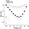
The TRPV4 antagonist (GSK219) does not alter Ca2+ currents in guinea pig airway smooth muscle cells. A protocol of depolarizing pulses from −60 to +50 mV in 10 mV steps produced Ba2+ inward currents that peaked at about 0.21 nanoamperes (nA). Ba2+ was used as an ion carrier instead of Ca2+ to enhance current responses through Ca2+ channels during the stimulus of depolarization. Application of 100 nM GSK219 did not alter Ba2+ currents. However, nifedipine, a blocker of L-type voltage-dependent Ca2+ channels, almost abolished them. Symbols and bars depict mean ± S.E.M. Repeated-measures analysis of variance followed by Dunnett’s test was performed. * p < 0.05, ** p < 0.01, n = 5.
Figure 11
Figure 11
The role of TRPV4 in sarcoplasmic reticulum Ca2+ refilling was indirectly evaluated by an experimental method of repeated stimulation with caffeine in guinea pig tracheal myocytes. (A) Original fluorometric recording showing S1 as the first stimulation with caffeine (10 mM) in a Ca2+-free medium for 10 min (to promote depletion of SR) and S2 as the second response in a Ca2+-free medium. Replenishment of RS with Ca2+ was enabled by perfusing the cell with Krebs solution containing 2 mM Ca2+ after S1 and before S2 stimulation. (B) Blocking TRPV4 for 4 min with 100 nM GSK219 before stimulation with S2 decreased Ca2+ replenishment, while activation (C) with 32 nM GSK101 increased Ca2+ replenishment of SR. (D) Bar graph illustrating the decrease and increase in Ca2+ replenishment rate of SR, expressed as S2/S1 ratio, caused by TRPV4 blockade or activation, respectively. One-way analysis of variance, followed by Dunnett’s test, was performed. * p < 0.05, n = 4–5.
Figure 12
Figure 12
Schematic representation of the proposed role of TRPV4 in airway smooth muscle contraction. Bronchoconstrictor agonists such as carbachol or histamine occupy their respective G protein-coupled receptor (GPCR), inducing the disassembly of the trimeric Gq protein into subunits α and β/γ. Later, Gqα activates phospholipase C (PLC) to induce the formation of inositol triphosphate (IP3) and diacylglycerol (DAG). IP3 stimulates its receptor (IP3R) in the sarcoplasmic reticulum (SR), driving the release of Ca2+ into the cytoplasm, while DAG activates protein kinase C (PKC), which phosphorylates targets involved in smooth muscle contraction. In the cytoplasm, Ca2+ binds to calmodulin (CAM), triggering myosin light chain kinase (MLCK) action. This kinase phosphorylates the regulatory chain of myosin, inducing the cross-bridge cycle with actin and smooth muscle contraction in the airway. Chemical agents such as KCl also produce smooth muscle contraction by causing membrane depolarization and opening the L-type voltage-dependent Ca2+ channel (L-VDCC). This channel allows the influx of Ca2+ into the cytoplasm and the subsequent contraction. Contractile stimulus is detected by TRPV4, which in turn triggers the influx of Ca2+ and Na+. This latter ion favors membrane depolarization and the opening of the L-VDCC. Ca2+ entering through TRPV4 and L-VDCC are taken up into the SR via the sarcoplasmic Ca2+ ATPase (SERCA), contributing to the refilling of Ca2+ of this organelle. The activation of TRPV4 and Ca2+ and Na+ influx through this polymodal channel participates in the optimal contraction of airway smooth muscle.
Figure 13
Figure 13
Chemical structure of the TRPV4 agonist, GSK1016790A (A) and the TRPV4 antagonist GSK2193874 (B).

Similar articles

References

    1. Lam M., Lamanna E., Bourke J.E. Regulation of airway smooth muscle contraction in health and disease. Adv. Exp. Med. Biol. 2019;1124:381–422. doi: 10.1007/978-981-13-5895-1_16. - DOI - PubMed
    1. Abohalaka R. Bronchial epithelial and airway smooth muscle cell interactions in health and disease. Heliyon. 2023;9:e19976. doi: 10.1016/j.heliyon.2023.e19976. - DOI - PMC - PubMed
    1. Shetty S.M., Archana S. In: Anatomy and Physiology of the Airway Applied Aspects. Ubaradka R.S., Gupta N., Bidkar P.U., Tripathy D.K., Gupta A., editors. Springer; Singapore: 2023. pp. 21–43. The Airway Manual. - DOI
    1. Montaño L.M., Flores-Soto E., Reyes-García J., Solis-Chagoyán H., Sommer B. Airway smooth muscle functioning in basal, agonists stimulated conditions and novel androgen asthma therapy. Adv. Med. Biol. 2020;157:1–64.
    1. Xiong D.J.P., Martin J.G., Lauzon A.M. Airway smooth muscle function in asthma. Front. Physiol. 2022;13:993406. doi: 10.3389/fphys.2022.993406. - DOI - PMC - PubMed