. 2024 Mar 6;8(1):10.
doi: 10.3390/epigenomes8010010.

Exploring Transcriptional Regulation of Beta Cell SASP by Brd4-Associated Proteins and Cell Cycle Control Protein p21

Affiliations

Exploring Transcriptional Regulation of Beta Cell SASP by Brd4-Associated Proteins and Cell Cycle Control Protein p21

Jasmine Manji et al. Epigenomes. .

Abstract

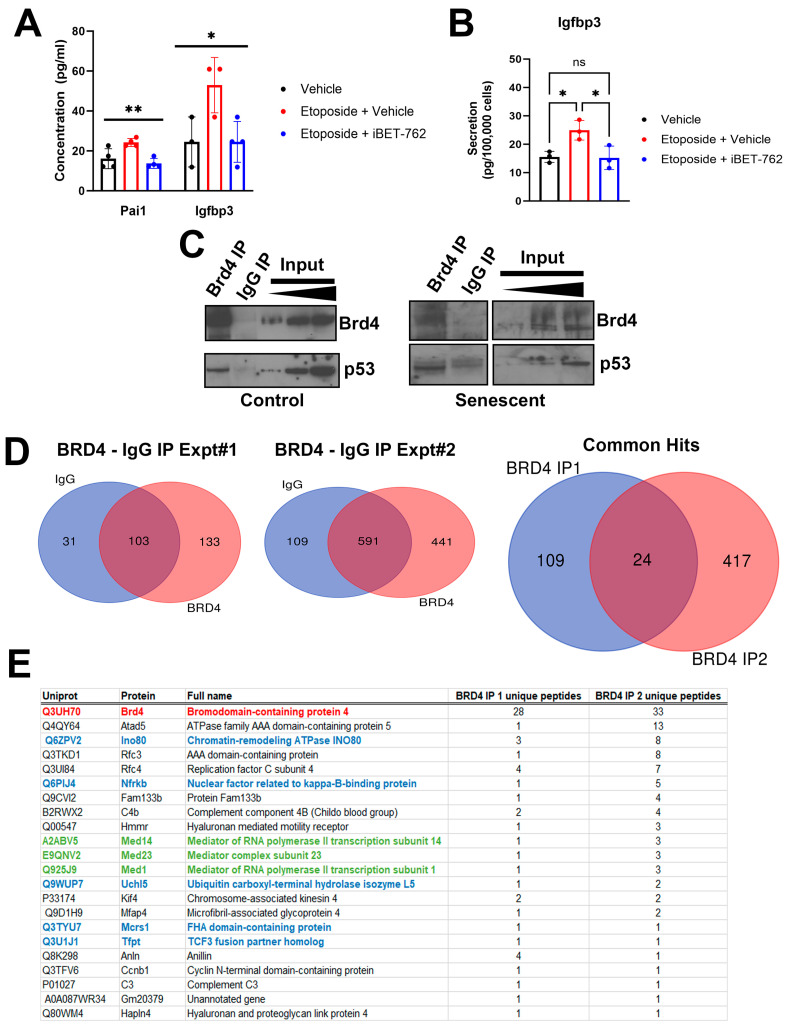
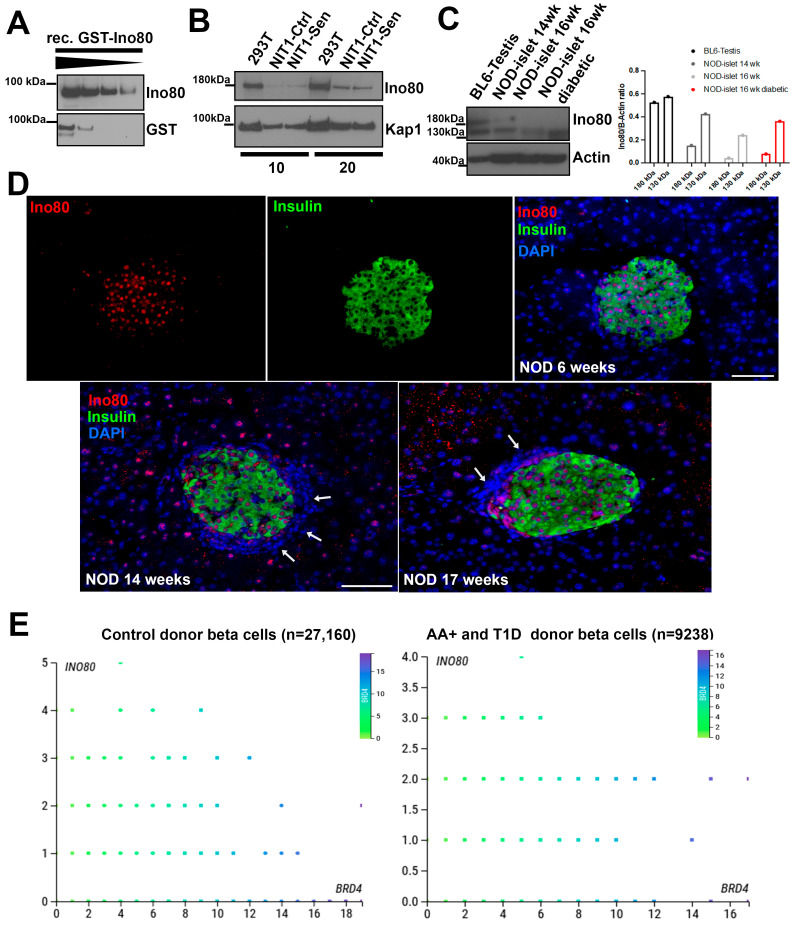
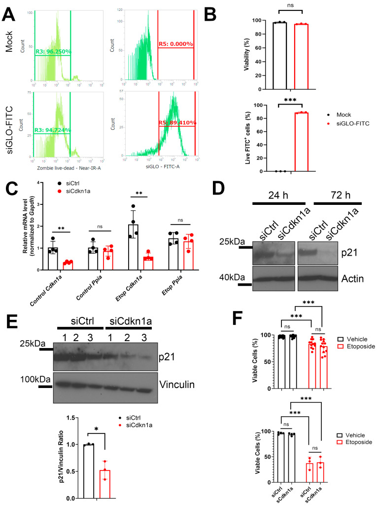
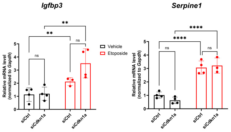
Type 1 diabetes (T1D) is a metabolic disease resulting from progressive autoimmune destruction of insulin-producing pancreatic beta cells. Although the majority of beta cells are lost in T1D, a small subset undergoes senescence, a stress response involving growth arrest, DNA damage response, and activation of a senescence-associated secretory phenotype (SASP). SASP in beta cells of the nonobese diabetic (NOD) mouse model of T1D and primary human islets is regulated at the level of transcription by bromodomain extra-terminal (BET) proteins, but the mechanisms remain unclear. To explore how SASP is transcriptionally regulated in beta cells, we used the NOD beta cell line NIT-1 to model beta cell SASP and identified binding partners of BET protein Brd4 and explored the role of the cyclin-dependent kinase inhibitor p21. Brd4 interacted with a variety of proteins in senescent NIT-1 cells including subunits of the Ino80 chromatin remodeling complex, which was expressed in beta cells during T1D progression in NOD mice and in human beta cells of control, autoantibody-positive, and T1D donors as determined from single-cell RNA-seq data. RNAi knockdown of p21 during senescence in NIT-1 cells did not significantly impact viability or SASP. Taken together, these results suggest that Brd4 interacts with several protein partners during senescence in NIT-1 cells, some of which may play roles in SASP gene activation and that p21 is dispensable for the SASP in this beta cell model.

Keywords: SASP; bromodomain extra-terminal domain proteins; cellular senescence; pancreatic beta cells; type 1 diabetes.


Conflict of interest statement

The authors declare no conflict of interest. The funders had no role in the design of the study; in the collection, analyses, or interpretation of data; in the writing of the manuscript; or in the decision to publish the results.

Figures

Figure 1
Figure 1
Regulation of SASP by BET proteins and identification of Brd4-associated proteins during senescence in NIT-1 cells. (A) Luminex assays measuring the concentration of SASP factors Pai1 and Igfbp3 in the 24 h conditioned media collected from NIT-1 cells control (vehicle, DMSO), or senescent cells (0.25 µM etoposide treatment) at 11 days post-etoposide washout. Following drug washout, cells were treated once with vehicle or iBET-762 (5 µM) until harvest. (B) A repeated experiment as in (A) except for assaying Igfpb3 secretion by normalizing the amount in the conditioned media to the viable cell counts at the end of the assay at day 7 post-etoposide washout. (C) Immunoprecipitation (IP) of Brd4 followed by Western blots for Brd4 and p53 from 100 µg of nuclear extract protein of control or senescent NIT-1 cells at day 6 post-etoposide treatment. Input is 3, 5, and 10 µg of nuclear extract and rabbit IgG was the species matched control IP. (D) Mass spectrometry was performed on IgG control and Brd4 IPs from senescent NIT-1 cell nuclear extracts in two independent experiments on senescent NIT-1 cells at day 6 post-etoposide treatment. The number of proteins identified uniquely or in common in the datasets are shown. Brd4 IP experiment 1 and 2 identified a total of 24 common accessions (22 different proteins). (E) Table listing the 22 different proteins in the Brd4 IP1 and IP2. Brd4 is highlighted in red, subunits of the Ino80 complex are in blue, and the Mediator complex is in green. ns, not significant, * p < 0.05, ** p < 0.005, one-way ANOVA.
Figure 2
Figure 2
Characterization of Ino80 protein and INO80 gene expression during T1D in NOD mice and humans, respectively. (A) Western blot analysis validating Ino80 antibody on a recombinant GST-tagged partial Ino80 protein (~90 kDa in size) with 200, 100, 50, 25 ng loaded per lane. (B) Western blot of Ino80 on 10 or 20 µg of nuclear extracts from human 293T cells as a control, or control and senescent NIT-1 cells at day 5 post-etoposide washout. Kap1 was a loading control. (C) Western blot of Ino80 on 10 µg of total RIPA protein extracts from 293T cells, C57BL6 3-month-old male testis and isolated islets from female NOD mice at indicated ages, including a 16-week-old diabetic NOD female. Beta-actin was a loading control. The 180 kDa and 130 kDa Ino80 bands from each sample were quantified relative to the beta-actin bands and plotted. (D) Immunohistochemistry and fluorescence staining of Ino80 along with insulin and DAPI as a nuclear counterstain on euglycemic NOD female mouse pancreas sections at 6 weeks, 14 weeks, and 17 weeks of age. Scale bars indicate 50 µm. (E) Analysis of publicly available single-cell RNA-seq data from PANC-DB (Human Pancreas Analysis Project) using CellxGene, comparing expression of INO80 with BRD4 across all beta cells in control donors (n = 27,160) or in autoantibody-positive (AA+) and T1D donors (n = 9238). Dots are colored according to BRD4 expression levels. Y-axis reports binned expression level of INO80 read-counts while the X-axis reports binned expression level of BRD4 read-counts.
Figure 3
Figure 3
RNAi knockdown of Cdkn1a during senescence in NIT-1 cells. (A,B) Viability (gated R3) and transfection efficiency (gated R5) histograms and quantifications of NIT-1 cells transfected with 25 nM siGLO-FITC or mock transfected, 24 h post-transfection. (C) qRT-PCR analysis of Cdkn1a or Ppia as a control gene in control or etoposide induced senescent NIT-1 cells (72 h post-etoposide treatment) at 24 h post-transfection with control non-targeting siRNA (siCtrl), or Cdkn1a siRNA. N = 3 or 4 biological replicates per KD, error bars are S.D. ns = not significant, ** p < 0.005, one-way ANOVAs. (D) Western blot analysis of p21 on whole cell protein extracts prepared from NIT-1 cells transfected with the indicated siRNAs at 24 h or 72 h post-transfection. Beta-actin was as a loading control. (E) Western blot analysis of p21 levels after transfection with control or Cdkn1a siRNA at 72 h post-transfection in control/non-senescent NIT-1 cells in n = 3 biological replicates per transfection. Vinculin was a loading control. Plot shows quantification of p21 normalized to vinculin, error bars are S.D. * p < 0.05 two-tailed t-test. (F) Upper plot: viability assay using trypan blue staining and automated cell counting (Bio-Rad TC-20) of control or senescent NIT-1 cells transfected with the indicated siRNAs at 24 h post-transfection. Data are n = 10–12 biological replicates (upper plot), error bars are S.D. Lower plot: viability assay as in upper plot except 48 h following a second transfection which was conducted 2 days after the first transfection in control or senescent NIT-1 cells. Data are n = 3 or 4 biological replicates, error bars are S.D. ns = not significant, ** p < 0.005, *** p < 0.0005, two-tailed T-tests with multiple comparisons corrections (C,E) or two-way ANOVA in (F).
Figure 4
Figure 4
SASP gene activation is unaffected by KD of Cdkn1a in NIT-1 cells Control or senescent NIT-1 cells were transfected with indicated siRNAs and harvested 24 h post-transfection for qRT-PCR analysis of SASP genes Igfbp3 and Serpine1 (encoding Pai1). Data are n = 3 or 4 biological replicates, error bars are S.D. ns = not significant, ** p < 0.005, **** p < 0.0001, two-way ANOVA.

Similar articles

References

    1. Lawrence J.M., Divers J., Isom S., Saydah S., Imperatore G., Pihoker C., Marcovina S.M., Mayer-Davis E.J., Hamman R.F., Dolan L., et al. Trends in Prevalence of Type 1 and Type 2 Diabetes in Children and Adolescents in the US, 2001–2017. JAMA. 2021;326:717–727. doi: 10.1001/jama.2021.11165. - DOI - PMC - PubMed
    1. Vived C., Lee-Papastavros A., Aparecida da Silva Pereira J., Yi P., MacDonald T.L. Beta cell stress and endocrine function during T1D: What’s next to discover? Endocrinology. 2023;165:bqad162. doi: 10.1210/endocr/bqad162. - DOI - PubMed
    1. Li Q., Xu B., Michie S.A., Rubins K.H., Schreriber R.D., McDevitt H.O. Interferon-α initiates type 1 diabetes in nonobese diabetic mice. Proc. Natl. Acad. Sci. USA. 2008;105:12439–12444. doi: 10.1073/pnas.0806439105. - DOI - PMC - PubMed
    1. Engin F., Yermalovich A., Nguyen T., Hummasti S., Fu W., Eizirik D.L., Mathis D., Hotamisligil G.S. Restoration of the unfolded protein response in pancreatic β cells protects mice against type 1. Sci. Transl. Med. 2013;5:211ra156. doi: 10.1126/scitranslmed.3006534. - DOI - PMC - PubMed
    1. Ovalle F., Grimes T., Xu G., Patel A.J., Grayson T.B., Thielen L.A., Li P., Shalev A. Verapamil and beta cell function in adults with recent-onset type 1 diabetes. Nat. Med. 2018;24:1108–1112. doi: 10.1038/s41591-018-0089-4. - DOI - PMC - PubMed

LinkOut - more resources