. 2024 Mar 26;21(1):75.
doi: 10.1186/s12974-024-03068-w.

Wnt5a/β-catenin-mediated epithelial-mesenchymal transition: a key driver of subretinal fibrosis in neovascular age-related macular degeneration

Affiliations

Wnt5a/β-catenin-mediated epithelial-mesenchymal transition: a key driver of subretinal fibrosis in neovascular age-related macular degeneration

Dandan Liu et al. J Neuroinflammation. .

Abstract

Background: Neovascular age-related macular degeneration (nAMD), accounts for up to 90% of AMD-associated vision loss, ultimately resulting in the formation of fibrotic scar in the macular region. The pathogenesis of subretinal fibrosis in nAMD involves the process of epithelial-mesenchymal transition (EMT) occurring in retinal pigment epithelium (RPE). Here, we aim to investigate the underlying mechanisms involved in the Wnt signaling during the EMT of RPE cells and in the pathological process of subretinal fibrosis secondary to nAMD.

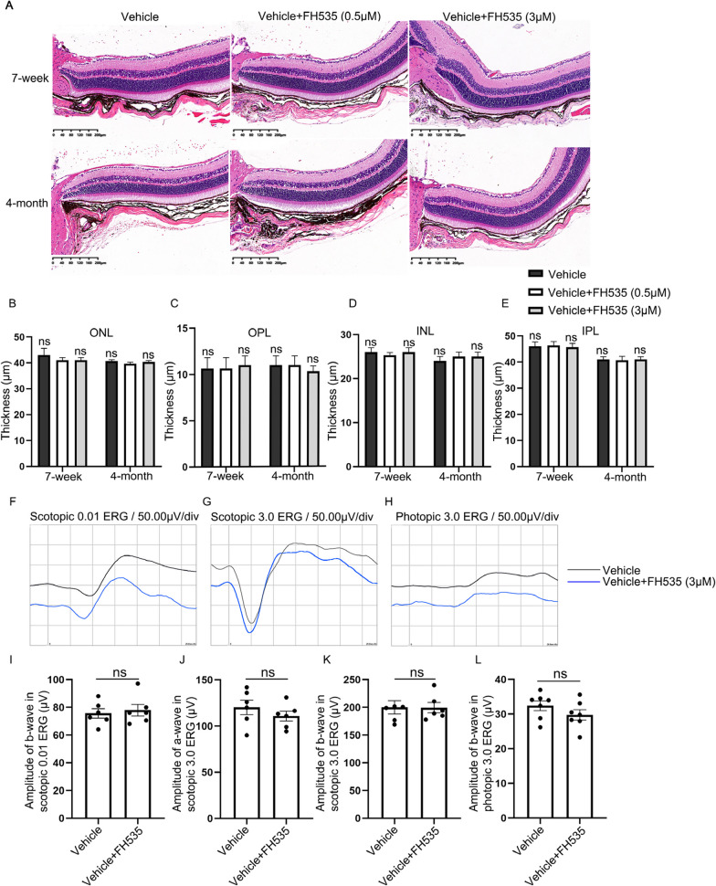
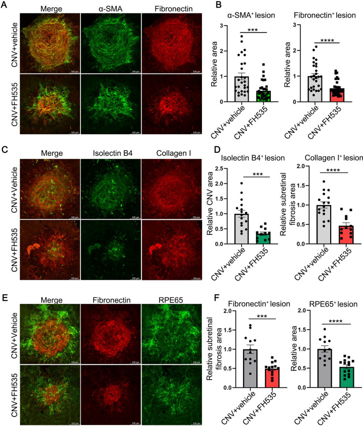
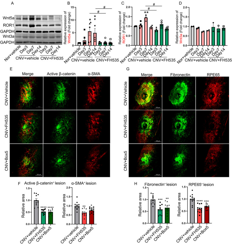
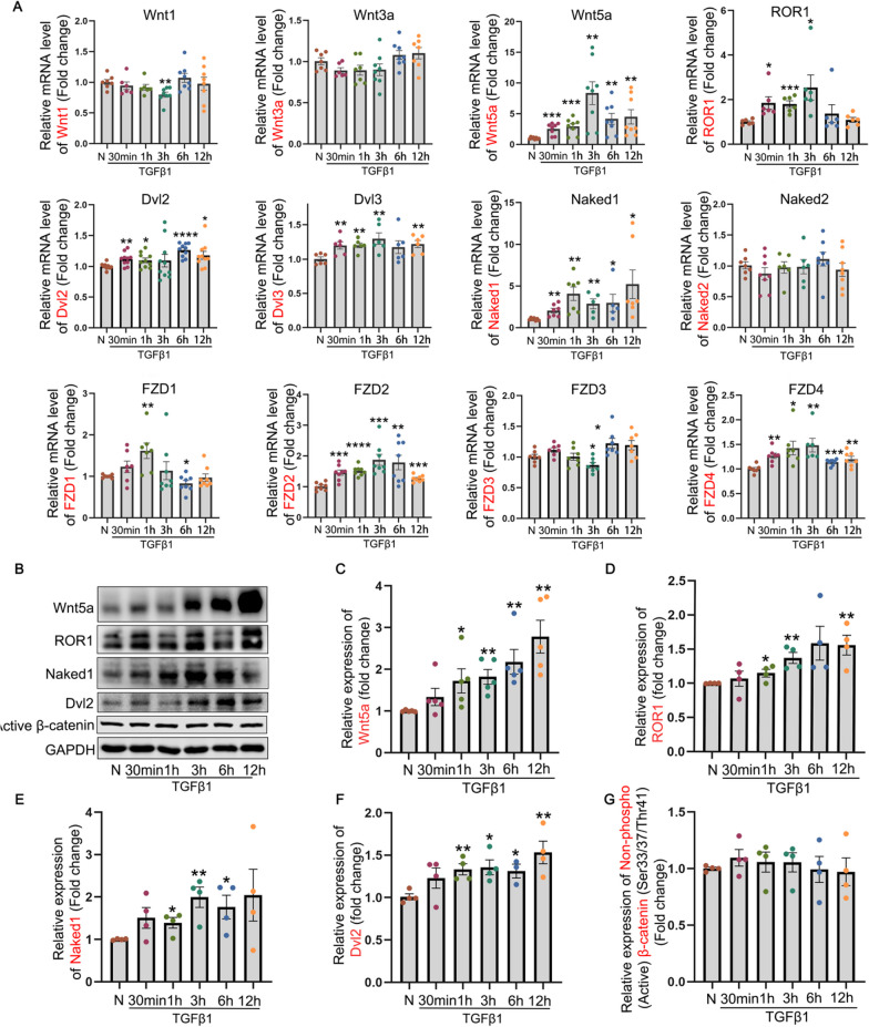
Methods: In vivo, the induction of subretinal fibrosis was performed in male C57BL/6J mice through laser photocoagulation. Either FH535 (a β-catenin inhibitor) or Box5 (a Wnt5a inhibitor) was intravitreally administered on the same day or 14 days following laser induction. The RPE-Bruch's membrane-choriocapillaris complex (RBCC) tissues were collected and subjected to Western blot analysis and immunofluorescence to examine fibrovascular and Wnt-related markers. In vitro, transforming growth factor beta 1 (TGFβ1)-treated ARPE-19 cells were co-incubated with or without FH535, Foxy-5 (a Wnt5a-mimicking peptide), Box5, or Wnt5a shRNA, respectively. The changes in EMT- and Wnt-related signaling molecules, as well as cell functions were assessed using qRT-PCR, nuclear-cytoplasmic fractionation assay, Western blot, immunofluorescence, scratch assay or transwell migration assay. The cell viability of ARPE-19 cells was determined using Cell Counting Kit (CCK)-8.

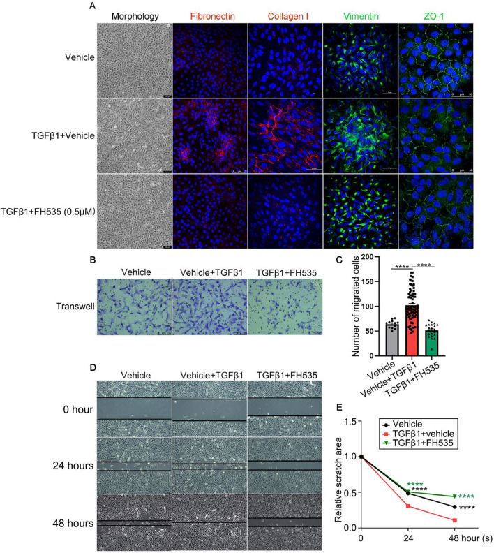
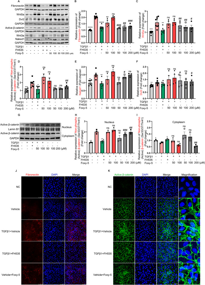
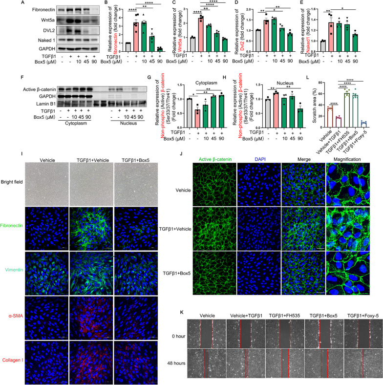
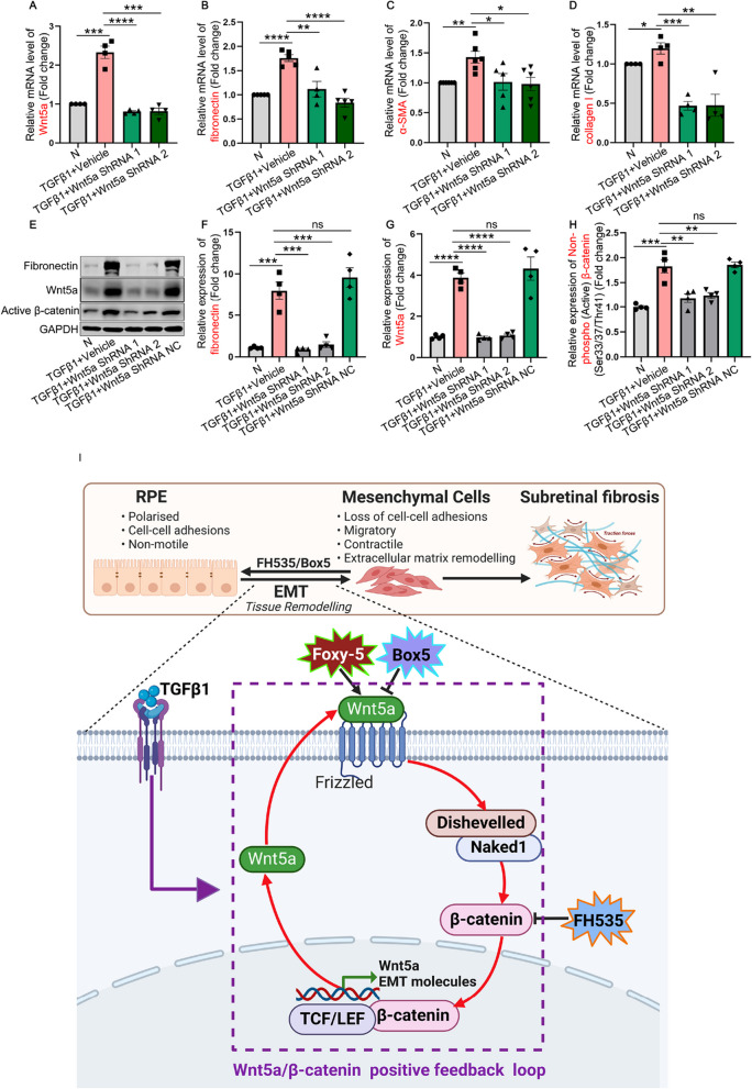
Results: The in vivo analysis demonstrated Wnt5a/ROR1, but not Wnt3a, was upregulated in the RBCCs of the laser-induced CNV mice compared to the normal control group. Intravitreal injection of FH535 effectively reduced Wnt5a protein expression. Both FH535 and Box5 effectively attenuated subretinal fibrosis and EMT, as well as the activation of β-catenin in laser-induced CNV mice, as evidenced by the significant reduction in areas positive for fibronectin, alpha-smooth muscle actin (α-SMA), collagen I, and active β-catenin labeling. In vitro, Wnt5a/ROR1, active β-catenin, and some other Wnt signaling molecules were upregulated in the TGFβ1-induced EMT cell model using ARPE-19 cells. Co-treatment with FH535, Box5, or Wnt5a shRNA markedly suppressed the activation of Wnt5a, nuclear translocation of active β-catenin, as well as the EMT in TGFβ1-treated ARPE-19 cells. Conversely, treatment with Foxy-5 independently resulted in the activation of abovementioned molecules and subsequent induction of EMT in ARPE-19 cells.

Conclusions: Our study reveals a reciprocal activation between Wnt5a and β-catenin to mediate EMT as a pivotal driver of subretinal fibrosis in nAMD. This positive feedback loop provides valuable insights into potential therapeutic strategies to treat subretinal fibrosis in nAMD patients.

Keywords: Epithelial–mesenchymal transition; Neovascular age-related macular degeneration; Retinal pigment epithelium; Subretinal fibrosis; Wnt5a/β-catenin.


Conflict of interest statement

The authors have no conflicting financial interests.

Figures

Fig. 1
Fig. 1
Safety assessment of intravitreal administration of FH535 in C57 mice. A Hematoxylin eosin (HE) staining of the retinas in 7-week and 4-month mice was shown after intravitreal injection of different concentrations of FH535 (0.5 or 3.0 μmol/L) for 7 days. BE Quantitative analysis of ONL, OPL, INL, and IPL thickness in retinas among abovementioned groups. FH Representative results of scotopic ERG at 0.01 or 3.0 log cds/m2 and photopic ERG at 3.0 log cds/m2 in 4-month-old male C57BL/6J mice 7 days after intravitreal injection of FH535 (3.0 μmol/L), compared to the Vehicle group injected with PBS. I The mean scotopic ERG b-wave amplitudes elicited by 0.01 log cds/m2 white-light stimuli. J The mean scotopic ERG a-wave amplitudes elicited by 3.0 log cds/m2 white-light stimuli. K The mean scotopic ERG b-wave amplitudes elicited by 3.0 log cds/m2 white-light stimuli.   The mean photopic ERG b-wave amplitudes elicited by 3.0 log cds/m2 white-light stimuli. Scale bars, 200 μm. ERG, Electroretinography; INL, inner nuclear layer; IPL, inner plexiform layer; OPL, outer plexiform layer; ONL, outer nuclear layer. Data are expressed as mean ± SEM. ns, no significant
Fig. 2
Fig. 2
The effects of FH535 on subretinal fibrosis, EMT and CNV in laser-induced CNV mice. A, C Immunofluorescence of the whole RBCC flat-mounts showing intravitreal injection of FH535 (0.5 μmol/L) inhibited the degree of subretinal fibrosis and CNV in mice at day 7 post laser induction. The fibrosis and neovascularization were marked by α-SMA, fibronectin, collagen I and IB4. B, D Quantitative analysis of the effects of FH535 on the subretinal fibrotic area and neovascularization area in laser-induced CNV mice. E Immunofluorescence staining of the whole RBCC flat-mounts showing the effects of FH535 on the co-staining of fibrotic indicator fibronectin and RPE marker RPE65 in CNV model mice. F Quantitative analysis of the effects of FH535 on the subretinal fibrotic area and RPE65-positive immunofluorescence area in CNV model mice. Scale bars, 100 µm. α-SMA, alpha-smooth muscle actin; CNV, choroidal neovascularization. Data are expressed as mean ± SEM. ***p < 0.001, ****p < 0.0001 compared with the CNV group
Fig. 3
Fig. 3
The impact of intravitreal administration of FH535 or Box5 on Wnt-signaling, EMT and subretinal fibrosis in laser-induced CNV mice. AD The changes of Wnt-related molecules (Wnt5a, ROR1, and Wnt3a) examined with Western blot in RBCCs from day 3 to day 14 after laser induction in mice with or without FH535 treatment (0.5 μmol/L). E and F The effects of intravitreal injections of FH535 or Box5 (90 μmol/L) on β-catenin and α-SMA expression within the CNV lesions in mice at day 21 after laser induction, along with the corresponding quantitative analysis. G and H The immunofluorescence of fibronectin and RPE65 in RBCC flat-mounts of mice among above different groups. Scale bars, 100 µm. α-SMA, alpha-smooth muscle actin; CNV, choroidal neovascularization; Nor, Normal; ROR1, receptor tyrosine kinase-like orphan receptor 1; RBCC, RPE-Bruch’s membrane-choriocapillaris complex. Data are expressed as mean ± SEM. *p < 0.05, **p < 0.01, ***p < 0.001, ****p < 0.0001 compared with the Nor or CNV group; #p < 0.05 compared with CNV group (B and C)
Fig. 4
Fig. 4
The influence of TGFβ1 on the Wnt-signaling molecules in ARPE-19 cells. The expression of Wnt signaling molecules was assessed by A RT-qPCR and BG Western blot in ARPE-19 cells treated with TGFβ1 (10 ng/mL) for different time points within a time duration of 12 h. Dvl, dishevelled; FZD, frizzled receptor; h, hour (s); min, minutes; N, normal; ROR1, receptor tyrosine kinase-like orphan receptor 1; TGFβ1, transforming growth factor beta 1. All results performed above are presented as mean ± SEM from three independent experiments. *p < 0.05, **p < 0.01, ***p < 0.001, ****p < 0.0001 compared with untreated ARPE-19 cells
Fig. 5
Fig. 5
The impact of FH535 co-incubation on the EMT and migratory capacity of ARPE-19 cells treated with TGFβ1. A Representative bright field images depicting cellular morphology, as well as immunostainings for fibronectin, collagen I, vimentin and ZO-1 proteins in ARPE-19 cells among Vehicle (0.005% DMSO), TGFβ1 (10 ng/mL) + Vehicle (0.005% DMSO), and TGFβ1 + FH535 (0.5 μmol/L) groups after treatment for 48 h. DAPI (blue). Scale bars = 100, 75 or 50 μm. B Representative images and C quantitative analysis of the number of migrating ARPE-19 cells subjected to the transwell migration assay after treatment of TGFβ1 with or without FH535 for 48 h. Scale bar = 100 µm. D Representative images depicting the scratch wound healing assay of ARPE-19 cells were captured at 0 h, 24 h, and 48 h after incubation of TGFβ1 in the presence or absence of FH535. The black lines showed the edge of the wound. E Statistical graph of scratch wound healing assay of ARPE-19 cells among above different groups. TGFβ1, transforming growth factor beta 1; ZO-1, zonula occludens-1. All results performed above are presented as mean ± SE from three independent experiments. ****p < 0.0001 compared with the TGFβ1-treated group
Fig. 6
Fig. 6
The effects of FH535 (a canonical Wnt signaling inhibitor) and Foxy-5 (a Wnt5a agonist) on Wnt signaling pathway and EMT in ARPE-19 cells. A Western blot analysis of fibronectin, Wnt5a, active β-catenin, Naked1, Dvl2, and Wnt3a in ARPE-19 cells treated with TGFβ1 (10 ng/mL) alone or in combination with FH535 (0.5 μmol/L) or Foxy-5 (50 and 100 μmol/L), as well as in untreated ARPE-19 cells with or without Foxy-5 (50, 100, or 200 μmol/L). BF Quantitative analysis of Western blot analysis of fibronectin, Wnt5a, active β-catenin, Naked1, and Dvl2 was performed in indicated groups with GAPDH served as loading control. GI Expression and quantitative analysis of active β-catenin protein in the nuclear and cytoplasmic extracts of ARPE-19 cells under different conditions; Lamin B1 and GAPDH were used as nuclear- or cytoplasmic-specific protein loading controls, respectively. JK The representative images of immunofluorescence for fibrotic marker fibronectin (red) or active β-catenin (green), with DAPI (blue) labeling all nuclei, were obtained from Vehicle (0.005% DMSO), TGFβ1 + Vehicle (0.005% DMSO), TGFβ1 + FH535 (0.5 μmol/L), Vehicle + Foxy-5 (200 μmol/L) groups. Scale bars = 75 μm in (J) and Scale bars = 50 μm in (K). Dvl, dishevelled; NC, negative control; TGFβ1, transforming growth factor beta 1. All results performed above are presented as mean ± SE from three independent experiments. *p < 0.05, **p < 0.01, ***p < 0.001, ****p < 0.0001 compared with TGFβ1-treated ARPE-19 cells; #p < 0.05, ##p < 0.01, ###p < 0.001 compared with untreated ARPE-19 cells
Fig. 7
Fig. 7
The impact of Box5 (a Wnt5a antagonist) on the expression profiles of EMT- and Wnt signaling-related molecules, as well as its influence on the migratory capacity in TGFβ1-treated ARPE-19 cells. AE The protein levels of fibronectin, Wnt5a, Dvl2, and Naked1 in TGFβ1 (10 ng/mL)-treated ARPE-19 cells with or without different concentrations of Box5 (10, 45 and 90 μmol/L) for 48 h. FH The expression and quantitative analysis of active β-catenin protein in the nuclear and cytoplasmic extracts of ARPE-19 cells among indicated groups; Lamin B1 and GAPDH were employed as nuclear- or cytoplasmic-specific protein loading controls, respectively. IJ Bright field images and immunofluorescence analysis of fibronectin, vimentin, α-SMA, collagen I and active β-catenin proteins in ARPE-19 cells among Vehicle (0.9% DMSO), TGFβ1 + Vehicle (0.9% DMSO), and TGFβ1 + Box5 (90 μmol/L) groups. Scale bars, 50 μm. K and L Cell scratch assay was performed to examine the migration and invasion of ARPE-19 cells among different groups. Scale bars = 250 μm. α-SMA, alpha-smooth muscle actin; Dvl, dishevelled; TGFβ1, transforming growth factor beta 1. All results performed above are presented as mean ± SE from three independent experiments. *p < 0.05; **p < 0.01, ***p < 0.001, ****p < 0.0001 compared with TGFβ1-treated ARPE-19 cells
Fig. 8
Fig. 8
Validation of the role of Wnt5a in regulation of EMT and β-catenin signaling. AH ARPE-19 cells expressing control shRNA, Wnt5a shRNA-1 or -2 were treated with TGFβ1 (10 ng/mL) for 48 h. AD qRT-PCR and/or EH Western blot was employed to examine the mRNA and protein expressions of Wnt5a, fibronectin, α-SMA, collagen I and active β-catenin. I Schematic diagram illustrating the positive feedback loop of Wnt5a/β-catenin involved in the process of EMT of RPE cells and subretinal fibrosis secondary to nAMD. The TGFβ1-induced upregulation of the non-canonical Wnt ligand Wnt5a and activation of canonical β-catenin signaling, forming a reciprocal activation, potentially amplifying disease signals and contributing to EMT and subretinal fibrosis. Line arrows indicate activation, whereas connector lines imply inhibition. I was created with https://www.biorender.com/. Dvl, Dishevelled; EMT, epithelial–mesenchymal transition; LEF, lymphoid enhancer-binding factor; ROR1, receptor tyrosine kinase-like orphan receptor 1; TCF, T-cell factor; TGFβ1, transforming growth factor beta 1

Similar articles

Cited by

References

    1. Mitchell P, Liew G, Gopinath B, Wong TY. Age-related macular degeneration. Lancet. 2018;392:1147–1159. doi: 10.1016/S0140-6736(18)31550-2. - DOI - PubMed
    1. ElSheikh RH, Chauhan MZ, Sallam AB. Current and novel therapeutic approaches for treatment of neovascular age-related macular degeneration. Biomolecules. 2022;12:1629. doi: 10.3390/biom12111629. - DOI - PMC - PubMed
    1. Cheong KX, Cheung CMG, Teo KYC. Review of fibrosis in neovascular age-related macular degeneration. Am J Ophthalmol. 2023;246:192–222. doi: 10.1016/j.ajo.2022.09.008. - DOI - PubMed
    1. Papadopoulos Z. Recent developments in the treatment of wet age-related macular degeneration. Curr Med Sci. 2020;40:851–857. doi: 10.1007/s11596-020-2253-6. - DOI - PubMed
    1. Ishikawa K, Kannan R, Hinton DR. Molecular mechanisms of subretinal fibrosis in age-related macular degeneration. Exp Eye Res. 2016;142:19–25. doi: 10.1016/j.exer.2015.03.009. - DOI - PMC - PubMed