

How much do we know about the metastatic process?
- PMID: 38520475
- PMCID: PMC11374507
- DOI: 10.1007/s10585-023-10248-0
How much do we know about the metastatic process?
Abstract
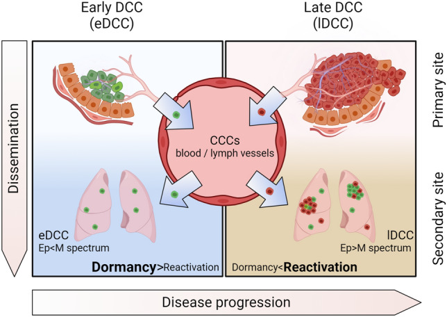
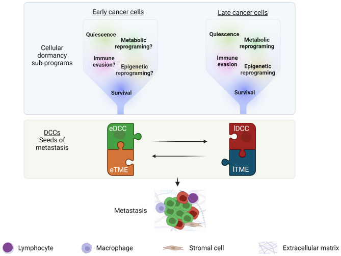
Cancer cells can leave their primary sites and travel through the circulation to distant sites, where they lodge as disseminated cancer cells (DCCs), even during the early and asymptomatic stages of tumor progression. In experimental models and clinical samples, DCCs can be detected in a non-proliferative state, defined as cellular dormancy. This state can persist for extended periods until DCCs reawaken, usually in response to niche-derived reactivation signals. Therefore, their clinical detection in sites like lymph nodes and bone marrow is linked to poor survival. Current cancer therapy designs are based on the biology of the primary tumor and do not target the biology of the dormant DCC population and thus fail to eradicate the initial or subsequent waves of metastasis. In this brief review, we discuss the current methods for detecting DCCs and highlight new strategies that aim to target DCCs that constitute minimal residual disease to reduce or prevent metastasis formation. Furthermore, we present current evidence on the relevance of DCCs derived from early stages of tumor progression in metastatic disease and describe the animal models available for their study. We also discuss our current understanding of the dissemination mechanisms utilized by genetically less- and more-advanced cancer cells, which include the functional analysis of intermediate or hybrid states of epithelial-mesenchymal transition (EMT). Finally, we raise some intriguing questions regarding the clinical impact of studying the crosstalk between evolutionary waves of DCCs and the initiation of metastatic disease.
Keywords: Disseminated cancer cells; Dormancy; EMT; Early dissemination; Metastasis.
© 2024. The Author(s).
Conflict of interest statement
The authors declare no competing interests.
Figures
Similar articles
-
Disseminated cancer cells in breast cancer: Mechanism of dissemination and dormancy and emerging insights on therapeutic opportunities.Semin Cancer Biol. 2022 Jan;78:78-89. doi: 10.1016/j.semcancer.2021.02.004. Epub 2021 Feb 21. Semin Cancer Biol. 2022. PMID: 33626407 Review.
-
Minimal residual disease in advanced or metastatic solid cancers: The G0-G1 state and immunotherapy are key to unwinding cancer complexity.Semin Cancer Biol. 2022 Feb;79:68-82. doi: 10.1016/j.semcancer.2020.03.009. Epub 2020 Mar 19. Semin Cancer Biol. 2022. PMID: 32201368 Review.
-
Epigenetic and Pluripotency Aspects of Disseminated Cancer Cells During Minimal Residual Disease.Adv Exp Med Biol. 2018;1100:1-18. doi: 10.1007/978-3-319-97746-1_1. Adv Exp Med Biol. 2018. PMID: 30411257 Review.
-
The force awakens: metastatic dormant cancer cells.Exp Mol Med. 2020 Apr;52(4):569-581. doi: 10.1038/s12276-020-0423-z. Epub 2020 Apr 16. Exp Mol Med. 2020. PMID: 32300189 Free PMC article. Review.
-
Prostate Cancer Dormancy and Reactivation in Bone Marrow.J Clin Med. 2021 Jun 16;10(12):2648. doi: 10.3390/jcm10122648. J Clin Med. 2021. PMID: 34208521 Free PMC article. Review.
References
-
- Hanahan D (2022) Hallmarks of cancer: new dimensions. Cancer Discov 12(1):31–46. 10.1158/2159-8290.CD-21-1059 10.1158/2159-8290.CD-21-1059 - DOI - PubMed
Publication types
MeSH terms
Grants and funding
LinkOut - more resources
Full Text Sources
