. 2024 Jul 9;73(3):336-346.
doi: 10.1538/expanim.23-0148. Epub 2024 Mar 22.

Transient receptor potential vanilloid 1 interacts with Toll-like receptor 4 (TLR4)/cluster of differentiation 14 (CD14) signaling pathway in lipopolysaccharide-mediated inflammation in macrophages

Affiliations

Transient receptor potential vanilloid 1 interacts with Toll-like receptor 4 (TLR4)/cluster of differentiation 14 (CD14) signaling pathway in lipopolysaccharide-mediated inflammation in macrophages

Julia Chu-Ning Hsu et al. Exp Anim. .

Abstract

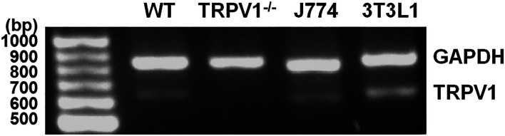
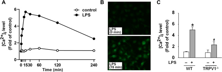
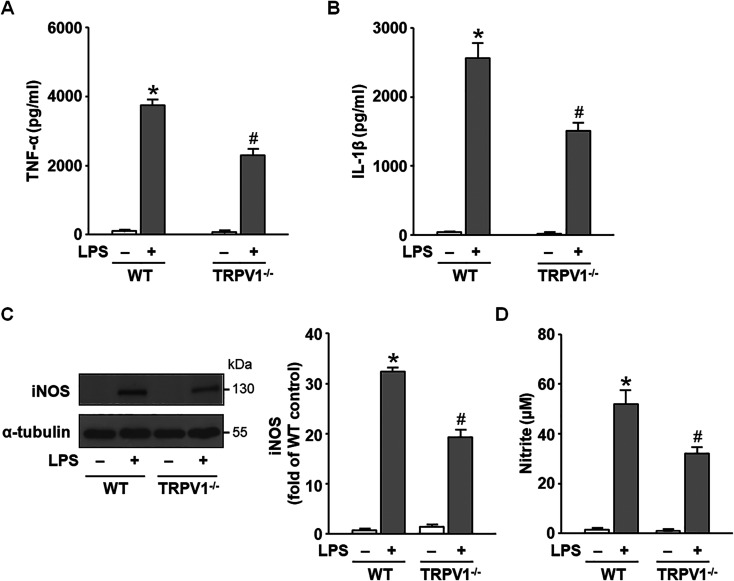
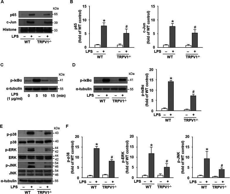
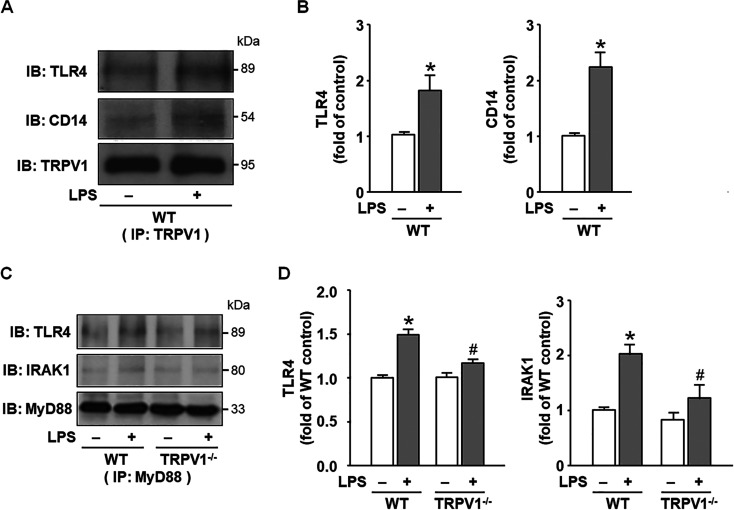
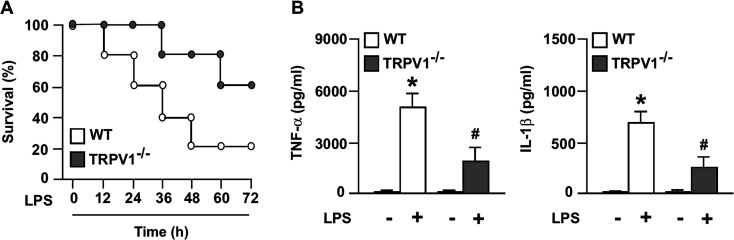
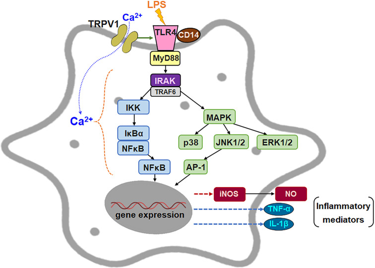
Transient receptor potential vanilloid 1 (TRPV1), a ligand-gated cation channel, is a receptor for vanilloids on sensory neurons and is also activated by capsaicin, heat, protons, arachidonic acid metabolites, and inflammatory mediators on neuronal or non-neuronal cells. However, the role of the TRPV1 receptor in pro-inflammatory cytokine secretion and its potential regulatory mechanisms in lipopolysaccharide (LPS)-induced inflammation has yet to be entirely understood. To investigate the role and regulatory mechanism of the TRPV1 receptor in regulating LPS-induced inflammatory responses, bone marrow-derived macrophages (BMDMs) harvested from wild-type (WT) and TRPV1 deficient (Trpv1-/-) mice were used as the cell model. In WT BMDMs, LPS induced an increase in the levels of tumor necrosis factor-α, IL-1β, inducible nitric oxide synthase, and nitric oxide, which were attenuated in Trpv1-/- BMDMs. Additionally, the phosphorylation of inhibitor of nuclear factor kappa-Bα and mitogen-activated protein kinases, as well as the translocation of nuclear factor kappa-B and activator protein 1, were all decreased in LPS-treated Trpv1-/- BMDMs. Immunoprecipitation assay revealed that LPS treatment increased the formation of TRPV1-Toll-like receptor 4 (TLR4)-cluster of differentiation 14 (CD14) complex in WT BMDMs. Genetic deletion of TRPV1 in BMDMs impaired the LPS-triggered immune-complex formation of TLR4, myeloid differentiation protein 88, and interleukin-1 receptor-associated kinase, all of which are essential regulators in LPS-induced activation of the TLR4 signaling pathway. Moreover, genetic deletion of TRPV1 prevented the LPS-induced lethality and pro-inflammatory production in mice. In conclusion, the TRPV1 receptor may positively regulate the LPS-mediated inflammatory responses in macrophages by increasing the interaction with the TLR4-CD14 complex and activating the downstream signaling cascade.

Keywords: Toll-like receptor 4 (TLR4); inflammation; lipopolysaccharide (LPS); macrophage; transient receptor potential vanilloid 1 (TRPV1).


Conflict of interest statement

The authors declare no conflict of interest.

Figures

Fig. 1.
Fig. 1.
Expression of transient receptor potential vanilloid 1 (TRPV1) in macrophages of wild-type (WT) and Trpv1−/− mice. The mRNA was isolated from WT, Trpv1−/− bone marrow-derived macrophages, murine J774.A1 macrophage-like cells, and murine 3T3L1 preadipocytes. The mRNA expression of the TRPV1 receptor and GAPDH was determined by reverse transcriptase-PCR (RT-PCR). The band of 644 bp in length was TRPV1 receptor, while the band of 802 bp was GAPDH.
Fig. 2.
Fig. 2.
Treatment with lipopolysaccharide (LPS) activates the transient receptor potential vanilloid 1 (TRPV1) receptor in macrophages. (A) Bone marrow-derived macrophages (BMDMs) were incubated with phosphate-buffered saline (control) or LPS (1 µg/ml) challenge for indicated times (0, 1, 2, 5, 10, 15, 30, 60, 120, and 240 min). Intracellular levels of Ca2+ ([Ca2+]i) were quantified by measuring the intensity of Ca2+-sensitive Fluo-8 fluorescence. (B) Representative microscopy images of Ca2+-binding Fluo-8 fluorescence at 15 min after incubation with or without LPS in BMDMs. (C) Intracellular levels of [Ca2+]i at 15 min after incubation with LPS in wild-type (WT) or Trpv1−/− BMDMs. Data are mean ± SEM from four independent experiments. *P<0.05 versus time 0 or WT control group, #P<0.05 versus WT LPS-treated group.
Fig. 3.
Fig. 3.
The production of lipopolysaccharide (LPS)-induced tumor necrosis factor-α (TNF-α), IL-1β, and nitric oxide was decreased in Trpv1−/− macrophages. Bone marrow-derived wild-type (WT) and Trpv1−/− macrophages were challenged with or without LPS (1 µg/ml) or control (phosphate-buffered saline (PBS)) for (A) 2 h or (B) 6 h, and then cell cultured media were collected for determining levels of (A) TNF-α and (B) IL-1β by ELISA. Moreover, bone marrow-derived WT and Trpv1−/− macrophages were treated with or without LPS (1 µg/ml) or control (PBS) for 24 h. (C) The protein levels of inducible nitric oxide synthase (iNOS) and α-tubulin were assessed with western blot analysis. (D) The levels of nitrite in cell culture media were determined by Griess’s assay. Data are mean ± SEM from four independent experiments. *P<0.05 versus WT control group, #P<0.05 versus WT LPS-treated group.
Fig. 4.
Fig. 4.
In lipopolysaccharide (LPS)-induced Trpv1−/− macrophages, there was a reduction in the translocation of nuclear factor kappa-B (NF-κB) into the nucleus, a decrease in the phosphorylation levels of inhibitor of NF-κBα (IκBα), and a decline in mitogen-activated protein kinase (MAPK) activation. (A) Nuclear protein was isolated from bone marrow-derived wild-type (WT) and Trpv1−/− macrophages with or without lipopolysaccharide (LPS) treatment (1 µg/ml) for 20 min. The levels of the p65 subunit of NF-κB, c-Jun subunit of AP-1, and histone H1 were examined by western blot analysis. (B) Quantitative results of panel A. (C) Bone marrow-derived WT macrophages were treated with LPS (1 µg/ml) or control (phosphate-buffered saline (PBS)) for indicated times (0, 5, 10, and 15 min). (D) Bone marrow-derived WT and Trpv1−/− macrophages were treated with or without LPS (1 µg/ml) for 5 min. Phosphorylated levels of IκBα and α-tubulin were examined by western blot analysis. (E) Bone marrow-derived WT and Trpv1−/− macrophages were treated with or without LPS (1 µg/ml) or control (PBS) for 15 min. The levels of phosphorylated and total p38, ERK1/2, JNK1/2, and α-tubulin were measured by western blot analysis. (F) Quantitative results of panel E. Data are mean ± SEM from four independent experiments. *P<0.05 versus WT control group, #P<0.05 versus WT LPS-treated group.
Fig. 5.
Fig. 5.
Upon lipopolysaccharide (LPS) challenge in macrophages, the interaction of transient receptor potential vanilloid 1 (TRPV1) with Toll-like receptor 4 (TLR4) complex was increased with the formation of TLR4–myeloid differentiation protein 88 (MyD88)–IL-1 receptor-associated kinase 1 (IRAK1). (A) Whole-cell lysates were obtained from bone marrow-derived wild-type (WT) macrophages in the presence or absence of LPS (1 µg/ml) for 5 min. Total cell lysates were immunoprecipitated with an anti-TRPV1 antibody. The blot was then immunoblotted with an anti-TLR4 antibody and an anti-CD14 antibody. (B) Quantitative results of panel A. (C) Whole-cell lysates prepared from bone marrow-derived WT and Trpv1−/− macrophages with or without LPS treatment (1 µg /ml) for 5 min were immunoprecipitated with anti-MyD88 antibody. The blot was then immunoblotted with an anti-TLR4 antibody and an anti-IRAK1 antibody. (D) Quantitative results of panel A. Data are mean ± SEM from four independent experiments. *P<0.05 versus WT control group, #P<0.05 versus WT LPS-treated group.
Fig. 6.
Fig. 6.
The role of transient receptor potential vanilloid 1 (TRPV1) in lipopolysaccharide (LPS)-induced inflammation in vivo. At eight weeks old, wild-type (WT) and Trpv1−/− mice (n=10) received intraperitoneal administration of sterile PBS or a lethal dose of LPS (20 mg/kg body weight) for the indicated times (12, 24, 36, 48, 60, and 72 h). (A) WT and Trpv1−/− mice received LPS at the indicated times, and the lethality within 72 h was determined. (B) The serum levels of tumor necrosis factor-α (TNF-α) and IL-1β were assessed by ELISA at 2 h or 4 h after LPS administration, respectively. The two-way ANOVA followed by the Holm-Sidak test was used for multiple comparisons, and then an unpaired t-test was applied to compare two independent groups.
Fig. 7.
Fig. 7.
The proposed mechanism for the involvement of transient receptor potential vanilloid 1 (TRPV1) receptor in lipopolysaccharide (LPS)-induced induction of pro-inflammatory mediators in macrophages. As shown, genetic ablation of the TRPV1 receptor causes a reduction in the formation of Toll-like receptor 4 (TLR4)/cluster of differentiation 14 (CD14)/myeloid differentiation protein 88 (MyD88)/IL-1 receptor-associated kinase 1 (IRAK1) complex and the phosphorylation of inhibitor of nuclear factor kappa-Bα (IκBα) and mitogen-activated protein kinases (MAPKs) (p38, ERK1/2, and JNK1/2), and thus decreases activation of nuclear factor kappa-B (NF-κB) and activator protein 1 (AP-1), leading to downregulation of pro-inflammatory mediators including tumor necrosis factor-α (TNF-α), IL-1β, and inducible nitric oxide synthase (iNOS).

Similar articles

References

    1. Pop-Began V, Păunescu V, Grigorean V, Pop-Began D, Popescu C. Molecular mechanisms in the pathogenesis of sepsis. J Med Life. 2014; 7 Spec No. 2: 38–41. - PMC - PubMed
    1. Nedeva C, Menassa J, Puthalakath H. Sepsis: inflammation is a necessary evil. Front Cell Dev Biol. 2019; 7: 108. doi: 10.3389/fcell.2019.00108 - DOI - PMC - PubMed
    1. Tucureanu MM, Rebleanu D, Constantinescu CA, Deleanu M, Voicu G, Butoi E, et al. Lipopolysaccharide-induced inflammation in monocytes/macrophages is blocked by liposomal delivery of Gi-protein inhibitor. Int J Nanomedicine. 2017; 13: 63–76. doi: 10.2147/IJN.S150918 - DOI - PMC - PubMed
    1. Miller YI, Choi SH, Wiesner P, Bae YS. The SYK side of TLR4: signalling mechanisms in response to LPS and minimally oxidized LDL. Br J Pharmacol. 2012; 167: 990–999. doi: 10.1111/j.1476-5381.2012.02097.x - DOI - PMC - PubMed
    1. Gegner JA, Ulevitch RJ, Tobias PS. Lipopolysaccharide (LPS) signal transduction and clearance. Dual roles for LPS binding protein and membrane CD14. J Biol Chem. 1995; 270: 5320–5325. doi: 10.1074/jbc.270.10.5320 - DOI - PubMed