. 2024 Mar 19;14(1):6541.
doi: 10.1038/s41598-024-56712-z.

5-HT_FAsTR: a versatile, label-free, high-throughput, fluorescence-based microplate assay to quantify serotonin transport and release

Affiliations

5-HT_FAsTR: a versatile, label-free, high-throughput, fluorescence-based microplate assay to quantify serotonin transport and release

Lina Bukowski et al. Sci Rep. .

Abstract

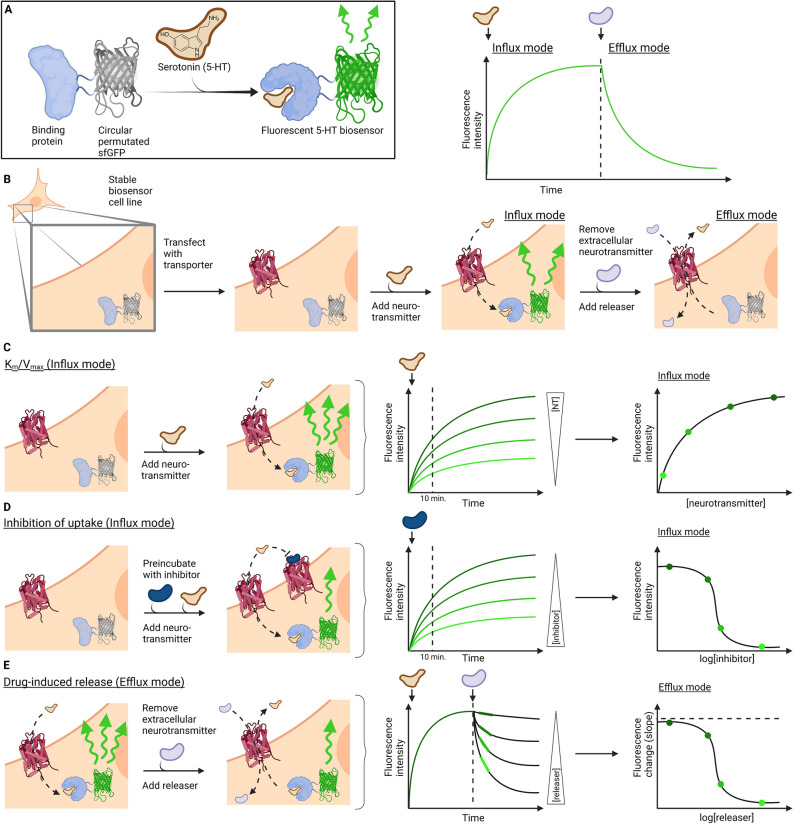
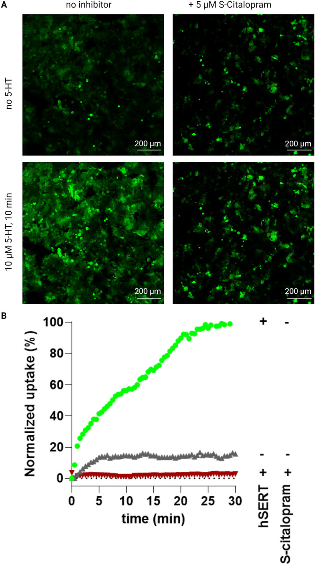
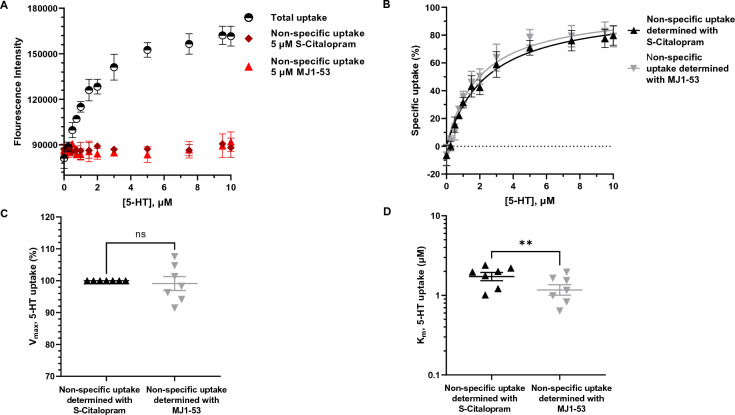
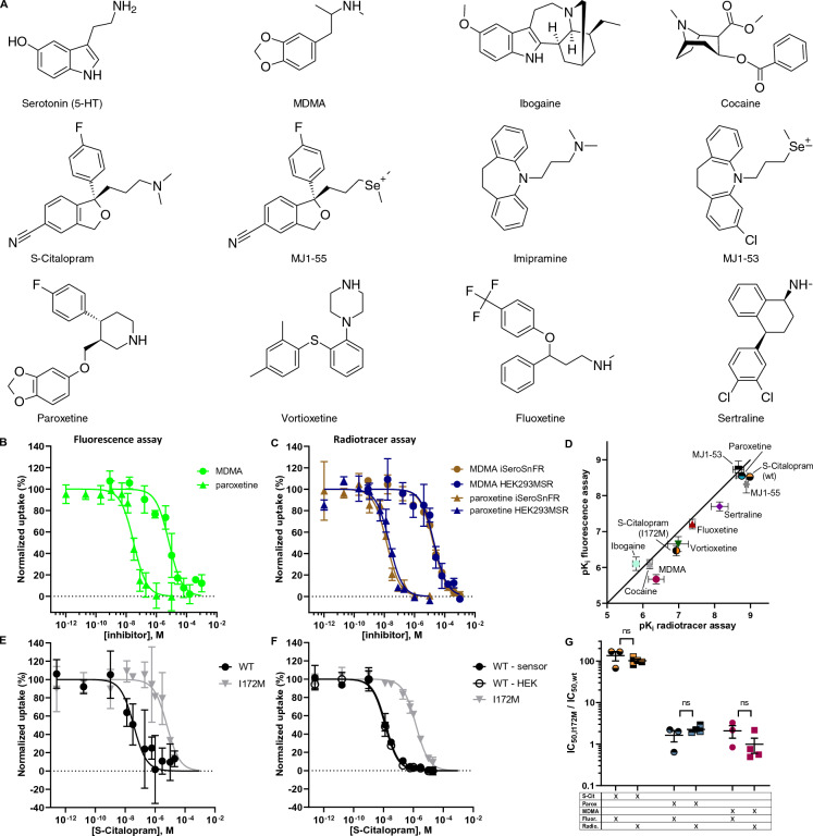
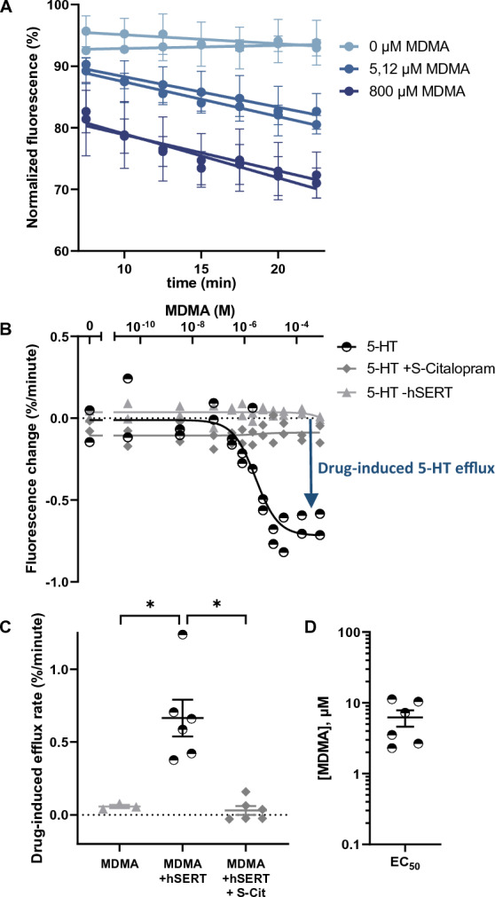
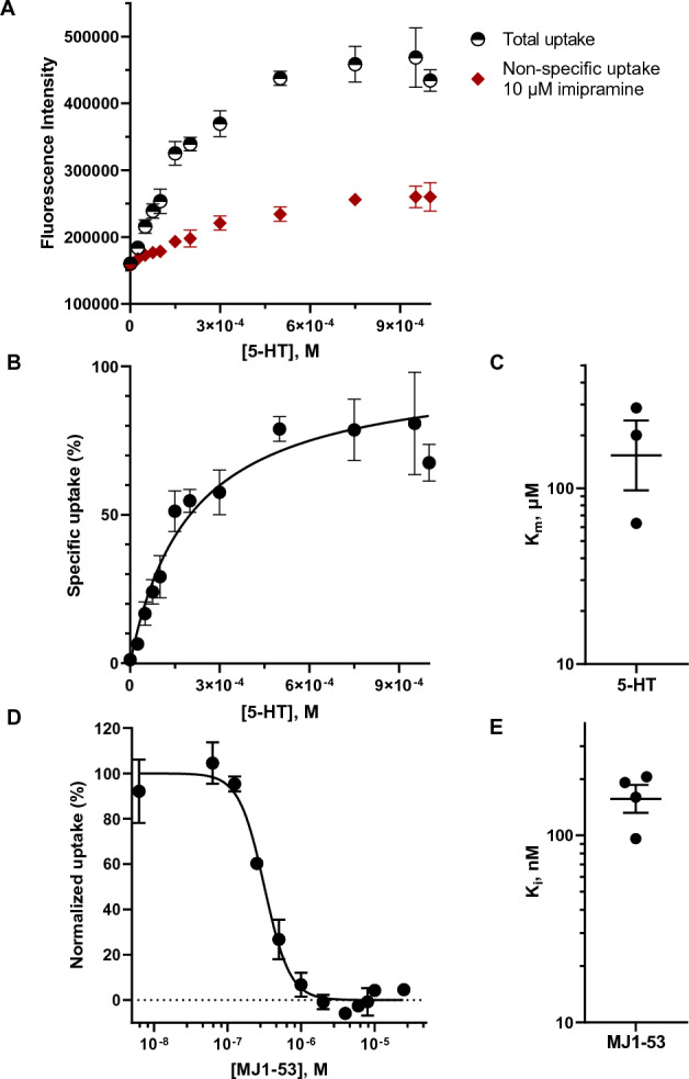
The neurotransmitter serotonin plays a pivotal role in mood and depression. It also acts as a vasoconstrictor within blood vessels and is the main neurotransmitter in the gastrointestinal system. In neurotransmission, released serotonin is taken up by serotonin transporters, which are principal targets of antidepressants and the psychostimulant, ecstasy. The investigation of serotonin transporters have relied almost exclusively on the use of radiolabeled serotonin in heterogenous end-point assays. Here we adapt the genetically encoded fluorescent biosensor, iSeroSnFR, to establish and validate the Serotonin (5-HT) Fluorescence Assay for Transport and Release (5-HT_FAsTR) for functional and pharmacological studies of serotonin transport and release. We demonstrate the applicability of the method for the study of a neuronal, high-affinity, low-capacity serotonin transporter (SERT) as well as an extraneuronal low-affinity, high-capacity organic cation transporter and mutants thereof. 5HT_FAsTR offers an accessible, versatile and reliable semi-homogenous assay format that only relies on a fluorescence plate reader for repeated, real-time measurements of serotonin influx and efflux. 5HT_FAsTR accelerates and democratizes functional characterization and pharmacological studies of serotonin transporters and genetic variants thereof in disease states such as depression, anxiety and ADHD.

Keywords: Biosensor; Depression; Organic cation transporter; Serotonin; Transporter.


Conflict of interest statement

The authors declare no competing interests.

Figures

Figure 1
Figure 1
Scheme for 5-HT_FAsTR. (A) The biosensor iSeroSnFR consists of a periplasmic binding protein (PBP) able to bind 5-HT and a circular permutated super folder green fluorescent protein (cpsfGFP). Conformational changes upon binding of 5-HT initiate a change of environment around the chromophore and lead to increased GFP fluorescence intensity. (B) HEK293MSR cells stably transfected with the biosensor (HEK293MSR_iSeroSnFR cells) express the 5-HT biosensor in the cytoplasm and can be transiently transfected with a monoamine transporter of choice. Transport of 5-HT into the cell via the membrane transporter and subsequent binding of 5-HT to the biosensor results in an increased GFP signal (influx mode). Adding a releasing drug to HEK293MSR_iSeroSnFR cells preloaded with 5-HT leads to 5-HT transport out of the cell (efflux) and thus a decrease in GFP signal (efflux mode). (C) Influx mode of the assay that can be used to determine Km and Vmax. Addition of neurotransmitter (NT) to HEK293MSR_iSeroSnFR cells expressing a monoamine transporter leads to a NT concentration dependent increase in fluorescence over time. Plotting the fluorescence intensity measured after a certain incubation time as a function of the NT concentration results in an uptake curve with Michaelis–Menten kinetics. (D) Influx mode of the assay used to determine inhibitory potencies. Pre-incubation of HEK293MSR_iSeroSnFR cells expressing a monoamine transporter with increasing inhibitor concentrations and subsequent addition of a constant concentration of NT leads to an increased fluorescence over time negatively dependent on inhibitor concentrations. Plotting the fluorescence intensity measured after a certain incubation time as a function of the inhibitor concentration, results in a classical sigmoidal dose–response curve. (E) Efflux mode of the assay used to determine drug-induced neurotransmitter release. Pre-loading of HEK293MSR_iSeroSnFR cells expressing a monoamine transporter with NT in a saturating concentration and subsequent addition of increasing concentrations of a NT releasing drug leads to a drug concentration dependent decrease in fluorescence over time. Plotting the fluorescence change (corresponding to the slope of the fluorescence decrease) as a function of drug concentrations, results in an efflux curve, where the potency of the releaser can be determined.
Figure 2
Figure 2
Fluorescence microscopy of HEK293MSR_iSeroSnFR cells. (A) Microscopy images of HEK293MSR_iSeroSnFR cells expressing hSERT before (top) and after (bottom) incubation with 10 µM 5-HT for 10 min. For control experiments, hSERT was blocked with 5 µM S-Citalopram for 15 min or HEK293MSR_iSeroSnFR cells were mock transfected. (B) Normalized fluorescence changes over time as a result of 5-HT uptake measured with the fluorescence microscope. Green points represents the fluorescence signal from fluorescent biosensor cells also expressing hSERT, whereas red points represent the nonspecific uptake in fluorescent biosensor cells expressing hSERT but inhibited with S-Citalopram. Grey points represent the negative control from fluorescent biosensor not expressing hSERT. Images were analysed in ImageJ by using built-in threshold algorithms on the image stacks.
Figure 3
Figure 3
5-HT_FAsTR reliably determines transport kinetic parameters in a high throughput 96-well microplate setup. (A) Graph showing fluorescence intensity in response to increasing concentrations of unlabeled 5-HT added to HEK293MSR_iSeroSnFR cells in a representative experiment. Shown is the total uptake of 5-HT (black, half open circles) and the non-specific uptake of 5-HT when blocking hSERT with the SSRI S-Citalopram (dark red diamonds) and when blocking hSERT with the membrane-impermeant imipramine analogue MJ1-53 (light red triangles). Shown are mean ± SD of four technical replicates for the total uptake and technical duplicates for the non-specific uptake. Each technical replicate constitutes the mean of four measurements conducted at four different positions in the same microplate well after 10 min incubation with 5-HT. (B) Representative graph showing the specific uptake of 5-HT via hSERT normalized to the maximal uptake calculated from A). Each point show the specific uptake calculated by subtracting either nonspecific uptake using S-citalopram (black traingles) or MJ1-53 (grey inverted triangles) from total uptake values. Shown are mean ± SD of four replicates and a Michaelis–Menten fit with robust regression. (C) Comparison of Vmax values determined by a Michaelis–Menten fit shows no difference between the inhibitor used to determine non-specific uptake. Shown are mean ± SEM from seven independent experiments. D) MJ1-53 appears to yield slightly lower KM values than S-Citalopram when used as a hSERT inhibitor to determine non-specific uptake. Shown is a comparison of mean KM values ± SEM from seven independent experiments determined by a Michaelis–Menten fit using either S-Citalopram or MJ1-53 to determine non-specific uptake. Statistical analysis of the difference is performed with a paired t-test (**P = 0.0032).
Figure 4
Figure 4
The performance of 5-HT_FAsTR in uptake inhibition assays is comparable to radiotracer uptake inhibition assays. (A) Chemical structures of hSERT substrates and inhibitors. (B) and (C) Representative graphs showing normalized 5-HT uptake in response to increasing MDMA (circle) and paroxetine (triangle) concentrations in (B) the fluorescence assay and (C) the radiotracer assay. Shown is the normalized uptake of 5-HT in HEK293MSR_iSeroSnFR cells stably expressing the biosensor (green or brown) and HEK293MSR cells not expressing the biosensor (blue). Shown are mean ± SD of duplicates for MDMA and four replicates for paroxetine and a sigmoidal dose–response fit using non-linear regression. (D) Comparison of pKi values of 10 hSERT inhibitors determined with the fluorescence assay versus pKi values determined with the radiotracer assay. Each data point represents the mean ± SD of three to five individual IC50 experiments. (E) and (F) Representative graph of normalized 5-HT uptake in response to increasing S-Citalopram concentrations in (E) the fluorescence assay and (F) the radiotracer assay. Shown is the normalized uptake of 5-HT in HEK293MSR_iSeroSnFR cells expressing either hSERT wild type (wt) (black circles) or the hSERT mutant I172M (grey triangles). Shown are mean ± SD of at least duplicates. (G) IC50 ratios for hSERT wt and the transporter variant I172M in response to S-Citalopram, paroxetine and MDMA were found to be identical when IC50 values were obtained in both the fluorescence and the radiotracer assay. Shown are mean ± SEM, N = 3, unpaired t-test.
Figure 5
Figure 5
5-HT_FAsTR fluorescence-based efflux assay to characterize ecstasy (MDMA) action on the serotonin transporter. (A) Representative graph showing the change in normalized fluorescence in response to three different MDMA concentrations over time. MDMA was added at time point 0 and a time window showing efflux in the linear range was chosen for further analysis. The fluorescence intensity measured in response to MDMA was normalized to the mean fluorescent intensity measured 2.5 min, 5 min and 7.5 min before the addition of MDMA. The response to MDMA was measured in duplicates at four different positions in each well. Shown are mean ± SD of four measurements per well for both duplicates and a simple linear regression performed with GraphPad Prism. (B) Representative dose–response curve for efflux induced by MDMA. The slope of the linear decrease in fluorescence in response to MDMA, calculated with the simple linear regression shown in (A), was plotted as a function of increasing MDMA concentrations added to the HEK293MSR_iSeroSnFR cells. A non-linear regression fit to a sigmoidal dose–response curve was used to calculate EC50, the top and the bottom plateau. Shown is the efflux rate in response to MDMA for cells transfected with hSERT and preloaded with 5-HT (black, half open circles), transfected with hSERT, preloaded with 5-HT and incubated with the hSERT inhibitor S-Citalopram (dark grey squares) or transfected with an empty vector and preloaded with 5-HT (light grey triangles). (C) Maximum drug-induced efflux rate of six individual experiments, calculated by subtracting the top plateau from the bottom plateau of the dose–response curve in B). Shown are mean ± SEM, N = 6, *P = 0.037 for MDMA vs. MDMA + hSERT and *P = 0.017 for MDMA + hSERT vs. MDMA + hSERT + S-Cit, one-way Anova with multiple comparisons (Tukey test). (D) EC50 values for efflux induced by MDMA from six individual experiments calculated with a non-linear fit as shown in figure B. Shown are mean ± SEM.
Figure 6
Figure 6
5-HT_FAsTR can be used to quantify uptake and pharmacology of a high-capacity, low-affinity organic cation transporter. (A) Representative graph showing fluorescence intensity in response to increasing concentrations of unlabeled 5-HT added to HEK293MSR_iSeroSnFR cells transfected with hOCT2. Shown is the total uptake of 5-HT (black, half open circles) and the non-specific uptake of 5-HT when blocking hOCT2 with the TCA imipramine (dark red squares) as mean ± SD of four replicates. Each replicate constitutes the mean of nine measurements conducted at different positions in the same well. (B) Representative graph showing the specific and saturable uptake of 5-HT via hOCT2 normalized to the maximal uptake. Shown are mean ± SD of four replicates and a Michaelis–Menten fit using non-linear regression. (C) Km values determined for three independent experiments by a Michaelis–Menten fit with robust regression, shown are mean ± SEM. (D) Representative graph showing normalized 5-HT uptake via hOCT2 in response to increasing concentrations of the imipramine analogue MJ1-53. Shown are mean ± SD of duplicates and a non-linear fit ([inhibitor] vs response variable slope – (four parameters). (E) Ki values of four independent experiments, shown are mean ± SEM.

Similar articles

References

    1. Gasser PJ. Organic cation transporters in brain catecholamine homeostasis. Handb. Exp. Pharmacol. 2021;266:187–197. doi: 10.1007/164_2021_470. - DOI - PubMed
    1. Rudnick G, Sandtner W. Serotonin transport in the 21st century. J. Gen. Physiol. 2019;151(11):1248–1264. doi: 10.1085/jgp.201812066. - DOI - PMC - PubMed
    1. Kristensen AS, et al. SLC6 neurotransmitter transporters: structure, function, and regulation. Pharmacol. Rev. 2011;63(3):585–640. doi: 10.1124/pr.108.000869. - DOI - PubMed
    1. Gasser PJ. Roles for the uptake(2) transporter OCT3 in regulation of dopaminergic neurotransmission and behavior. Neurochem. Int. 2019;123:46–49. doi: 10.1016/j.neuint.2018.07.008. - DOI - PMC - PubMed
    1. Koepsell H. Organic cation transporters in health and disease. Pharmacol. Rev. 2020;72(1):253–319. doi: 10.1124/pr.118.015578. - DOI - PubMed

MeSH terms

Substances