. 2024 Apr 4;111(4):791-804.
doi: 10.1016/j.ajhg.2024.02.013. Epub 2024 Mar 18.

Expanding the PRAAS spectrum: De novo mutations of immunoproteasome subunit β-type 10 in six infants with SCID-Omenn syndrome

Affiliations

Expanding the PRAAS spectrum: De novo mutations of immunoproteasome subunit β-type 10 in six infants with SCID-Omenn syndrome

Caspar I van der Made et al. Am J Hum Genet. .

Abstract

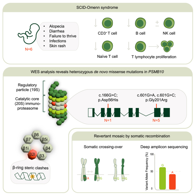
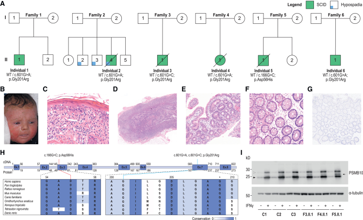
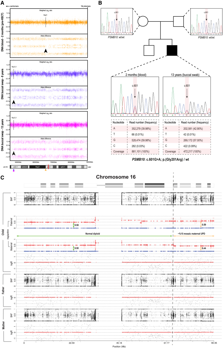
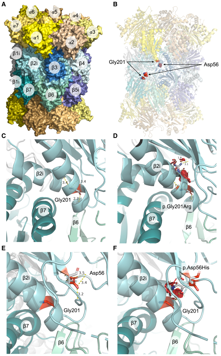
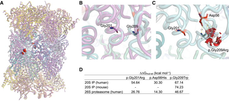
Mutations in proteasome β-subunits or their chaperone and regulatory proteins are associated with proteasome-associated autoinflammatory disorders (PRAAS). We studied six unrelated infants with three de novo heterozygous missense variants in PSMB10, encoding the proteasome β2i-subunit. Individuals presented with T-B-NK± severe combined immunodeficiency (SCID) and clinical features suggestive of Omenn syndrome, including diarrhea, alopecia, and desquamating erythematous rash. Remaining T cells had limited T cell receptor repertoires, a skewed memory phenotype, and an elevated CD4/CD8 ratio. Bone marrow examination indicated severely impaired B cell maturation with limited V(D)J recombination. All infants received an allogeneic stem cell transplant and exhibited a variety of severe inflammatory complications thereafter, with 2 peri-transplant and 2 delayed deaths. The single long-term transplant survivor showed evidence for genetic rescue through revertant mosaicism overlapping the affected PSMB10 locus. The identified variants (c.166G>C [p.Asp56His] and c.601G>A/c.601G>C [p.Gly201Arg]) were predicted in silico to profoundly disrupt 20S immunoproteasome structure through impaired β-ring/β-ring interaction. Our identification of PSMB10 mutations as a cause of SCID-Omenn syndrome reinforces the connection between PRAAS-related diseases and SCID.

Keywords: Omenn syndrome; PSMB10; immunoproteasome; revertant somatic mosaicism; severe combined immune deficiency; uniparental disomy.


Conflict of interest statement

Declaration of interests The authors declare no competing interests.

Figures

None
Graphical abstract
Figure 1
Figure 1
Clinical features and identification of PSMB10 de novo missense variants (A) PSMB10 variants in six infants with SCID. (B) Erythematosquamous rash in individual 1 at 12 weeks after birth. (C) Histology of the initial skin biopsy of individual 1 showed a graft-versus-host-disease-like pattern with vacuolar interface inflammation, multiple scattered apoptotic keratinocytes, and involvement of the adnexal structures, in the presence of a limited lymphocytic infiltrate (hematoxylin and eosin staining; original magnification ×41). (D) Histopathological evaluation of an inguinal lymph node extracted from individual 5 showed a paucicellular, stroma-rich lymph node. (E) A jejunal biopsy from individual 3 was hallmarked by partial villous atrophy and crypt hyperplasia with relatively few lymphocytes. (F) Colonic mucosa biopsied from individual 5 showed preserved crypt architecture with an empty lamina propria with few lymphocytes, in keeping with an immunodeficiency-related enteropathy. (G) Immunohistochemistry showed the absence of CD3+ and CD20+ positive cells in the colon samples from individual 5, although significant numbers of CD4+ cells were observed that may be of a macrophage/monocyte lineage. (H) Visualization of the three identified PSMB10 variants at the cDNA and protein level. The conservation across species is shown and scaled by color. The asterisk indicates the position of a previously studied Psmb10 variant in TUB6 mice. (I) Immunoblot for PSMB10 in dermal fibroblasts of 3 controls, individual 3 (F3.II.1; p.Gly201Arg), individual 4 (F4.II.1; p.Gly201Arg), and individual 5 (F5.II.1; p.Asp56His), with or without prior IFN-gamma induction. Upper band (white arrowhead) represents immature and lower band (black arrowhead), mature, PSMB10; subject samples also show an additional, intermediate band (black arrow). Representative of 4 independent experiments.
Figure 2
Figure 2
Acquired segmental UPD overlapping the PSMB10 locus shows evidence of RM (A) Aberrant BAF profile of the genome-wide SNP-array analysis in individual 1 reveals two independent UPD events at the q arm of chromosome 16 in blood (pre-HSCT at 2 months of age; 16q12.1) and in the buccal mucosa (16q12.1) spanning to the terminal end of 16q (16qter). Arrows indicate the respective breakpoints of UPD and are color-coded for each tissue; the red bar in the ideogram represents the distinct location of PSMB10. (B) Exome inclusion of the unaffected parents of individual 1 resulted in the identification of a unique heterozygous de novo missense mutation in PSMB10 (c.601G>A [GenBank: NM_002801.3] [p.Gly201Arg]). De novo status was verified by Sanger sequencing, while deep amplicon sequencing using the Ion Torrent accurately determined the respective mosaic levels in both tissues. (C) Results from haplarithmisis on blood-derived DNA from individual 1 (pre-HSCT at 2 months of age) and parents. From top to bottom we depict BAF, paternal haplarithm, maternal haplarithm, and logR (relative copy number) values of the child, followed by BAF and logR-values of the parents. BAF of a single-nucleotide variant (SNV) is the number of allele B over the number of alleles A and B for that SNV, and logR is the base 2 logarithm of the summed normalized number of both alleles in a window of 100 kb over the expected signal intensity values.
Figure 3
Figure 3
Predicted structural consequences of the PSMB10 variants (A) Crystal structure of human IP 20S particle (PDB: 6E5B) is shown with its alpha subunits in shades of yellow/orange and beta subunits in shades of blue/green. (B) Positions of interest, Gly201 and Asp56, in the β2i subunits are highlighted as red spheres. The local structural environment of Gly201 (sticks) is depicted in (C) including distances from its interacting residues in the α-helix, which is located close to the β-ring interface. (D) Local structural clashes (in red discs) potentially brought about by p.Gly201Arg are shown. Together with the drastic changes in free energy of the complex, p.Gly201Arg appears to be structurally damaging. Similar structural representations are illustrated for the variant p.Asp56His in (E) and (F), which also include the position Gly201 for visual reference.
Figure 4
Figure 4
Comparison of predicted structural impact of variants on human and mouse PSMB10 (A) Superposition of human (PDB: 6E5B) and mouse (PDB: 3UNH, purple) 20S IP crystal structures is shown (root-mean-square deviation [RMSD] = 0.59 Å), with the positions of interest highlighted in red spheres. The local structural environment of Gly201 and Gly209 is depicted in (B), which highlights the clear overlap of residues between human (cyan) and mouse (purple) PSMB10. (C) The steric clashes potentially brought about by Gly209 in human 20S IP are illustrated as red discs, suggesting a structurally damaging outcome of the p.Gly209Trp variant. The table in (D) lists the predicted differences in free energies of the proteasome complexes for each variant type for each protein system.

Similar articles

References

    1. Murata S., Takahama Y., Kasahara M., Tanaka K. The immunoproteasome and thymoproteasome: functions, evolution and human disease. Nat. Immunol. 2018;19:923–931. doi: 10.1038/s41590-018-0186-z. - DOI - PubMed
    1. McCarthy M.K., Weinberg J.B. The immunoproteasome and viral infection: a complex regulator of inflammation. Front. Microbiol. 2015;6 doi: 10.3389/fmicb.2015.00021. - DOI - PMC - PubMed
    1. Kincaid E.Z., Che J.W., York I., Escobar H., Reyes-Vargas E., Delgado J.C., Welsh R.M., Karow M.L., Murphy A.J., Valenzuela D.M., et al. Mice completely lacking immunoproteasomes show major changes in antigen presentation. Nat. Immunol. 2011;13:129–135. doi: 10.1038/ni.2203. - DOI - PMC - PubMed
    1. Van den Eynde B.J., Morel S. Differential processing of class-I-restricted epitopes by the standard proteasome and the immunoproteasome. Curr. Opin. Immunol. 2001;13:147–153. doi: 10.1016/S0952-7915(00)00197-7. - DOI - PubMed
    1. Heink S., Ludwig D., Kloetzel P.-M., Krüger E. Vol. 102. 2005. IFN-γ-induced immune adaptation of the proteasome system is an accelerated and transient response; pp. 9241–9246. - DOI - PMC - PubMed

Publication types

MeSH terms

Substances