. 2024 Jan 23;4(1):ycae016.
doi: 10.1093/ismeco/ycae016. eCollection 2024 Jan.

Zooming in on the intracellular microbiome composition of bacterivorous Acanthamoeba isolates

Affiliations

Zooming in on the intracellular microbiome composition of bacterivorous Acanthamoeba isolates

Binod Rayamajhee et al. ISME Commun. .

Abstract

Acanthamoeba, a free-living amoeba in water and soil, is an emerging pathogen causing severe eye infection known as Acanthamoeba keratitis. In its natural environment, Acanthamoeba performs a dual function as an environmental heterotrophic predator and host for a range of microorganisms that resist digestion. Our objective was to characterize the intracellular microorganisms of phylogenetically distinct Acanthamoeba spp. isolated in Australia and India through directly sequencing 16S rRNA amplicons from the amoebae. The presence of intracellular bacteria was further confirmed by in situ hybridization and electron microscopy. Among the 51 isolates assessed, 41% harboured intracellular bacteria which were clustered into four major phyla: Pseudomonadota (previously known as Proteobacteria), Bacteroidota (previously known as Bacteroidetes), Actinomycetota (previously known as Actinobacteria), and Bacillota (previously known as Firmicutes). The linear discriminate analysis effect size analysis identified distinct microbial abundance patterns among the sample types; Pseudomonas species was abundant in Australian corneal isolates (P < 0.007), Enterobacteriales showed higher abundance in Indian corneal isolates (P < 0.017), and Bacteroidota was abundant in Australian water isolates (P < 0.019). The bacterial beta diversity of Acanthamoeba isolates from keratitis patients in India and Australia significantly differed (P < 0.05), while alpha diversity did not vary based on the country of origin or source of isolation (P > 0.05). More diverse intracellular bacteria were identified in water isolates as compared with clinical isolates. Confocal and electron microscopy confirmed the bacterial cells undergoing binary fission within the amoebal host, indicating the presence of viable bacteria. This study sheds light on the possibility of a sympatric lifestyle within Acanthamoeba, thereby emphasizing its crucial role as a bunker and carrier of potential human pathogens.

Keywords: Acanthamoeba; environmental predator; eye infection; sympatric lifestyle; training ground.


Conflict of interest statement

The authors have declared that no competing interests exist.

Figures

Graphical Abstract
Graphical Abstract
Figure 1
Figure 1
Venn-diagram showing unique and shared ASVs (relative abundance >0) among different Acanthamoeba groups as per source of isolation and origin of country (A). The top 20 most abundant ASVs clustered into six different bacterial families cross all Acanthamoeba isolates as per source of isolation and origin of country (B).
Figure 2
Figure 2
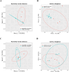
Beta diversity of bacterial microbiome composition in Acanthamoeba corneal isolates was compared between the countries of origin, India (n = 8) and Australia (n = 7), and also within Australian isolates based on their sources: corneal (n = 7) and water isolates (n = 5). Two-dimensional PCoA plots comparing Bray–Curtis dissimilarity index (A) and weighted UniFrac distance metric (B) show significant differences (P < 0.05) between Indian and Australian corneal isolates of Acanthamoeba spp., but no significant differences (P > 0.05) between Australian water and corneal isolates (C, D). The axes represent the first two principal coordinates of the PCoA plot, with each point on the plot representing the bacterial microbiome of an individual Acanthamoeba strain. The ASVs data were transformed to relative abundance before plotting to account for differences in sequencing depth and some of the sample points are overlapped on the plots due to the very similar bacterial microbiome composition.
Figure 3
Figure 3
Alpha diversity of bacterial microbiome composition of Acanthamoeba strains by group; (A) Shannon index, and (B) number of observed ASVs. A global Kruskal–Wallis test was used to perform statistical analysis among the three groups, whereas a Wilcoxon rank sum test was performed between the two groups. Acanthamoeba isolates; Australia water (n = 5), Australia cornea (n = 7), and India cornea (n = 8). The boxplots show the smallest and largest values (the 25th and 75th quartiles), the median, and outliers.
Figure 4
Figure 4
Intracellular bacterial microbiome composition of Acanthamoeba isolates by groups; Indian corneal isolates, Australian corneal, and water isolates. Stacked bar plots visually represent the average relative abundance (%) of 16S V1–3 rRNA gene sequences assigned to bacterial phyla (A), families (B), and genera (C). For visualization, taxa with <1% relative abundance have been grouped together. In cases where the genus level classification was not possible, a higher taxonomic level is mentioned and “Candidatus” was mentioned for Candidatus Jidaibacter acanthamoeba. (D) Heatmap representing the top 20 most abundant ASVs (log10). ASVs (genus level) are shown in y-axis and x-axis represents individual samples included for intracellular microbiome profiling of Acanthamoeba isolates targeting 16S rRNA, V1–3 (refer Table S1 for details of Acanthamoeba isolates). White cells correspond no ASVs detected. For visualization, “Candidatus” was labelled for Candidatus Jidaibacter acanthamoeba in B, C, and D.
Figure 5
Figure 5
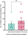
Bar plot showing the relative count of ASVs in recent and stock isolates. ASVs with <1% of the total sequence count of each isolate were excluded for calculation and visualization. Unpaired t-test was used to compare the counts between two groups.
Figure 6
Figure 6
Representative FISH micrographs showing the presence of intracellular bacteria in Acanthamoeba trophozoites investigated in this study. Probes EUK516 conjugated with Cy5, targeting Acanthamoeba, and EUB conjugated with Cy3, targeting most of bacterial strains were used for all Acanthamoeba strains positive for bacterial 16S rRNA. DAPI was used in mounting medium when visualized by a fluorescence microscope. Probe pB-914 labelled with 6-FAM was used for isolates containing high abundance of bacteria belong to Enterobacteriaceae family. (A) Rod shaped bacteria were observed throughout the cytoplasm of Acanthamoeba trophozoites (Indian corneal isolate) and a few cocci bacteria were also observed (yellow arrows). The white arrow represents bacterium cell undergoing binary fission. (B) Bacteria showing binary fission (white arrows) in vacuole like structure of Acanthamoeba recovered from water sample (R3). (C and D) Corneal isolates of Acanthamoeba spp. (Ac-112 and L-579/20, respectively) with intracellular bacteria. (E) Intracellular bacteria labelled with probes EUB and pB-914 simultaneously in Acanthamoeba sp. isolated from an AK patient. (F) Clinical (Ac-102) isolate of Acanthamoeba trophozoite depicting rod shaped intracellular bacteria. Indicators: white arrow, bacterial cell undergoing binary fission; yellow arrow: cocci shaped bacteria. Scale bar in each panel represents 10 μm.
Figure 7
Figure 7
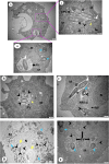
Representative images of TEM showing Acanthamoeba isolates containing intracellular bacteria. (A) Overview of an Acanthamoeba trophozoite (Indian corneal isolate) harbouring intracellular bacteria. (A.i-ii) Higher magnification showing rod (white arrow) and cocci (blue arrow) shaped bacteria inside early phagocytic (i) or PV (ii), and bacterial cells were also observed in trophozoite cytoplasm (ii). A bacterium undergoing binary fission (asterisk) and digested bacteria (arrowhead) appear disintegrated surrounded by multiple layers (yellow arrow) (ii). (B) Engulfed bacteria appeared disintegrated and digested inside PV surrounded by multiple layers (Australian water isolate). (C) Rod and spherical shaped bacterial cells close to host NM appears enclosed by double-membranous vacuole and disintegrated (arrowhead). And a bacterial cell is undergoing binary fission (Australian corneal isolate). (D) Engulfed bacteria appeared disintegrated and digested inside PV close to host NM. Both digested and undigested bacteria in the same PV consisting multiple layers of membrane. (E) Digested and undigested cocci bacteria in the same PV. Symbols = EPV: early phagocytic vacuole; PV: phagocytic vacuole; M: mitochondria; N: nucleus; NM: nuclear membrane; NP: nuclear pore; DV: digestive vacuole; CV: contractile vacuole; white arrow: rod bacteria; blue arrow: spherical bacteria; arrowhead: digested bacteria; yellow arrow: surrounded by multiple layers; asterisk (*): binary fission; alpha (α): electron translucent space. The lengths of bars in the bottom right corner of each image represent 500 nm except A (1 μm), and D (1 μm).

Similar articles

Cited by

References

    1. Antonelli A, Favuzza E, Galano Aet al. . Regional spread of contact lens-related Acanthamoeba keratitis in Italy. New Microbiol 2018;41:83–5. - PubMed
    1. Carnt N, Hoffman JM, Verma Set al. . Acanthamoeba keratitis: confirmation of the UK outbreak and a prospective case-control study identifying contributing risk factors. Br J Ophthalmol 2018;102:1621–8. 10.1136/bjophthalmol-2018-312544. - DOI - PubMed
    1. Damhorst GL, Watts A, Hernandez-Romieu Aet al. . Acanthamoeba castellanii encephalitis in a patient with AIDS: a case report and literature review. Lancet Infect Dis 2022;22:e59–65. 10.1016/s1473-3099(20)30933-6. - DOI - PMC - PubMed
    1. Randag AC, van Rooij J, van Goor ATet al. . The rising incidence of Acanthamoeba keratitis: a 7-year nationwide survey and clinical assessment of risk factors and functional outcomes. PLoS One 2019;14:e0222092. 10.1371/journal.pone.0222092.. - DOI - PMC - PubMed
    1. Chin J, Young AL, Hui Met al. . Acanthamoeba keratitis: 10-year study at a tertiary eye care center in Hong Kong. Cont Lens Anterior Eye 2015;38:99–103. 10.1016/j.clae.2014.11.146. - DOI - PubMed