. 2024 Aug;29(8):2408-2423.
doi: 10.1038/s41380-024-02509-5. Epub 2024 Mar 18.

Scanning ultrasound-mediated memory and functional improvements do not require amyloid-β reduction

Affiliations

Scanning ultrasound-mediated memory and functional improvements do not require amyloid-β reduction

Gerhard Leinenga et al. Mol Psychiatry. 2024 Aug.

Erratum in

Abstract

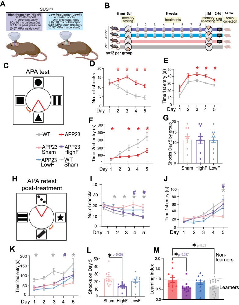
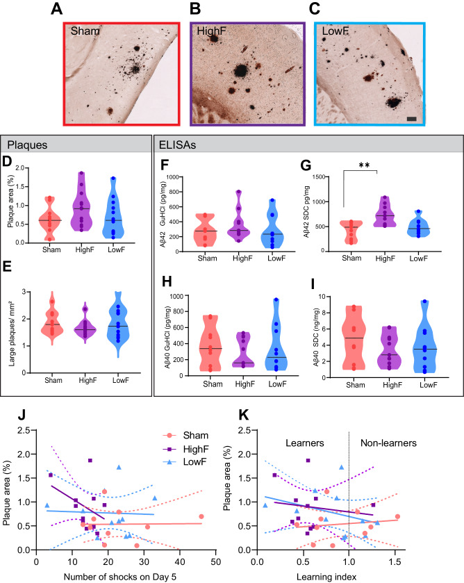
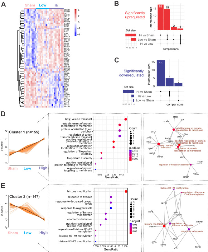
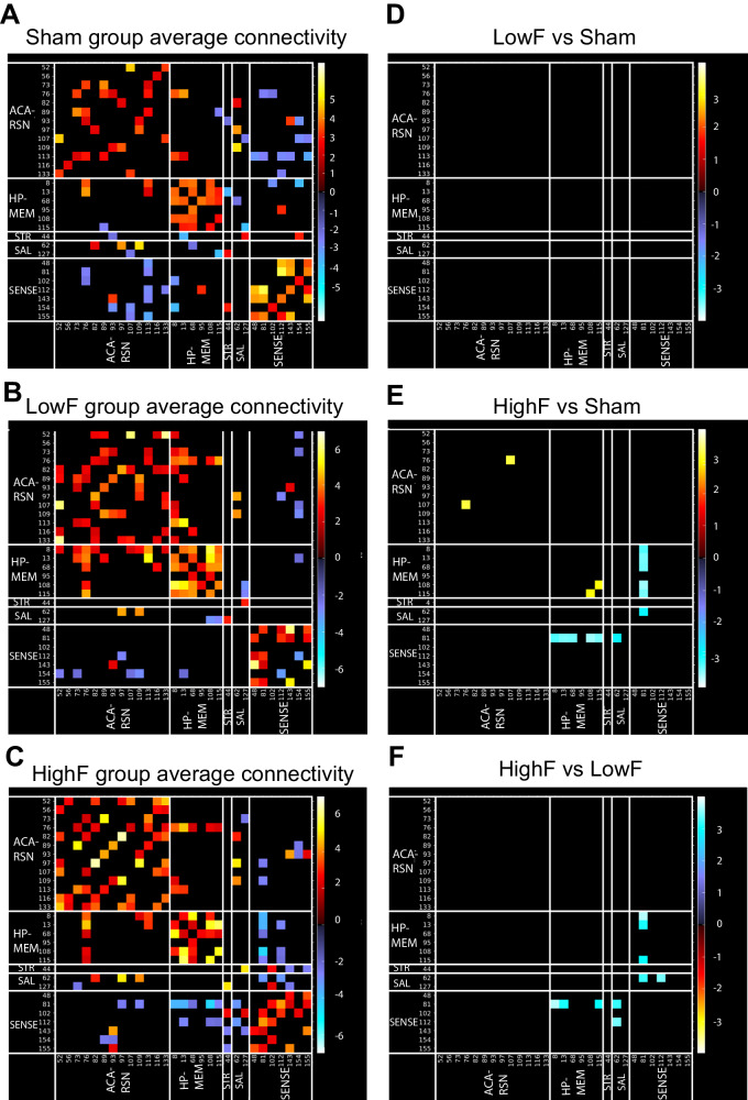
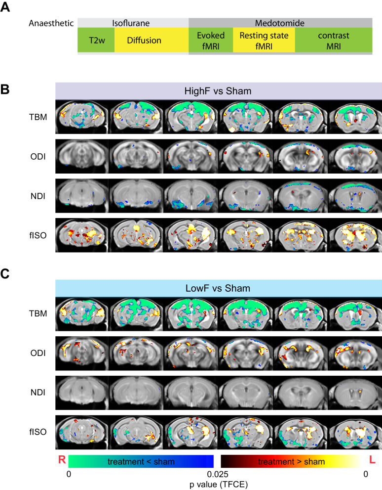
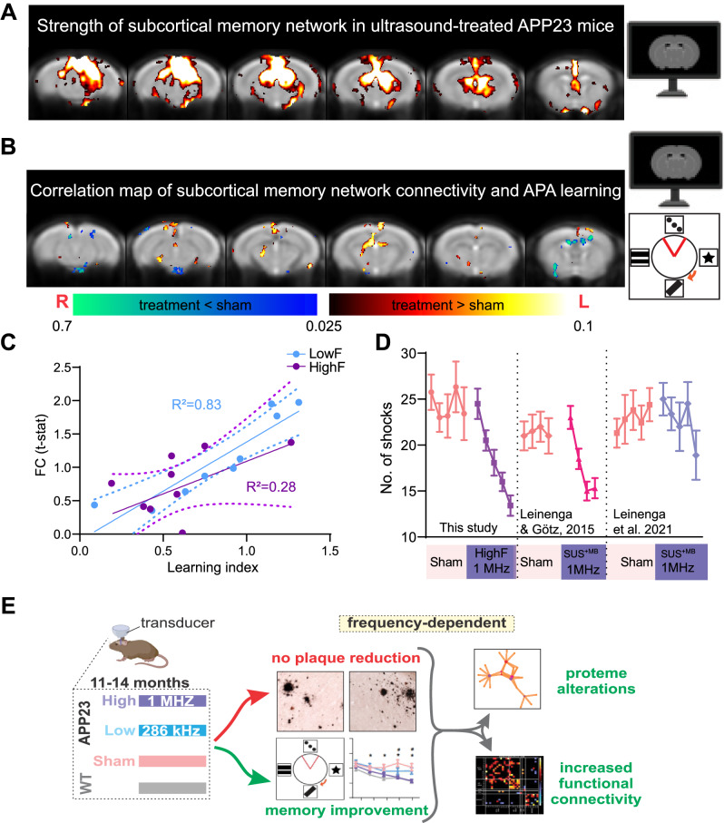
A prevalent view in treating age-dependent disorders including Alzheimer's disease (AD) is that the underlying amyloid plaque pathology must be targeted for cognitive improvements. In contrast, we report here that repeated scanning ultrasound (SUS) treatment at 1 MHz frequency can ameliorate memory deficits in the APP23 mouse model of AD without reducing amyloid-β (Aβ) burden. Different from previous studies that had shown Aβ clearance as a consequence of blood-brain barrier (BBB) opening, here, the BBB was not opened as no microbubbles were used. Quantitative SWATH proteomics and functional magnetic resonance imaging revealed that ultrasound induced long-lasting functional changes that correlate with the improvement in memory. Intriguingly, the treatment was more effective at a higher frequency (1 MHz) than at a frequency within the range currently explored in clinical trials in AD patients (286 kHz). Together, our data suggest frequency-dependent bio-effects of ultrasound and a dissociation of cognitive improvement and Aβ clearance, with important implications for the design of trials for AD therapies.


Conflict of interest statement

The authors declare no competing interests.

Figures

Fig. 1
Fig. 1. Study design and spatial memory improvements in APP23 mice in response to SUSonly at 1 MHz (HighF).
A APP23 mice aged 11 months were treated with scanning ultrasound (SUSonly) at 1 MHz center frequency (HighF) or 286 kHz center frequency (LowF) with conditions arriving at the same pressure inside the skull, accounting for the higher attenuation of high frequency ultrasound. Exposure of the whole brain was achieved by treating a 5 × 6 grid of spots with the HighF device, and 4 spots with the LowF device, taking into account the different −6 dB widths of the ultrasound focus at HighF (1 MHz, 1.5 mm) versus LowF (286 kHz, 6 mm). B APP23 mice and wild-type (WT) littermate controls were tested in the active place avoidance (APA) test over 5 days of training. APP23 mice then received eight once-per-week ultrasound treatments with either the HighF or LowF device. Controls received a sham treatment consisting of anesthesia and being placed under the ultrasound transducer, but the transducer was not turned on. WT mice were sham treated. Three days after the last ultrasound treatment, mice were retested in the APA (retest) in which the extra-maze cues were changed, the shock zone was placed in the opposite quadrant, and the arena rotated in a different direction. Between 2 and 7 days after the conclusion of the APA, mice received magnetic resonance imaging (MRI) scans and were sacrificed at the conclusion of the scans. C Schematic of the arena for the APA test, in which mice must use spatial cues to learn to avoid a shock zone within a rotating arena. APP23 mice show impaired performance compared to their WT littermates on the measures (D) number of shocks, (E) time to first entry of the shock zone, and (F) time to second entry to the shock zone. (Two-way ANOVA, *p < 0.05, APP23 compared to WT). G All mice were ranked based on their performance on the last day (day 5) of the APA and allocated to groups based on matching performance. H Following eight ultrasound treatments (1 MHz ultrasound (HighF), 286 kHz ultrasound (LowF) or sham), the mice were retested in the APA with the shock zone location, extra-maze cues and the direction of the arena rotation altered. There were significant differences between the treatment groups, such that compared to sham treated APP23 mice, (I) HighF-ultrasound treated APP23 mice and sham treated WT mice received fewer shocks, (J) had an increased time to first entry on day five (K), and had an increased time to second entry of the shock zone. L HighF treated APP23 mice received significantly fewer shocks on day five of the APA retest compared to sham treated APP23 mice. (Two-way ANOVA with follow-up Holm–Sidak test, #p < 0.05 HighF compared to sham, *p < 0.05 WT compared to sham treated APP23 mice, #p < 0.05 HighF treated APP23 mice compared to sham treated APP23 mice). M A learning index was calculated by dividing the number of shocks received on day five compared to day one of the APA retest, with better learning indicated by a lower ratio on the learning index measure. HighF mice demonstrated better learning on this index (One-way ANOVA with Holm–Sidak multiple comparisons test, *p < 0.05).
Fig. 2
Fig. 2. Repeated SUSonly treatments at either 1 MHz (HighF) or 286 kHz (LowF) does not reduce plaque burden and amyloid-β levels.
A Plaque burden and morphology appeared similar when comparing sham treated APP23 mice with (B) HighF treated and (C) LowF treated APP23 mice. The Campbell-Switzer silver staining method was used which stains both diffuse and compact plaques equally well. Plaque burden and number of large plaques were analyzed by automated thresholding in ImageJ. (Scale bar: 100 µm). D Plaque burden expressed as % area was not significantly different between the three groups (one-way ANOVA p = 0.16). E There was also no difference in the number of large plaques per mm2 of cortex. Cortical tissue was sequentially lysed to generate a detergent-soluble (SDC: sodium deoxycholate) fraction and a detergent-insoluble (GuHCl: guanidine hydrochloride) fraction. F Enzyme-linked immunosorbent assays (ELISAs) for Aβ40 and Aβ42 revealed no difference in detergent-insoluble Aβ42 levels, but (G) the detergent-soluble Aβ42 was higher in the HighF group. H Levels of Aβ40 were not different between the groups in the detergent-insoluble GuHCl fraction, or (I) in the detergent-soluble SDC fraction (I). Violin plots with the median value indicated with a line. J There was no correlation between amyloid-β plaque burden and the number of shocks the mice received on day 5 of the APA retest, and (K) no correlation between amyloid-β plaque burden and the learning index, calculated as the ratio of the number of shocks received on day one day five of the APA retest (Simple linear regression, slopes did not significantly differ from zero, dashed line is 95% confidence intervals).
Fig. 3
Fig. 3. Proteomic analysis of the cortex of APP23 mice subjected to either HighF, LowF or sham treatment reveals treatment-dependent changes.
A Heatmap of the top significant proteins (n = 59) grouped by ultrasound treatment regime. B Upset plot revealing the shared and unique number of proteins that were significantly upregulated (left) or downregulated (C) when compared between treatment regimes. D Analysis of the proteomic data identifies expression patterns (left), top significant biological processes (middle) and expression networks (right) induced by the HighF and LowF ultrasound treatment. Cluster 1 shows an ultrasound treatment-dependent increase in expression of proteins related to Golgi vesicle transport and dynamics in membrane-associated proteins. E Cluster 2 is defined by an ultrasound treatment-dependent decrease in expression. The cluster is characterized by biological processes associated with histone modification.
Fig. 4
Fig. 4. HighF ultrasound treated mice show differences in averaged resting-state functional connectivity.
Average resting state functional connectivity for selected brain regions for (A) sham treated APP23 mice, (B) LowF treated APP23 mice, and (C) HighF treated APP23 mice. No significant differences in functional connectivity were found when comparing the LowF and sham treated APP23 mice (D); however, HighF treated APP23 mice showed significant differences in functional connectivity when compared to sham treated APP23 mice (E), and LowF treated APP23 mice (F). (Color scaled by Z test statistics; non-black cells were defined as component-component connectivity deemed statistically significant. One sample t-tests, corrected for multiple comparison correction with False Discovery Rate, thresholded at Q < 0.05. Red–Yellow cells: positive correlation. Blue–Green cells: negative correlation).
Fig. 5
Fig. 5. Changes to brain morphometry and NODDI diffusion measures by group.
A Time-course of MRI and anesthesia. Diffusion MRI reveals structural changes to the brain following HighF (B) and LowF ultrasound treatment (C). Red–yellow voxels show significant increases comparing treatment to sham, and blue–green voxels indicate significant reductions when comparing ultrasound treatment to sham. Tensor-based morphometry (TBM) visualized voxels that underwent statistically significant changes indicative of volumetric effects in HighF treated APP23 mice. Neurite orientation dispersion and density imaging (NODDI) revealed changes in neurite density index (NDI), orientation dispersion index (ODI), and isotropic diffusion volume fraction (fISO) obtained from the diffusion model fitting. Similar changes were found in the LowF group. (2 samples t-test results, implemented as randomized test of General Linear Model; statistical maps were corrected for multiple comparisons with Threshold-free Cluster Enhancement (TFCE) at P value < 0.05 (two-tailed). ODI Orientation Dispersion Index, NDI Neurite Density Index, fISO isotropic diffusion volume fraction. Red anatomical orientation indicators: L Left, R Right).
Fig. 6
Fig. 6. Functional connectivity is increased in HighF and LowF treated mice and correlates with memory performance in the APA test.
A A subcortical memory network was identified using ICA analysis involving the posterior hippocampus, thalamus and the retrosplenial-anterior congulate cortex which is involved in spatial learning. B In ultrasound treated mice, performance in the APA memory test was positively correlated with the subcortical memory network connectivity, particularly the connectivity between the anterior thalamus and the retrosplenial cortex. (Heat map indicates voxels with p < 0.05 correlation). C Linear regression of functional connectivity in the subcortical memory circuit (FC t-stat) and the learning index revealed a significant correlation in LowF treated mice (R2 = 0.83, p = 0.0006), and a correlation in HighF treated mice (R2 = 0.28, p = 0.1). D The magnitude of the improvement in learning and memory after ultrasound treatment is equivalent to those obtained in APP23 mice treated with ultrasound plus microbubbles that opens the BBB (SUS+MB) reported in two earlier studies. E Graphical representation of the findings of this study: Improved memory, enhanced functional connectivity and an altered proteome were found in APP23 mice in the absence of Aβ reduction after repeated treatments with scanning ultrasound without microbubbles (SUSonly) at 1 MHz (HighF) and, to a lesser extent, at 286 kHz (LowF).

Similar articles

References

    1. Goedert M, Spillantini MG. A century of Alzheimer’s disease. Science. 2006;314:777–81. - PubMed
    1. Jack CR Jr. Advances in Alzheimer’s disease research over the past two decades. Lancet Neurol. 2022;21:866–9. - PubMed
    1. Padmanabhan P, Götz J. Clinical relevance of animal models in aging-related dementia research. Nat Aging. 2023;3:481–93. - PubMed
    1. Sturchler-Pierrat C, Abramowski D, Duke M, Wiederhold KH, Mistl C, Rothacher S, et al. Two amyloid precursor protein transgenic mouse models with Alzheimer disease-like pathology. Proc Natl Acad Sci USA. 1997;94:13287–92. - PMC - PubMed
    1. Blackmore DG, Razansky D, Götz J. Ultrasound as a versatile tool for short- and long-term improvement and monitoring of brain function. Neuron. 2023;111:1174–90. - PubMed

Substances

LinkOut - more resources