[Preprint]. 2024 Mar 8:2024.03.07.583986.
doi: 10.1101/2024.03.07.583986.

A tale of two fusion proteins: understanding the metastability of human respiratory syncytial virus and metapneumovirus and implications for rational design of uncleaved prefusion-closed trimers

Affiliations

A tale of two fusion proteins: understanding the metastability of human respiratory syncytial virus and metapneumovirus and implications for rational design of uncleaved prefusion-closed trimers

Yi-Zong Lee et al. bioRxiv. .

Update in

Abstract

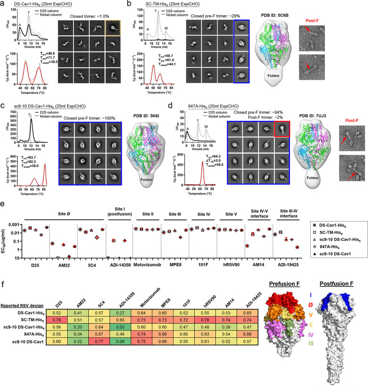
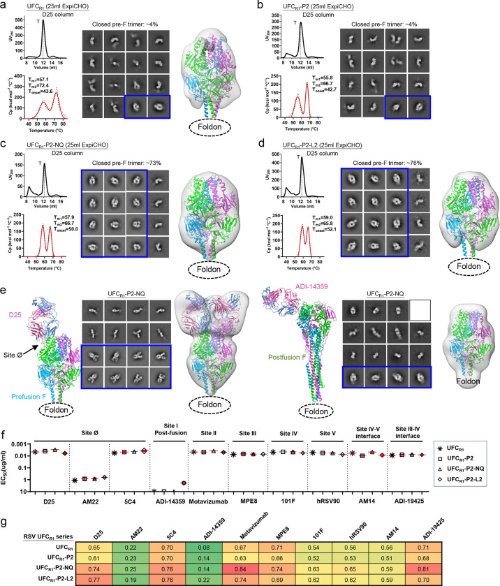
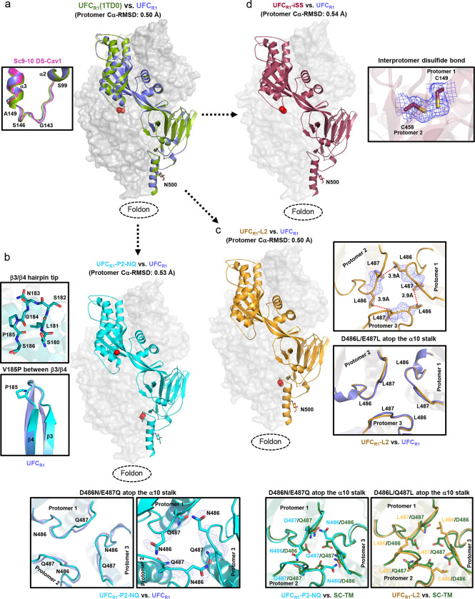
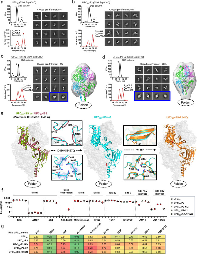
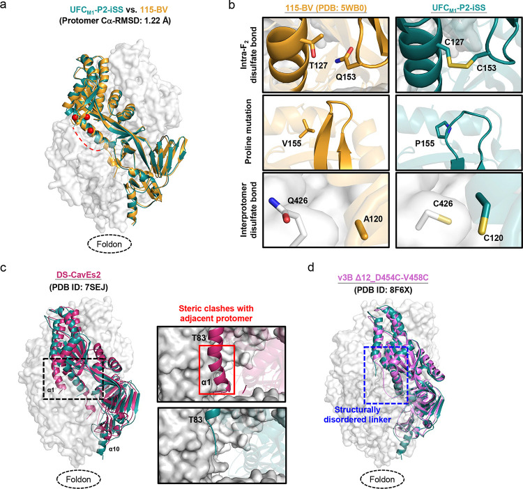
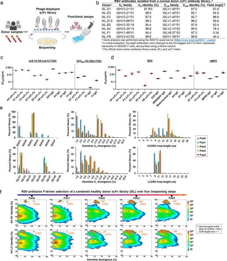
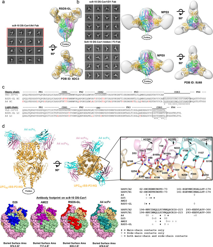
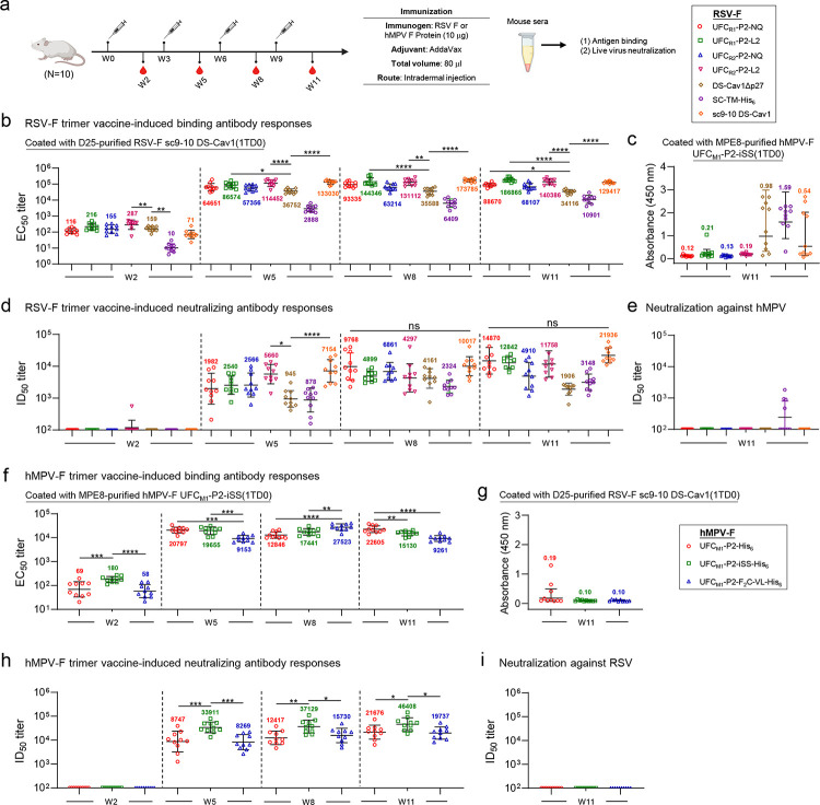
Respiratory syncytial virus (RSV) and human metapneumovirus (hMPV) cause human respiratory diseases and are major targets for vaccine development. In this study, we designed uncleaved prefusion-closed (UFC) trimers for the fusion (F) proteins of both viruses by examining mutations critical to F metastability. For RSV, we assessed four previous prefusion F designs, including the first and second generations of DS-Cav1, SC-TM, and 847A. We then identified key mutations that can maintain prefusion F in a native-like, closed trimeric form (up to 76%) without introducing any interprotomer disulfide bond. For hMPV, we developed a stable UFC trimer with a truncated F2-F1 linkage and an interprotomer disulfide bond. Tens of UFC constructs were characterized by negative-stain electron microscopy (nsEM), x-ray crystallography (11 RSV-F and one hMPV-F structures), and antigenic profiling. Using an optimized RSV-F UFC trimer as bait, we identified three potent RSV neutralizing antibodies (NAbs) from a phage-displayed human antibody library, with a public NAb lineage targeting sites Ø and V and two cross-pneumovirus NAbs recognizing site III. In mouse immunization, rationally designed RSV-F and hMPV-F UFC trimers induced robust antibody responses with high neutralizing titers. Our study provides a foundation for future prefusion F-based RSV and hMPV vaccine development.


Conflict of interest statement

Competing interests: The authors declare that they have no competing interests.

Figures

Fig. 1.
Fig. 1.. Comparative in vitro characterization of previously reported RSV-F designs.
Size exclusion chromatography (SEC) profile (left top), differential scanning calorimetry (DSC) profile (left bottom), representative 2D classification images (middle, or right when no 3D models shown), and 3D reconstruction from the nsEM analysis (right) for (a) DS-Cav1, (b) SC-TM, (c) Sc9–10 DS-Cav1, and (d) 847A. All constructs contain a foldon trimerization motif with or without a C-terminal His6 tag, as indicated in the construct name with a suffix (-His6). A total of eight constructs were transiently expressed in 25 ml of ExpiCHO cells and purified using a Nickel column or a D25 antibody column. Major SEC peaks, such as aggregation (A), trimer (T), and monomer (A), are marked on the profile. The SEC profiles after Nickel and D25 antibody purification are shown as dotted and solid lines, respectively. For the 2D classification analysis, prefusion-closed trimers are circled in a blue line box, and closed trimers in the postfusion and non-prefusion states are circled in a red line box and a dotted orange line box, respectively. For SC-TM-His6 and 847A-His6, two micrographs are enlarged to show shapes characteristic of the postfusion F trimer. Prefusion RSV-F trimer structures (PDB ID: 5C6B, 5K6I, and 7UJ3) are used for structural fitting into the nsEM densities. (e) ELISA-derived EC50 (μg/ml) values of five RSV-F trimers binding to 10 antibodies that target six antigenic sites and two interface epitopes, which are labeled on the plots. (f) Biolayer interferometry (BLI) antigenic profiles of five RSV-F trimers binding to the same antibodies. Sensorgrams were obtained from an Octet RED96 instrument using an antigen titration series of six concentrations (starting at 600 nM followed by two-fold dilutions) and are shown in Fig. S1g. Peak values at the highest concentration are shown in a matrix, in which cells are colored in green (weak binding) to red (strong binding) (left). In (e) and (f), the four His-tagged RSV-F trimers were purified using a Nickel column, whereas sc9–10 DS-Cav1 was purified using a D25 antibody column. (g) Footprints of six antigenic sites colored on the surface representation of prefusion (PDB ID: 4JHW) and postfusion (PDB ID: 3RRR) RSV-F trimers (right).
Fig. 2.
Fig. 2.. Design and in vitro characterization of RSV-F UFCR1 series.
SEC profile (left top), DSC profile (left bottom), representative 2D classification images (middle, or right when no 3D models shown), and 3D reconstruction from nsEM analysis (right) for (a) UFCR1, (b) UFCR1-P2, (c) UFCR1-P2-NQ, and (d) UFCR1-P2-L2. All UFCR1 constructs were transiently expressed in 25 ml of ExpiCHO cells and purified using a D25 antibody column. The trimer (T) peak is marked on the profile. (e) The nsEM analysis of UFCR1-P2-NQ in the presence of prefusion-specific antibody D25 (left panel) or postfusion-specific antibody ADI-14359 (right panel). Each panel shows the ribbon model of the RSV-F/antibody complex (left), representative 2D classification images (middle), and 3D reconstruction (right). The 2D classes corresponding to prefusion-closed trimers (either ligand-free or antibody-bound) are circled in blue, and a prefusion RSV-F trimer (PDB ID: 4JHW) was used for structural fitting into the nsEM densities. (f) ELISA-derived EC50 (μg/ml) values of four UFCR1 constructs binding to 10 antibodies, as in Fig. 1e. (g) BLI-derived antigenic profiles of four UFCR1 constructs binding to 10 antibodies. Sensorgrams were obtained using the same protocol as in Fig. 1f and are shown in Fig. S2e. The matrix of peak values at the highest antigen concentration is shown, as in Fig. 1f.
Fig. 3.
Fig. 3.. Crystallographic analysis of RSV-F UFCR1 series and variants.
(a) Crystal structures of UFCR1 and UFCR1(1TD0) (2.26 and 2.28 Å) are superimposed and shown as green and blue ribbon models, respectively, within the gray trimer surface. The F2-F1 linkage is shown for UFCR1 and UFCR1(1TD0) with respect to sc9–10 DS-Cav1 (pink) in the left inset. (b) The crystal structure of UFCR1-P2-NQ (2.70 Å) is shown as a cyan ribbon model within the gray trimer surface. The atomic model of the β3/β4 hairpin tip and a close-up view of the V185P mutation between β3 and β4 are shown in the left insets, and the backbone and side chains of acidic patch mutations in UFCR1-P2-NQ (D486N-E487Q) are compared with UFCR1 in the bottom insets. (c) The crystal structure of UFCR1-L2 (2.30 Å) is shown as a gold ribbon model within the gray trimer surface. The backbone and side chains of acidic patch mutations in UFCR1-L2 (D486L-E487L) are compared with UFCR1 in the right insets, and structural details of this region in UFCR1-P2-NQ and UFCR1-L2 are compared with SC-TM in the bottom insets. (d) The crystal structure of UFCR1-iSS (2.3 Å) is shown as a red ribbon model within the gray trimer surface. A close-up view of the interprotomer disulfide bond (A149C-Y458C) within the density is shown in the right inset.
Fig. 4.
Fig. 4.. Design, in vitro characterization, and crystallographic analysis of RSV-F UFCR2 series.
SEC profile (left top), DSC profile (left bottom), representative 2D classification images (middle, or right when no 3D models shown), and 3D reconstruction from nsEM analysis (right) for (a) UFCR2, (b) UFCR2-P2, (c) UFCR2-P2-NQ, and (d) UFCR2-P2-L2. All UFCR2 constructs were transiently expressed in 25 ml of ExpiCHO cells and purified using a D25 antibody column. The trimer (T) peak is marked on the profile. The 2D classification images corresponding to prefusion-closed trimers are circled in blue, and a prefusion RSV-F trimer (PDB ID: 4JHW) is used for structural fitting into the nsEM densities. (e) Crystallographic analysis of three UFCR2-derived constructs. Left: Crystal structures of UFCR2-iSS and UFCR1-iSS (2.83 and 2.30 Å) are superimposed and shown as green and red ribbon models, respectively, within the gray trimer surface. Middle: The crystal structure of UFCR2-iSS-NQ (2.30 Å) is shown as a cyan ribbon model within the gray trimer surface. The backbone and side chains of acidic patch mutations in UFCR3-iSS-NQ (D486N-E487Q) are compared with UFCR2-iSS in the insets to the left of the protomer/surface model. Right: The crystal structure of UFCR2-iSS-P2-NQ (2.30 Å) is shown as a gold ribbon model within the gray trimer surface. Details of the V185P mutation and acidic patch are shown in the insets to the left of the protomer/surface model. (f) ELISA-derived EC50 (μg/ml) values of five UFCR2 constructs binding to 10 antibodies, as in Fig. 1e. (g) BLI-derived antigenic profiles of five UFCR2 constructs binding to 10 antibodies. Sensorgrams were obtained using the same protocol as in Fig. 1f and are shown in Fig. S4e. The matrix of peak values at the highest antigen concentration is shown, as in Fig. 1f.
Fig. 5.
Fig. 5.. Design and in vitro characterization of hMPV-F UFCM1 series.
SEC profile (left top), DSC profile (left bottom), representative 2D classification images (middle, or right when no 3D models shown), and 3D reconstruction from nsEM analysis (right) for (a) UFCM1, (b) UFCM1-P2, (c) UFCM1-P2-iSS, and (d) UFCM1-P2-F2C-VL. All UFCM1 constructs were transiently expressed in 25 ml of ExpiCHO cells and purified using an MPE8 antibody column and a Nickel column, as all constructs contain a His6 tag. The trimer (T) peak is marked on the profile. (e) The nsEM analysis of UFCM1-P2-iSS bound to antibody MPE8. Representative 2D classification images are shown on the top, and side and top views of 3D reconstruction of the complex are shown on the bottom left and right, respectively. A 3.25 Å-resolution cryo-EM model of MPE8 scFv-bound v3B Δ12_D454C-V458C (PDB ID: 8F6X) was used for density fitting. (f) The nsEM analysis of UFCM1-P2-iSS bound to antibody 101F. Representative 2D classification images are shown on the left, and side and top views of 3D reconstruction of the complex are shown on the right. A model of 101F Fab modeled onto a prefusion hMPV-F trimer (PDB ID: 5WB0) was used for density fitting. (g) ELISA-derived EC50 (μg/ml) values of four UFCM1 constructs binding to four antibodies, as in Fig. 1e. (h) BLI-derived antigenic profiles of four UFCM1 constructs binding to four antibodies. Sensorgrams were obtained using the same protocol as in Fig. 1f and are shown in Fig. S6e. The matrix of peak values at the highest antigen concentration is shown as in Fig. 1f.
Fig. 6.
Fig. 6.. Crystallographic analysis of hMPV-F UFCM1-P2-iSS.
(a) The crystal structure of UFCM1-P2-iSS (6.0 Å) is superimposed onto that of 115-BV (PDB ID: 5WB0), which are shown as green and gold ribbon models, respectively, within the gray trimer surface. Due to the limited resolution, structural details cannot be determined for the F2-F1 linker, A344-S347, and V442-E457. A red dotted line is added to show the expected approximate location of the missing F2-F1 linker. (b) Structural details of the intra-F2 disulfide bond T127C-Q153C, the V155P (P2) mutation inserted into the β3/β4 hairpin tip for destabilizing the postfusion state, and the interprotomer disulfide bond A120C-Q426C (iSS) are shown in the top, middle, and bottom insets, respectively. The crystal structure of 115-BV is included for comparison. (c) The crystal structures of UFCM1-P2-iSS and DS-CavEs2 (PDB ID: 7SEJ) are superimposed and shown as green and rouge pink ribbon models, respectively, within the gray trimer surface. The extended α1 helix in DS-CavEs2 that will clash with an adjacent protomer in a prefusion-closed trimer is circled in a black dotted line box. Close-up views of this region in DS-CavEs2 and UFCM1-P2-iSS are shown in the right insets. (d) Crystal structures of UFCM1-P2-iSS and v3B Δ12_D454C-V458C (PDB ID: 8F6X) are superimposed and shown as green and pink ribbon models, respectively, within the gray trimer surface. The F2-F1 linker region is circled in a blue dotted line box.
Fig. 7.
Fig. 7.. Potent pneumovirus neutralizing antibodies identified from a human antibody library.
(a) Schematic representation of the phage display workflow. (b) Gene family analysis of eight scFv clones identified from a phage-displayed human antibody (scFv) library using RSV-F UFCR1-P2-NQ as a biopanning antigen. (c) ELISA-derived EC50 (μg/ml) values of library-derived antibodies binding to RSV-F sc9–10 DS-Cav1 and hMPV-F UFCM1-P2-iSS. (d) IC50 (μg/ml) values derived from live RSV and hMPV neutralization assays. (e) Distribution of germline gene usage, somatic hypermutation, and CDR3 length plotted for heavy chains (HCs) and λ-light chains (λ-LCs) of the scFv library during the biopanning process. (f) Identity-divergence analysis of the IXL-A4 (or A4) within the scFv library during the biopanning process. The sequence datasets used in (e) and (f) were obtained from next-generation sequencing (NGS) of the scFv libraries on an Ion GeneStudio S5 platform. For the heatmaps in (f), after data processing using an Antibodyomics pipeline, each sequence is plotted as a function of sequence identity from a reference antibody chain and sequence divergence from the assigned germline gene. Color indicates sequence density at a particular point on the 2D plot. Sequences with the same germline gene, a CDR3 identity ≥ 95%, and a 1-residue error margin of CDR length calculation with respect to the reference antibody chain are plotted as orange dots with the number of sequences and gene family percentage labeled. The schematic representation of phage-based antibody isolation was created with BioRender.com.
Fig. 8.
Fig. 8.. Structural characterization of potent pneumovirus neutralizing human antibodies.
(a) The nsEM analysis of sc9–10 DS-Cav1 in complex with A4. Representative 2D classification images are shown on the left, and side and top views of 3D reconstruction of the complex are shown on the left. A 3.50 Å-resolution crystal structure of RSD5-bound DS-Cav1 (PDB ID: 6DC3) was used for density fitting. The 2D classification images containing two or more than two bound A4 Fabs are circled in a red line box. (b) The nsEM analysis of sc9–10 DS-Cav1 in complex with D1 and F3. Representative 2D classification images are shown on the left, and side and top views of 3D reconstruction of the complex are shown on the right. A 3.08 Å-resolution crystal structure of MPE8-bound DS-Cav1 (PDB ID: 5U68) was used for density fitting. (c) Sequence analysis of A4 and RSD5 heavy and light chains with alignment to respective germline genes. Mature antibody residues that differ from the germline are colored in red. (d) Crystallographic analysis of UFCR2-iSS-P2-NQ in complex with A4 scFv and structural epitope mapping. Top left: A 4.0 Å-resolution crystal structure UFCR2-iSS-P2-NQ in complex with A4 scFv is shown as ribbon models, with UFCR2-iSS-P2-NQ in gold and A4 heavy and light chains in pink and cyan, respectively. Top right: Close-up view of the A4/RSV-F interface. Side chains are shown for residues involved in hydrogen bond interactions across the A4/RSV-F interface, which were identified based on the estimated donor-acceptor distances and are indicated by dotted black lines. HCDR1, HCDR3, LCDR3 and LCDR1 loops are indicated. Bottom left: Surface models of prefusion RSV-F trimer showing footprints of D25, AM22, RSD5, and A4 colored in blue, rouge pink, red, and teal blue, respectively. Bottom right: RSV-F sequence with antibody-interacting residues labeled for D25, AM22, RSD5, and A4. Three types of contact are considered using a cutoff distance of 5 Å: main-chain contacts, side-chain contacts, and both.
Fig. 9.
Fig. 9.. Antibody responses to rationally designed RSV and hMPV-F trimer vaccines in mice.
(a) Schematic representation of the mouse immunization regimen for both RSV-F and hMPV-F vaccines (n = 10 mice/group). (b, c) RSV-F vaccine-induced binding antibody responses against RSV-F sc9–10 DS-Cav1(1TD0) and hMPV-F UFCM1-P2-iSS(1TD0). (d, e) RSV-F vaccine-induced neutralizing antibody responses against live RSV-A2-GFP and live hMPV-GFP. In (b)-(e), DS-Cav1Δp27, SC-TM-His6, and sc9–10 DS-Cav1 are included for comparison. (f, g) HMPV-F vaccine-induced binding antibody responses against hMPV-F UFCM1-P2-iSS(1TD0) and RSV-F sc9–10 DS-Cav1(1TD0). (h, i) HMPV-F vaccine-induced neutralizing antibody responses against live hMPV-GFP and live RSV-A2-GFP. EC50 values (b, c, f, g) were derived from the ELISA analysis of mouse serum against coating antigens, with geometric mean EC50 values labeled on the plots. ID50 titers were derived from the live RSV and hMPV neutralization assays, with geometric mean ID50 values labeled on the plots. Notably, the ID50 values were derived by setting the lower/upper constraints of % neutralization at 0.0/100.0. The data were analyzed using one-way ANOVA, followed by Tukey’s multiple comparison post hoc test for each timepoint. The statistical significance is indicated as the following: ns (not significant), *p < 0.05, **p < 0.01, ***p < 0.001, and ****p < 0.0001. In (b) and (d), statistical analyses of EC50 and ID50 values were performed by comparing individual RSV-F vaccine with the control, DS-Cav1Δp27. Detailed ELISA and neutralization data and the complete statistical analysis are shown in Fig. S10. The schematic representation of the mouse immunization protocol was created with BioRender.com.

Similar articles

References

    1. Borchers A. T., Chang C., Gershwin M. E., Gershwin L. J., Respiratory Syncytial Virus - A Comprehensive Review. Clin. Rev. Allergy Immunol. 45, 331–379 (2013). - PMC - PubMed
    1. Schildgen V. et al., Human metapneumovirus: lessons learned over the first decade. Clin. Microbiol. Rev. 24, 734–754 (2011). - PMC - PubMed
    1. Falsey A. R., Hennessey P. A., Formica M. A., Cox C., Walsh E. E., Respiratory syncytial virus infection in elderly and high-risk adults. N. Engl. J. Med. 352, 1749–1759 (2005). - PubMed
    1. Stockman L. J., Curns A. T., Anderson L. J., Fischer-Langley G., Respiratory syncytial virus-associated hospitalizations among infants and young children in the United States, 1997–2006. Pediatr. Infect. Dis. J. 31, 5–9 (2012). - PubMed
    1. Shi T. et al., Global, regional, and national disease burden estimates of acute lower respiratory infections due to respiratory syncytial virus in young children in 2015: a systematic review and modelling study. Lancet 390, 946–958 (2017). - PMC - PubMed

Publication types