. 2024 Mar 1;25(5):2898.
doi: 10.3390/ijms25052898.

Human Macrophages Activate Bystander Neutrophils' Metabolism and Effector Functions When Challenged with Mycobacterium tuberculosis

Affiliations

Human Macrophages Activate Bystander Neutrophils' Metabolism and Effector Functions When Challenged with Mycobacterium tuberculosis

Dearbhla M Murphy et al. Int J Mol Sci. .

Abstract

Neutrophils are dynamic cells, playing a critical role in pathogen clearance; however, neutrophil infiltration into the tissue can act as a double-edged sword. They are one of the primary sources of excessive inflammation during infection, which has been observed in many infectious diseases including pneumonia and active tuberculosis (TB). Neutrophil function is influenced by interactions with other immune cells within the inflammatory lung milieu; however, how these interactions affect neutrophil function is unclear. Our study examined the macrophage-neutrophil axis by assessing the effects of conditioned medium (MΦ-CM) from primary human monocyte-derived macrophages (hMDMs) stimulated with LPS or a whole bacterium (Mycobacterium tuberculosis) on neutrophil function. Stimulated hMDM-derived MΦ-CM boosts neutrophil activation, heightening oxidative and glycolytic metabolism, but diminishes migratory potential. These neutrophils exhibit increased ROS production, elevated NET formation, and heightened CXCL8, IL-13, and IL-6 compared to untreated or unstimulated hMDM-treated neutrophils. Collectively, these data show that MΦ-CM from stimulated hMDMs activates neutrophils, bolsters their energetic profile, increase effector and inflammatory functions, and sequester them at sites of infection by decreasing their migratory capacity. These data may aid in the design of novel immunotherapies for severe pneumonia, active tuberculosis and other diseases driven by pathological inflammation mediated by the macrophage-neutrophil axis.

Keywords: Mycobacterium tuberculosis; glycolysis; granulocytes; immunometabolism; infection; neutrophil function; neutrophil metabolism; neutrophil priming and activation; polymorphonuclear cells; tuberculosis.


Conflict of interest statement

The authors declare no conflicts of interest. The funders had no role in the design of the study; in the collection, analyses, or interpretation of data; in the writing of the manuscript; or in the decision to publish the results.

Figures

Figure 1
Figure 1
Methods Summary. (A) Macrophage Stage: hMDMs, differentiated from PBMCs isolated from healthy blood donors over 7 days, were stimulated with Mtb for three hours and washed to remove unphagocytosed Mtb (hMDMs were also left unstimulated and treated with 100 ng/mL LPS in parallel). Then, 24 h post-stimulation, the hMDM-conditioned medium (MΦ-CM) was processed and stored at −80 °C for later use. MΦ-CM from a minimum of three independent donors was pooled and was used for all subsequent experimentation techniques. Neutrophil Stage: On a subsequent day, fresh CD15+ cells were isolated, purified, and treated with 20% MΦ-CM (80% fresh cRPMI) for one hour. Flow cytometry, real-time extracellular Seahorse flux analysis, and MSD ELISAs were subsequently carried out to determine the effect of the MΦ-CM on neutrophil function. (B) All treatments with MΦ-CM consisted of freshly isolated neutrophils being exposed to 20% cRPMI (RPMI supplemented with 10% human serum), 20% MΦ-CM from unstimulated hMDMs (UsCoM), 20% MΦ-CM from Mtb-stimulated hMDMs (MtbCoM), and 20% MΦ-CM from LPS-stimulated hMDMs (LPSCoM).
Figure 2
Figure 2
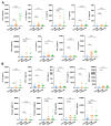
Characterizing secreted cytokine and chemokine levels in MΦ-CM (UsCoM, MtbCoM, and LPSCoM) from unstimulated, Mtb-stimulated, and LPS-stimulated hMDMs. hMDMs, differentiated from PBMCs isolated from healthy blood donors, were stimulated with iH37Rv Mtb for three hours and were washed to remove unphagocytosed Mtb. hMDMs were also left unstimulated and treated with LPS (100 ng/mL) in parallel. (A) 24 h post-stimulation, the secreted levels of the cytokines TNF-α, IL-1β, IL-10, IL-4, IL-2, CXCL8, IL-13, IFN-γ, and IL-6 were quantified using MSD Multi-Array technology, (B) along with the chemokines IP-10, MCP-4, TARC, MIP-1α, MDC, Eotaxin, Eotaxin-3, MIP-1β, and MCP-1. Bars denote mean ± SEM (n = 18 independent donors). * p < 0.05, ** p < 0.01, *** p < 0.001, and **** p < 0.0001, ns = no significance (Friedman ANOVA test with Dunn’s multiple comparisons tests).
Figure 3
Figure 3
Examining if MΦ-CM from Mtb-stimulated or LPS-stimulated hMDMs affects human neutrophil activation and migration. (A) Neutrophils exposed to 20% MΦ-CM were visualised under light microscopy (40×) to examine if the MΦ-CM affects neutrophil morphology. (B) Flow cytometric analyses were carried out to examine if neutrophils exposed to MΦ-CM could be activated, first by gating on single cells (FSC-A vs. FSC-H), granular cells (FSC-A vs. SSC-A), and finally viable CD15+ neutrophils (AmCyan vs. PE-Cy7). (C) Neutrophil activation was assessed by determining the expression of the granulation marker CD62L and (D) the degranulation marker CD63 in viable CD15+ neutrophils in response to cRPMI, UsCoM, MtbCoM, and LPSCoM (n = 5). (E) As activation status is a good surrogate indicator of migration in neutrophils, the ability of neutrophils to migrate was also assessed. Cells were treated with 20% MΦ-CM for one hour as before, placed into the top chamber of 5 µm transwell inserts, and allowed to migrate toward 1.25 ng/mL fMLP in the bottom chamber for 15 min. Samples were then stained for CD15 expression and viability, an equal number of PrecisionTM Counting Beads were added, and the numbers of migrated neutrophils (cells/µL) were quantified by flow cytometry (n = 6). (F) Expression of the cell surface migration marker CXCR2 was examined in neutrophils exposed to the MΦ-CM treatments (n = 5). * p < 0.05 and ** p < 0.01 (Friedman ANOVA test with Dunn’s multiple comparisons tests). MFI: Median fluorescence intensity.
Figure 4
Figure 4
Investigating if MΦ-CM from Mtb-stimulated or LPS-stimulated hMDMs affects baseline glycolysis and oxidative phosphorylation in unstimulated human neutrophils. The effect of MΦ-CM on the real-time baseline (A) OCR (n = 14) and (B) ECAR (n = 18), representing oxidative phosphorylation and glycolysis, respectively, was determined utilising Seahorse extracellular flux assays in neutrophils treated for one hour with 20% cRPMI, UsCoM, MtbCoM, and LPSCoM. (C) The immunometabolic shift from UsCoM treatment to MtbCoM and LPSCoM treatment can be illustrated by the metabolic phenograms. ** p < 0.01, *** p < 0.001, and **** p < 0.0001 (Friedman ANOVA test with Dunn’s multiple comparisons tests).
Figure 5
Figure 5
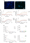
Assessing if MΦ-CM from Mtb-stimulated or LPS-stimulated hMDMs affects bioenergetics in fMPL-stimulated and Mtb-stimulated human neutrophils. (A) Neutrophils were seeded into Labtek II chamber slides, unstimulated or stimulated with iH37Rv-Mtb, washed, and phagocytosed Mtb was fluorescently visualised through auramine (FITC) and hoechst (DAPI) staining. (B) Baseline metabolic OCR (left) and ECAR (right) values were examined in neutrophils in response to iH37Rv-Mtb (MOI: 1–10), (C) prior to determining if 20% cRPMI, UsCoM, MtbCoM, and LPSCoM pre-treatment exhibited an effect on OCR and ECAR profiles in neutrophils stimulated with iH37Rv (n = 3). After neutrophils were treated for one hour with 20% cRPMI, UsCoM, MtbCoM, and LPSCoM, cells were also stimulated with fMLP (1.25 ng/mL) and real-time baseline (D) OCR and (E) ECAR values were determined utilising Seahorse extracellular flux assays (n = 5). (D,E) OCR and ECAR rates were plotted and graphed (right-hand side graphs) at the time point reflecting the highest metabolic response to fMLP stimulation (40 min). * p < 0.05, ** p < 0.01, *** p < 0.001, and **** p < 0.0001 ((A,B): Friedman ANOVA test with Dunn’s multiple comparisons tests; (D,E): Two-way RM ANOVA with Šídák’s multiple comparisons test).
Figure 6
Figure 6
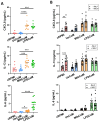
Elucidating if MΦ-CM from Mtb-stimulated or LPS-stimulated hMDMs alters cytokine release in primary human neutrophils. Human neutrophils were treated with 20% cRPMI, UsCoM, MtbCoM, and LPSCoM for 1 h. Following incubation, neutrophils were left unstimulated (A) and stimulated with 1.25 ng/mL fMLP (B). 4 h later, the secreted levels of CXCL8, IL-13, and IL-6 were assessed using MSD-ELISA (n = 13). * p < 0.05, ** p < 0.01, *** p < 0.001, and **** p < 0.0001 ((A): Friedman ANOVA test with Dunn’s multiple comparisons tests; (B): Two-way ANOVA with mixed effects model multiple comparisons test).
Figure 7
Figure 7
Determining if MΦ-CM from Mtb-stimulated or LPS-stimulated hMDMs alters NETosis, reactive oxygen species (ROS) production, glucose uptake, and apoptosis in primary human neutrophils. Flow cytometric analyses were carried out to examine if neutrophils exposed to MΦ-CM exhibited altered NETosis, ROS production, and glucose uptake. This was achieved by first gating on single cells (FSC-A vs. FSC-H), granular cells (FSC-A vs. SSC-A), and finally, viable CD15+ neutrophils (AmCyan vs. PE-Cy7). (A) Flow cytometric dot plots illustrating the effect of 20% MΦ-CM on NETosis in a representative donor (NETosis is characterized as being double positive in the Sytox red (APC) and DAPI (Pacific Blue) channels). Histogram shifts in (B) DHR-123 (FITC) and (C) 2-NBDG (FITC) expression are characterized by changes in ROS production and glucose uptake in MΦ-CM-treated CD15+ neutrophils, respectively. Using these flow cytometric tools, NETosis (n = 10), ROS production (n = 16), and glucose uptake (n = 4) were assessed in CD15+ neutrophils one hour post-treatment with 20% MΦ-CM. (D) Early apoptosis (characterized as CD15+/Annexin V+) and late apoptosis (characterized as CD15+/Annexin V+/Zombie+) were also determined to examine the effect of the MΦ-CM on cell viability. A representative flow cytometric dot plot is shown. * p < 0.05, ** p < 0.01, **** p < 0.0001 (Friedman ANOVA test with Dunn’s multiple comparisons tests).
Figure 8
Figure 8
A graphical summary of the study. Freshly isolated CD15+ neutrophils were exposed to four different conditions, complete RPMI media (cRPMI), media from unconditioned MDMs (UsCoM), media from MDMs infected with Mtb (MtbCoM), and media from MDMs stimulated with LPS (LPSCoM). Glucose uptake and apoptosis were unaffected by MtbCoM and LPSCoM. Effects on several aspects of neutrophil function were examined via several methods, including flow cytometry, Agilent Seahorse assays, MSD ELISA, and transwell migration assays. Our data suggests an augmentation of many neutrophil functions as a result of MtbCoM and LPSCoM, such as NETosis, ROS generation, and inflammatory cytokine production. Neutrophils exposed to the inflammatory conditioned media also exhibited a significant energetic shift, with glycolysis and oxidative phosphorylation significantly augmented. MtbCoM and LPSCoM also decrease neutrophil chemotaxis, as shown both via the quantification of the chemotactic receptor CXCR2 and via transwell migration assays. Overall, our data demonstrated the importance of the macrophage–neutrophil axis in infection.

Similar articles

References

    1. Mantovani A., Cassatella M.A., Costantini C., Jaillon S. Neutrophils in the activation and regulation of innate and adaptive immunity. Nat. Rev. Immunol. 2011;11:519–531. doi: 10.1038/nri3024. - DOI - PubMed
    1. Pechous R.D. With Friends Like These: The Complex Role of Neutrophils in the Progression of Severe Pneumonia. Front. Cell. Infect. Microbiol. 2017;7:160. doi: 10.3389/fcimb.2017.00160. - DOI - PMC - PubMed
    1. Lowe D.M., Redford P.S., Wilkinson R.J., O’Garra A., Martineau A.R. Neutrophils in tuberculosis: Friend or foe? Trends Immunol. 2012;33:14–25. doi: 10.1016/j.it.2011.10.003. - DOI - PubMed
    1. Martineau A.R., Newton S.M., Wilkinson K.A., Kampmann B., Hall B.M., Nawroly N., Packe G.E., Davidson R.N., Griffiths C.J., Wilkinson R.J. Neutrophil-mediated innate immune resistance to mycobacteria. J. Clin. Investig. 2007;117:1988–1994. doi: 10.1172/JCI31097. - DOI - PMC - PubMed
    1. Ong C.W.M., Elkington P.T., Brilha S., Ugarte-Gil C., Tome-Esteban M.T., Tezera L.B., Pabisiak P.J., Moores R.C., Sathyamoorthy T., Patel V., et al. Neutrophil-Derived MMP-8 Drives AMPK-Dependent Matrix Destruction in Human Pulmonary Tuberculosis. PLoS Pathog. 2015;11:e1004917. doi: 10.1371/journal.ppat.1004917. - DOI - PMC - PubMed