. 2024 Feb 29;16(5):1021.
doi: 10.3390/cancers16051021.

Intratumoral Delivery of Interleukin 9 via Oncolytic Vaccinia Virus Elicits Potent Antitumor Effects in Tumor Models

Affiliations

Intratumoral Delivery of Interleukin 9 via Oncolytic Vaccinia Virus Elicits Potent Antitumor Effects in Tumor Models

Junjie Ye et al. Cancers (Basel). .

Abstract

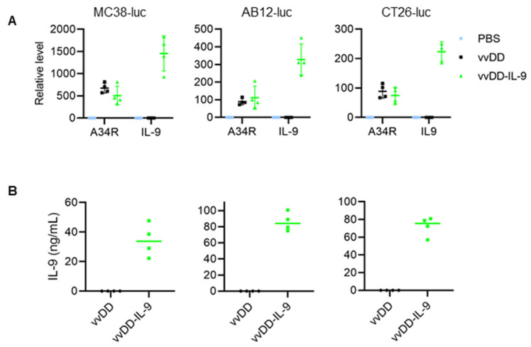
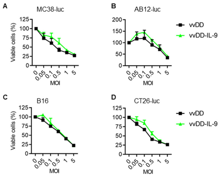
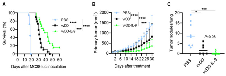
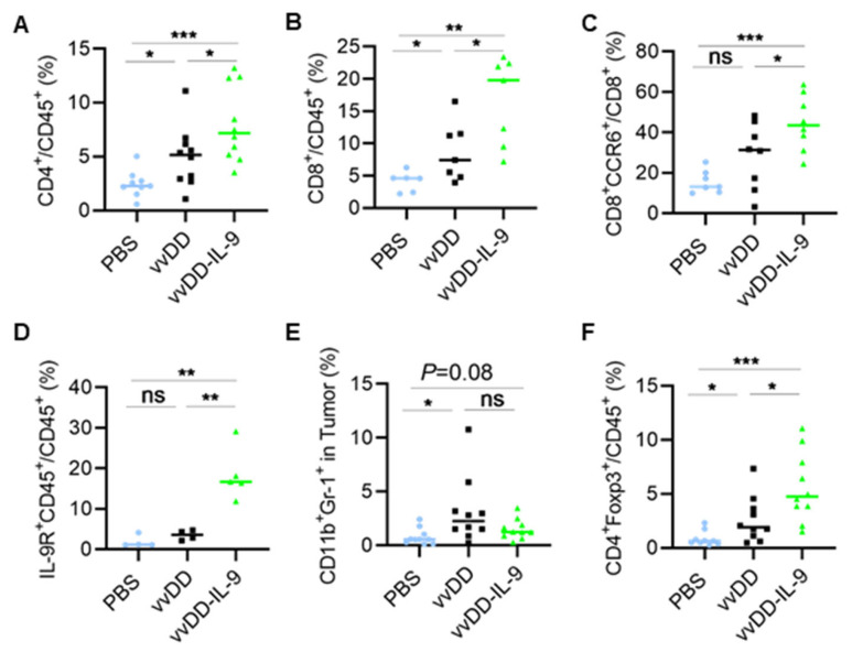
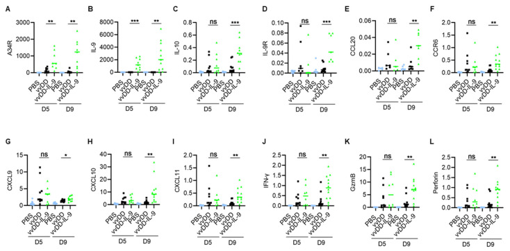
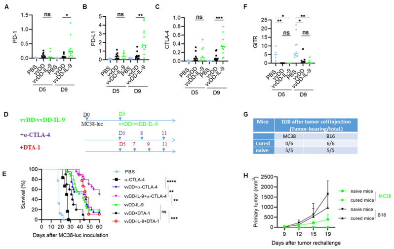
The success of cancer immunotherapy is largely associated with immunologically hot tumors. Approaches that promote the infiltration of immune cells into tumor beds are urgently needed to transform cold tumors into hot tumors. Oncolytic viruses can transform the tumor microenvironment (TME), resulting in immunologically hot tumors. Cytokines are good candidates for arming oncolytic viruses to enhance their function in this transformation. Here, we used the oncolytic vaccinia virus (oVV) to deliver interleukin-9 (IL-9) into the tumor bed and explored its antitumor effects in colon and lung tumor models. Our data show that IL-9 prolongs viral persistence, which is probably mediated by the up-regulation of IL-10. The vvDD-IL-9 treatment elevated the expression of Th1 chemokines and antitumor factors such as IFN-γ, granzyme B, and perforin. IL-9 expression increased the percentages of CD4+ and CD8+ T cells in the TME and decreased the percentage of oVV-induced immune suppressive myeloid-derived suppressor cells (MDSC), leading to potent antitumor effects compared with parental virus treatment. The vvDD-IL-9 treatment also increased the percentage of regulatory T cells (Tregs) in the TME and elevated the expression of immune checkpoint molecules such as PD-1, PD-L1, and CTLA-4, but not GITR. The combination therapy of vvDD-IL-9 and the anti-CTLA-4 antibody, but not the anti-GITR antibody, induced systemic tumor-specific antitumor immunity and significantly extended the overall survival of mice, indicating a potential translation of the IL-9-expressing oncolytic virus into a clinical trial to enhance the antitumor effects elicited by an immune checkpoint blockade for cancer immunotherapy.

Keywords: IL-10; IL-9; anti-CTLA-4 antibody; colon cancer; combination; immune checkpoint blockade; immunotherapy; modulation; oncolytic virus; tumor microenvironment.


Conflict of interest statement

The authors declare no conflicts of interest.

Figures

Figure 1
Figure 1
vvDD-IL-9 infection shows significantly higher IL-9 secretion and a similar viral replication compared vvDD in vitro. Tumor cells of MC38-luc (3.0 × 10⁵ cells), AB12-luc (3.0 × 10⁵ cells), or CT26-luc (3.0 × 10⁵ cells) were mock-infected or infected with vvDD or vvDD-IL-9 at an MOI of 1. At 24 h post infection, the cells were pelleted and harvested to measure A34R or IL-9 expression using RT-qPCR (A) and the supernatants were harvested 24 h after infection to measure IL-9 amount using ELISA (B), respectively.
Figure 2
Figure 2
vvDD-IL-9 has a similar viral cytotoxicity compared with vvDD in vitro. Tumor cells of MC38-luc (1.0 × 10⁴ cells) (A), AB12-luc (8.0 × 10³ cells) (B), B16 (8.0 × 10³ cells) (C), or CT-26-luc (8.0 × 10³ cells) (D), were infected with vvDD-IL-9 or vvDD at indicated MOIs. Cell viability was measured 48 h after viral infection.
Figure 3
Figure 3
vvDD-IL-9 treatment elicits potent therapeutic effects in murine tumor models. B6 mice were i.p. inoculated with 5.0 × 10⁵ MC38-luc cells and then treated with PBS, vvDD, or vvDD-IL-9 at 2.0 × 10⁸ PFU/mouse five days after tumor cell inoculation. The Kaplan–Meier survival curve is shown (A). B6 mice were s.c. inoculated with 1.0 × 10⁶ LLC cells in right flank and were i.t. treated with 60 µL PBS or 60 µL virus (5.0 × 10⁷ PFU) per mouse on day 9 after tumor cell inoculation. The primary tumor size was measured every two days, and all treated mice were euthanized after the first mouse with a tumor size over 2 cm in any direction was found to count lung metastatic tumor nodules. Tumor growth curves (B) and lung metastatic tumor nodules (C) are shown, respectively. A log-rank (Mantel–Cox) test was used to compare survival rates. A two-way ANOVA test was used to compare tumor growth cures. *: p < 0.05; ***: p < 0.001; and ****: p < 0.0001.
Figure 4
Figure 4
vvDD-IL-9 treatment transforms tumor-infiltrating leukocytes. B6 mice were i.p. inoculated with 5.0 × 10⁵ MC38-luc cells and treated with PBS, vvDD, or vvDD-IL-9 at 2.0 × 10⁸ PFU/mouse five days after tumor cell inoculation. Tumor-bearing mice were euthanized five days after treatment and primary tumors were collected. Tumor infiltrating leukocytes were analyzed using flow cytometry. The percentage of CD4+ T cells (A), CD8+ T cells (B), CCR6+CD8+ T cells (C), IL-9R+CD45+ cells (D), MDSCs (E) and Tregs (F) is shown, respectively. * p < 0.05; ** p < 0.01; *** p < 0.001; ns: not significant.
Figure 5
Figure 5
vvDD-IL-9 treatment transforms tumor immunity-associated factors in TME. B6 mice were i.p. inoculated with 5.0 × 10⁵ MC38-luc cells and treated with PBS, vvDD, or vvDD-IL-9 at 2.0 × 10⁸ PFU/mouse five days after tumor cell inoculation. Tumor-bearing mice were euthanized to collect primary tumors five days or nine days after treatment. The expression of factors (AL) associated with tumor effects in the TME was analyzed using RT-qPCR. * p < 0.05; ** p < 0.01 and *** p < 0.001. ns: not significant.
Figure 6
Figure 6
vvDD-IL-9 treatment elicits superior antitumor effects in the combination of anti-CTLA-4 antibody, but not DTA-1 antibody. B6 mice were i.p. inoculated with 5.0 × 10⁵ MC38-luc cells and, 5 days later, treated with PBS, vvDD, or vvDD-IL-9 at 2.0 × 10⁸ PFU/mouse. Tumor-bearing mice were euthanized to collect primary tumors five days or nine days after treatment. The expression of immune checkpoint molecules PD-1, PD-L1, CTLA-4 in tumors was analyzed using RT-qPCR (AC). B6 mice were i.p. inoculated with 5.0 × 10⁵ MC38-luc and treated with PBS, anti-CTLA-4 antibody (200 µg/injection), vvDD (2.0 × 10⁸ PFU/mouse) plus anti-CTLA-4 antibody (200 µg/injection), vvDD-IL-9 (2.0 × 10⁸ PFU/mouse) plus anti-CTLA-4 antibody (200 µg/injection), vvDD (2.0 × 10⁸ PFU/mouse) plus DTA-1 antibody (200 µg/injection), vvDD-IL-9 (2.0 × 10⁸ PFU/mouse) plus DTA-1 antibody (200 µg/injection), or vvDD-IL-9 at 2.0 × 10⁸ PFU/mouse five days after tumor cell inoculation (D) and the survival curves are shown (E). B6 mice were i.p. inoculated with 5.0 × 10⁵ MC38-luc cells and treated with PBS, vvDD, or vvDD-IL-9 at 2.0 × 10⁸ PFU/mouse five days after tumor cell inoculation. Tumor-bearing mice were euthanized to collect primary tumors five days or nine days after treatment. The expression of immune checkpoint molecule GITR was analyzed using RT-qPCR. (F). Naïve B6 mice or MC38-luc-tumor-bearing B6 mice treated with vvDD-IL-9 plus anti-CTLA-4 antibody mentioned above, which had survived for more than 120 days, were s.c. challenged with MC38 tumor cells (1.0 × 10⁶) in the left flanks and irrelative B16 tumor cells (5.0 × 10⁵) in the right flanks. The tumor-bearing mouse ratio is shown in (G) and the tumor growth curve is shown in (H). * p < 0.05; ** p < 0.01; *** p < 0.001; and **** p < 0.0001; ns: not significant.

Similar articles

References

    1. Van der Woude L.L., Gorris M.A., Halilovic A., Figdor C.G., de Vries I.J.M. Migrating into the Tumor: A Roadmap for T Cells. Trends Cancer. 2017;3:797–808. doi: 10.1016/j.trecan.2017.09.006. - DOI - PubMed
    1. Galon J., Bruni D. Approaches to treat immune hot, altered and cold tumours with combination immunotherapies. Nat. Rev. Drug Discov. 2019;18:197–218. doi: 10.1038/s41573-018-0007-y. - DOI - PubMed
    1. Chen D.S., Mellman I. Elements of cancer immunity and the cancer-immune set point. Nature. 2017;541:321–330. doi: 10.1038/nature21349. - DOI - PubMed
    1. Bartlett D.L., Liu Z., Sathaiah M., Ravindranathan R., Guo Z., He Y., Guo Z.S. Oncolytic viruses as therapeutic cancer vaccines. Mol. Cancer. 2013;12:103. doi: 10.1186/1476-4598-12-103. - DOI - PMC - PubMed
    1. Guo Z.S., Lu B., Guo Z., Giehl E., Feist M., Dai E., Liu W., Storkus W.J., He Y., Liu Z., et al. Vaccinia virus-mediated cancer immunotherapy: Cancer vaccines and oncolytics. J. Immunother. Cancer. 2019;7:6. doi: 10.1186/s40425-018-0495-7. - DOI - PMC - PubMed

Grants and funding

This work was supported by Allegheny Health Network Cancer Institute Chair Core fund (19467509).