. 2024 Mar 9;14(1):5799.
doi: 10.1038/s41598-024-56533-0.

STAP-2 facilitates insulin signaling through binding to CAP/c-Cbl and regulates adipocyte differentiation

Affiliations

STAP-2 facilitates insulin signaling through binding to CAP/c-Cbl and regulates adipocyte differentiation

Yuichi Sekine et al. Sci Rep. .

Abstract

Signal-transducing adaptor protein-2 (STAP-2) is an adaptor molecule involved in several cellular signaling cascades. Here, we attempted to identify novel STAP-2 interacting molecules, and identified c-Cbl associated protein (CAP) as a binding protein through the C-terminal proline-rich region of STAP-2. Expression of STAP-2 increased the interaction between CAP and c-Cbl, suggesting that STAP-2 bridges these proteins and enhances complex formation. CAP/c-Cbl complex is known to regulate GLUT4 translocation in insulin signaling. STAP-2 overexpressed human hepatocyte Hep3B cells showed enhanced GLUT4 translocation after insulin treatment. Elevated levels of Stap2 mRNA have been observed in 3T3-L1 cells and mouse embryonic fibroblasts (MEFs) during adipocyte differentiation. The differentiation of 3T3-L1 cells into adipocytes was highly promoted by retroviral overexpression of STAP-2. In contrast, STAP-2 knockout (KO) MEFs exhibited suppressed adipogenesis. The increase in body weight with high-fat diet feeding was significantly decreased in STAP-2 KO mice compared to WT animals. These data suggest that the expression of STAP-2 correlates with adipogenesis. Thus, STAP-2 is a novel regulatory molecule that controls insulin signal transduction by forming a c-Cbl/STAP-2/CAP ternary complex.


Conflict of interest statement

The authors declare no competing interests.

Figures
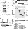
Figure 1
Figure 1
Interaction between STAP-2 and CAP (A) HEK293T cells were transfected with indicated plasmid. At 36 h after transfection, cells were lysed and pulled down with GSH-sepharose, and immunoblotted with anti-Myc and anti-GST antibodies. (B) At 6 days after adipocyte induced 3T3-L1 cells were lysed and immunoprecipitated with normal rabbit IgG or anti-STAP-2 antibody. Then, immunoprecipitants were immunoblotted with anti-CAP and anti-STAP-2 antibodies. (C) Schematic images of GST-fused STAP-2 deletion constructs. (D) The HEK293T cells were transfected with indicated plasmid. At 36 h after transfection, cells were lysed and pulled down with GSH-sepharose, and immunoblotted with anti-Myc and anti-GST antibodies.
Figure 2
Figure 2
Ternary complex of STAP-2 with CAP and c-Cbl (A) HEK293T cells were transfected with indicated plasmid. At 36 h after transfection, cells were lysed and immunoprecipitated with anti-c-Cbl antibody, and immunoblotted with anti-Myc and anti-GST antibodies. (B) The graph shows the quantification of CAP protein levels in the immunoprecipitates normalized to total cell lysate. Independent experiments from 3 replicates are summarized and presented as mean ± SEM. *p < 0.05, Student’s two-tailed t test.
Figure 3
Figure 3
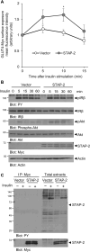
Enhancement of insulin signaling by STAP-2 expression (A) Hep3B cells transfected with empty vector (Vector) or stably expressing Myc-STAP-2 (STAP-2) in 6-well plates at 1 × 105 cells/well were transfected with GLUT4-Myc-GFP. Thirty 6 h after transfection, cells were starved for 12 h and stimulated with insulin (1 µg/ml) for the indicated periods. Cells were fixed and stained with anti-Myc antibody, then Myc stained cells in the GFP-positive cells at each time point was evaluated using a FACSCalibur. The surface-exposed GLUT4-Myc levels of each time point were normalized to those of untreated controls. quantification of CAP protein levels in the immunoprecipitates normalized to total cell lysate. Independent experiments from 4 replicates are summarized and presented as mean ± SEM. *p < 0.05, Student’s two-tailed t test. (B) Hep3B/Vector or /STAP-2 cells were stimulated with insulin (300 ng/ml) for the indicated periods. Cells were lysed and immunoblotted for individual antibodies. (C) Hep3B/Vector or /STAP-2 cells were stimulated with insulin (300 ng/ml) for 5 min. Cells were lysed and immunoprecipitated with anti-Myc antibody, then immunoblotted with anti-PY and anti-Myc antibodies.
Figure 4
Figure 4
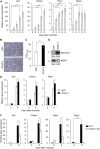
A functional role of STAP-2 in in vitro adipogenesis. (A) Mouse 3T3-L1 cells were induced for adipose differentiation. At 0, 2, 4, 6 and 8 days after differentiation, mRNA was extracted and analyzed the induction of Ap2, C/ebpα, Pparγ and Stap2 genes. Independent experiments from 5 replicates are summarized and presented as mean ± SEM. *p < 0.05, **p < 0.01, ***p < 0.005, one-way ANOVA followed by Dunnett's test (Day 0). (B, C) Mouse 3T3-L1 cells were retrovirally transduced with GFP or Myc-STAP-2 and induced into adipocyte. At 8 days after differentiation, cells were stained with Oil Red O (B). The Oil Red O dye was extracted and measured at OD490-595. Data are presented as the mean ± SEM, n = 3. *p < 0.05, Student’s two-tailed t test (C). (D) Retrovirally transduced GFP and STAP-2 in 3T3-L1 cells were detected by immunoblot using anti-Myc and anti-GFP antibodies. (E) GFP or STAP-2 was retrovirally transduced into 3T3-L1 cells and cells were cultured for adipose differentiation. At 0 or 8 days after differentiation, mRNA was extracted and analyzed the induction of Ap2, C/ebpα and Pparγ genes. Independent experiments from 5 replicates are summarized and presented as mean ± SEM. *p < 0.05, Student’s two-tailed t test. (F) MEFs were taken from WT and STAP-2 KO mice and cultured for adipose differentiation. At 0 or 8 days after differentiation, mRNA was extracted and analyzed the induction of Ap2, C/ebpα, Pparγ and Stap2 genes. Independent experiments from 3 replicates are summarized and presented as mean ± SEM. *p < 0.05, **p < 0.01, Student’s two-tailed t test.
Figure 5
Figure 5
Function of STAP-2 in high fat diet (HFD) mouse. (A, B) Age matched male (A) and female (B) from WT and STAP-2 KO mice were fed high fat diet and their weight was measured every week for the indicated periods. Error bars represent SEM, n = 5. *p < 0.05, **p < 0.01, ***p < 0.005, Student’s two-tailed t test. (C, D) Representative photos of HFD fed animals at 14 weeks after HFD feeding. (E, F) After 6 weeks HFD feeding, daily food intake was monitored for 7 days. Error bars represent SEM, n = 5. (G, H) At 14 weeks after HFD feeding, animals were sacrificed and dissected liver and adipose tissues. Representative photos of HFD fed animals in male (G) and female (H). (IL) White adipose tissues from male (I) and female (J), and livers from male (K) and female (L) were taken and weighed. Error bars represent SEM, n = 5. Student’s two-tailed t test.

Similar articles

References

    1. Minoguchi M, et al. STAP-2/BKS, an adaptor/docking protein, modulates STAT3 activation in acute-phase response through its YXXQ motif. J. Biol. Chem. 2003;278:11182–11189. doi: 10.1074/jbc.M211230200. - DOI - PubMed
    1. Sekine Y. Adaptor protein STAP-2 modulates cellular signaling in immune systems. Biol. Pharm. Bull. 2014;37:185–194. doi: 10.1248/bpb.b13-00421. - DOI - PubMed
    1. Sekine Y, et al. Physical and functional interactions between STAP-2/BKS and STAT5. J. Biol. Chem. 2005;280:8188–8196. doi: 10.1074/jbc.M411692200. - DOI - PubMed
    1. Sekine Y, et al. Leukemia inhibitory factor-induced phosphorylation of STAP-2 on tyrosine-250 is involved in its STAT3-enhancing activity. Biochem. Biophys. Res. Commun. 2007;356:517–522. doi: 10.1016/j.bbrc.2007.03.031. - DOI - PubMed
    1. Ikeda O, et al. STAP-2 regulates c-Fms/M-CSF receptor signaling in murine macrophage Raw 264.7 cells. Biochem. Biophys. Res. Commun. 2007;358:931–937. doi: 10.1016/j.bbrc.2007.05.030. - DOI - PubMed