. 2024 Mar 7;12(3):e008652.
doi: 10.1136/jitc-2023-008652.

Mucosal tumor vaccination delivering endogenous tumor antigens protects against pulmonary breast cancer metastases

Affiliations

Mucosal tumor vaccination delivering endogenous tumor antigens protects against pulmonary breast cancer metastases

Friederike Oltmanns et al. J Immunother Cancer. .

Abstract

Background: Generally, early-stage breast cancer has a good prognosis. However, if it spreads systemically, especially with pulmonary involvement, prospects worsen dramatically. Importantly, tumor-infiltrating T cells contribute to tumor control, particularly intratumoral T cells with a tissue-resident memory phenotype are associated with an improved clinical outcome.

Methods: Here, we use an adenoviral vector vaccine encoding endogenous tumor-associated antigens adjuvanted with interleukin-1β to induce tumor-specific tissue-resident memory T cells (TRM) in the lung for the prevention and treatment of pulmonary metastases in the murine 4T1 breast cancer model.

Results: The mucosal delivery of the vaccine was highly efficient in establishing tumor-specific TRM in the lung. Concomitantly, a single mucosal vaccination reduced the growth of pulmonary metastases and improved the survival in a prophylactic treatment. Vaccine-induced TRM contributed to these protective effects. In a therapeutic setting, the vaccination induced a pronounced T cell infiltration into metastases but resulted in only a minor restriction of the disease progression. However, in combination with stereotactic radiotherapy, the vaccine increased the survival time and rate of tumor-bearing mice.

Conclusion: In summary, our study demonstrates that mucosal vaccination is a promising strategy to harness the power of antitumor TRM and its potential combination with state-of-the-art treatments.

Keywords: adjuvant; breast cancer; memory; radiotherapy/radioimmunotherapy; vaccine.


Conflict of interest statement

Competing interests: None declared.

Figures

Figure 1
Figure 1
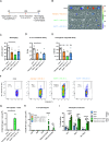
TAA-specific memory T cell subsets in the lung. BALB/c mice were vaccinated either intranasally (i.n.) or intramuscularly (i.m.) with Ad4T1 (107 IU) + AdIL1β or Adempty (2.5×10⁶ IU) as adjuvant component (A) and sacrificed 35 days later for T cell analysis. CD44 was used as a marker for antigen-experienced T cells with the addition of an intravascular CD45 staining to discriminate circulating (iv+) and tissue-resident (iv) memory cells. The bars represent the mean of each group±SEM (B). The phenotypic analysis of effector T cells (TEFF; CD127KLRG1+), effector memory T cells (TEM; CD127+KLRG1+), central memory T cells (TCM; CD127+KLRG1CD69CD103) and TRM cells (CD127+/−KLRG1CD103+CD69+; C) with AH1-specific TRM within the iv population (D). PD1 and TIM-3 expression within different T cell phenotypes. TEFF, TEM and TCM analyzed in Ad4T1+AdIL1β i.m. and TRM populations analyzed in Ad4T1+AdIL1β i.n. group (E). The gating strategies are shown in online supplemental figure 1. Bars display group means overlaid with individual data points (C–E); all groups n=6 (total 24 animals). Significances were determined using Kruskal-Wallis tests followed by Dunn‘s multiple comparisons test (B–E). P values indicate significant differences (*p<0.05; **p<0.005; ***p<0.0005; ****p<0.0001; only statistically significant comparisons shown). CMV, cytomegalovirus promotor; i.m., intramuscular; i.n., intranasal; iv, intravenous, PD1, programmed cell death protein 1; T cell immunoglobulin and mucin-domain containing-3, TIM-3; TRM, tissue-resident memory T cells.
Figure 2
Figure 2
Tumor antigen-specific CD8+ T cell responses. BALB/c mice were vaccinated either i.n. or i.m. as described in figure 1 and sacrificed 35 days later for T cell analysis. Representative flow cytometry plots for IFNγ production in iv+ and iv CD8+ T cells of lung homogenates restimulated with an AH1 peptide (A). Responding CD8+ T cells were determined by an intracellular cytokine staining in spleen and lung homogenates (B–D). The gating strategy is shown in online supplemental figure 2. Bars show group means overlaid with individual data points; all groups n=6 (total 24 animals). Significances were determined using Kruskal-Wallis tests followed by Dunn‘s multiple comparisons test (B–D). P values indicate significant differences (*p<0.05; **p<0.005; ***p<0.0005; ****p<0.0001; only statistically significant comparisons shown). poly, polyfunctional T cell population positive for all assessed markers. IFN, interferon; i.m., intramuscular; i.n., intranasal; iv, intravenous; TNFα, tumor necrosis factor α.
Figure 3
Figure 3
Vaccine efficacy in a prophylactic setting. BALB/c mice were vaccinated with Ad4T1 (107 IU) + AdIL1β (2.5×10⁶ IU) i.n. or i.m. or, alternatively, with Adempty (107 IU) + AdIL1β (2.5×10⁶ IU) i.n. and challenged with 4T1-luc cells intravenously 35 days later. Survival was monitored for a period of 8 weeks (A). Bioimaging was performed on day 14 after tumor induction in an IVIS imaging system after the injection of mice with D-luciferin (B). Signal intensities are shown for individual animals in each group (C). The background signal is shown as a dashed line, indicating the detection limit (DL). Survival analysis over an 8-week period (D). The individual time until animals reach humane endpoints is shown together with group means. The experiment was terminated at day 56 and animals surviving to that time point were set to a survival of 56 days (E). Bars display group means overlaid with individual data points, all groups n=6 (total 24 animals). Significances were determined using Kruskal-Wallis tests followed by Dunn‘s multiple comparisons test (C, E). Significances in survival were determined using Gehan-Breslow-Wilcoxon-test (D). P values indicate significant differences (*p<0.05; only statistically significant comparisons shown). i.m., intramuscular; i.n., intranasal; IU, infectious units; iv, intravenous.
Figure 4
Figure 4
Analysis of the lung in the prophylactic vaccination setting. BALB/c mice were vaccinated i.n. or i.m. as described in figure 3 and challenged with 4T1-luc cells intravenously 35 days later. Lungs were harvested 14 days later for tumor burden and T cell analyses (A). Bioimaging of the resected lungs in the endpoint analysis of the experiment (n=5 for Ad4T1+AdIL1β, n=6 for other groups; total 23 animals; 1 animal lost during housing) in an IVIS imaging system after the injection of mice with D-luciferin (B). Intensities are shown for individual animals in each group (C). Ex vivo luciferase assay (D) and clonogenic assay (E) were performed with lung homogenates. The background signal is shown as a dashed line, indicating the detection limit (DL). AH1-specific cells are shown within the CD44+CD8+ population in exemplary dot plots with group mean values and SEM (F) and as individual counts per animal with group mean values (G). Effector (TEFF; CD127KLRG1+), effector memory (TEM; CD127+KLRG1+) and central memory T cells (TCM; CD127+KLRG1CD69CD103) as well as KLRG1CD103+CD69+ TRM and KLRG1CD103CD69+ TRM within the CD44+CD8+AH1-spec. population (H). Gating strategies are shown in online supplemental figure 1 (no intravenous staining performed in this experiment). Programmed cell death protein 1 (PD1) and T cell immunoglobulin and mucin-domain containing-3 (TIM-3) expression within different T cell phenotypes. TEFF, TEM and TCM analyzed in Ad4T1+AdIL1β i.m. and TRM populations analyzed in Ad4T1+AdIL1β i.n. (I). Bars display group means overlaid with individual data points. Significances were determined using Kruskal-Wallis tests followed by Dunn‘s multiple comparisons test (C–E and G–I). P values indicate significant differences (*p<0.05; **p<0.005; ***p<0.0005; only statistically significant comparisons shown). i.m., intramuscular; i.n., intranasal; IU, infectious units; iv intravenous; spec., specific; TRM, tissue-resident memory T cells.
Figure 5
Figure 5
Protective efficacy in the absence of circulating T cells. BALB/c mice were vaccinated i.n. or i.m. with Ad4T1 (107 IU) + AdIL1β (2.5×10⁶ IU) and challenged with 4T1-luc cells intravenously 35 days later. The animals received FTY720-supplemented drinking water (2 µg/mL) 3 days before the tumor challenge to inhibit T cell circulation and the treatment was continued throughout the experiment (A). Number of CD8+ T cells in the blood of FTY720-treated animals compared with untreated animals was determined by flow cytometry analysis (B). Survival analysis over an 8-week period (C). Corresponding bioimaging (D) with quantification of luciferase signal at the 2-week time point (E). The background signal is shown as a dashed line. Statistical significances were determined using Mann-Whitney test (B, E) and Gehan-Breslow-Wilcoxon-test (C). All groups n=6 (total 12 animals). P values indicate significant differences (*p<0.05; **p<0.005; only statistically significant comparisons shown). i.m., intramuscular; i.n., intranasal; IU, infectious units; iv, intravenous.
Figure 6
Figure 6
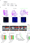
Vaccine efficacy after therapeutic vaccination. BALB/c mice were challenged with 4T1-luc cells intravenously and vaccinated i.n. with Ad4T1 (107 IU) + AdIL1β (2.5×10⁶ IU) or Adempty (107 IU) + AdIL1β (2.5×10⁶ IU) (A). Lungs of individual naive and i.n. immunized animals were resected 14 days later, embedded into O.C.T. and stained with anti-CD8 monoclonal antibody (green) for infiltrating T cells and Hoechst 33342 for nuclei (blue). Images were generated on a Leica SP5X confocal microscope (40×) with representative images shown. Tumor nodules are further displayed in H&E staining of respective lungs and are highlighted by indicating arrows (B). In vivo bioimaging on day 18 in an IVIS imaging system after the injection of mice with D-luciferin (C) with the corresponding quantification of the bioimaging (D) and an ex vivo luciferase assay was conducted to quantify the tumor burden (E). The background signal is shown as a dashed line. In a separate experiment, BALB/c mice were challenged with 4T1-luc cells intravenously and vaccinated i.n. with Ad4T1 (107 IU) + AdIL1β (2.5×10⁶ IU) 3 days later. Survival analysis over an 8-week period (F). The individual time until animals reach humane endpoints is shown together with group means (G). Bars display group means overlaid with individual data points. Significances were determined using Kruskal-Wallis tests followed by Dunn‘s multiple comparisons (D, E) test or Mann-Whitney test (G). Significances in survival were determined using Gehan-Breslow-Wilcoxon test (F). No statistically significant differences were found. Groups n=6 (C, D and E, total of 18 animals, 1 naïve animal reached the humane endpoint on day 17; one Ad4T1+AdIL1β i.n. died shortly before bioimaging, data included for this animal in E but not D) and n=7 (F and G; total 14 animals). DL, detection limit; IL, interleukin; i.n., intranasal; IU, infectious units; i.v., intravenous; IF, immunofluorescence.
Figure 7
Figure 7
Vaccine efficacy after therapeutic vaccination in combination with RT. BALB/c mice were challenged with 4T1-luc cells intravenously and received whole thorax radiation with a single dose of 5 Gy on day 5. One day later, they were vaccinated i.n. with Ad4T1 (107 IU) + AdIL1β (2.5×10⁶ IU) (A). Survival analysis over an 8-week period (B). The individual time until animals reach humane endpoints is shown together with group means. The experiment was terminated at day 56 and animals surviving to that time point were set to a survival of 56 days. Bars display group means overlaid with individual data points with significances determined using Kruskal-Wallis tests followed by Dunn‘s multiple comparisons test (C). Weekly in vivo bioimaging in an IVIS imaging system after the injection of mice with D-luciferin (D). Significances in survival were determined using Gehan-Breslow-Wilcoxon test (B). n=7 for Ad4T1+AdIL1β i.n., other groups n=14 (total 49 animals). P values indicate significant differences (*p<0.05; **p<0.005; ***p<0.0005). Gy, Gray; i.n., intranasal; IU, infectious units; i.v., intravenous; RT, radiotherapy.

Similar articles

Cited by

References

    1. Sung H, Ferlay J, Siegel RL, et al. . Global cancer Statistics 2020: GLOBOCAN estimates of incidence and mortality worldwide for 36 cancers in 185 countries. CA Cancer J Clin 2021;71:209–49. 10.3322/caac.21660 - DOI - PubMed
    1. Allemani C, Weir HK, Carreira H, et al. . Global surveillance of cancer survival 1995-2009: analysis of individual data for 25 676 887 patients from 279 population-based registries in 67 countries (CONCORD-2). The Lancet 2015;385:977–1010. 10.1016/S0140-6736(14)62038-9 - DOI - PMC - PubMed
    1. Rabbani SA, Mazar AP. Evaluating distant metastases in breast cancer: from biology to outcomes. Cancer Metastasis Rev 2007;26:663–74. 10.1007/s10555-007-9085-8 - DOI - PubMed
    1. Dafni U, Martín-Lluesma S, Balint K, et al. . Efficacy of cancer vaccines in selected gynaecological breast and ovarian cancers: A 20-year systematic review and meta-analysis. Eur J Cancer 2021;142:63–82. 10.1016/j.ejca.2020.10.014 - DOI - PubMed
    1. Luo N, Nixon MJ, Gonzalez-Ericsson PI, et al. . DNA methyltransferase inhibition Upregulates MHC-I to potentiate cytotoxic T lymphocyte responses in breast cancer. Nat Commun 2018;9:1–11. 10.1038/s41467-017-02630-w - DOI - PMC - PubMed