Review
. 2024 Mar-Apr;15(2):e1832.
doi: 10.1002/wrna.1832.

Target-directed microRNA degradation: Mechanisms, significance, and functional implications

Affiliations
Review

Target-directed microRNA degradation: Mechanisms, significance, and functional implications

Nicholas M Hiers et al. Wiley Interdiscip Rev RNA. 2024 Mar-Apr.

Abstract

MicroRNAs (miRNAs) are small non-coding RNAs that play a fundamental role in enabling miRNA-mediated target repression, a post-transcriptional gene regulatory mechanism preserved across metazoans. Loss of certain animal miRNA genes can lead to developmental abnormalities, disease, and various degrees of embryonic lethality. These short RNAs normally guide Argonaute (AGO) proteins to target RNAs, which are in turn translationally repressed and destabilized, silencing the target to fine-tune gene expression and maintain cellular homeostasis. Delineating miRNA-mediated target decay has been thoroughly examined in thousands of studies, yet despite these exhaustive studies, comparatively less is known about how and why miRNAs are directed for decay. Several key observations over the years have noted instances of rapid miRNA turnover, suggesting endogenous means for animals to induce miRNA degradation. Recently, it was revealed that certain targets, so-called target-directed miRNA degradation (TDMD) triggers, can "trigger" miRNA decay through inducing proteolysis of AGO and thereby the bound miRNA. This process is mediated in animals via the ZSWIM8 ubiquitin ligase complex, which is recruited to AGO during engagement with triggers. Since its discovery, several studies have identified that ZSWIM8 and TDMD are indispensable for proper animal development. Given the rapid expansion of this field of study, here, we summarize the key findings that have led to and followed the discovery of ZSWIM8-dependent TDMD. This article is categorized under: Regulatory RNAs/RNAi/Riboswitches > Regulatory RNAs RNA Turnover and Surveillance > Turnover/Surveillance Mechanisms RNA in Disease and Development > RNA in Development.

Keywords: RNAi; TDMD; ZSWIM8; development; microRNA.


Conflict of interest statement

Conflict of Interest

The authors declare no conflicts of interest.

Figures

Figure 1:
Figure 1:
A) An illustration summarizing canonical animal miRNA biogenesis. Briefly, primary miRNA (pri-miRNA) is transcribed within the nucleus and cleaved by Drosha (pink) in complex with DGCR8 (purple). The precursor miRNA (pre-miRNA) hairpin is exported out of the nucleus to the cytosol via XPO5 (violet), where the terminal loop is cleaved by Dicer (blue). AGO (cyan) selects a strand from the miRNA duplex, the more abundant species being referred to as the guide strand (red), whereas the less abundant is referred to as the passenger strand (blue line). The passenger strand is quickly degraded without a protein binding-partner. B) An illustration detailing miRNA-mediated target repression. AGO-bound miRNAs (cyan, black) will associate with target RNAs (maroon) via sequence complementarity. Upon association, the complex will recruit additional factors to mediate translational repression, mRNA deadenylation, and mRNA destabilization. The complex can then disassociate from the target to induce multiple rounds of destabilization. This image was produced using Biorender.com.
Figure 2:
Figure 2:
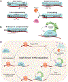
A) MiRNA base-pairing modalities with target RNAs. Canonical seed-match (g2-g8), 3′ supplement base pairing (g13-g16, in addition to seed), siRNA-like full complementarity, and non-canonical 3′ end base pairing. B) Extensive 3′ complementarity, with a central bulge separating seed-matching. This binding modality is charactisitic of and normally sufficient to induce ZSWIM8-dependent TDMD (green). C) A model of the current ZSWIM8-dependent TDMD mechanism. Expression of a trigger RNA (maroon) bearing extensive complementarity to an AGO-loaded miRNA (cyan, black), upon AGO association, will direct it for turnover via recruitment of the ZSWIM8 ubiquitin ligase complex (green). ZSWIM8 will catalyse polyubiquitination (purple) of AGO, leading to its proteasomal decay, exposing the miRNA to RNases. The trigger RNA is preserved to induce multiple rounds of miRNA destabilization. This image was produced using Biorender.com.
Figure 3:
Figure 3:
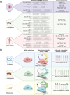
A) A list of validated naturally occurring TDMD triggers, their genetic origins, affected miRNAs, trigger-miRNA base pairing, and the publications initially characterizing them. B) A summary of developmentally timed TDMD in various animal models. C. elegans uses the AGO orthologs Alg1/2 and is directed for degradation via Ebax1. Most of the cel-miR-35 family (cel-miR-35–42) is derived from the same clustered transcript (cel-pri-miR-35~41) and is sharply degraded during worm development. D. melanogaster uses the Ago1 ortholog to bind most miRNAs and is directed for turnover via Dora. A portion of the dme-miR-3 family is derived from the dme-pri-miR-309~6–3 cluster, and their degradation by an unknown trigger is critical to fly embryo viability. Mus musculus uses Ago1-4 and is directed for turnover via ZSWIM8. Mmu-miR-322/503, derived from the mmu-pri-miR-322~450b transcript, degradation via an unknown trigger is critical to mouse growth. The ZSWIM8 (or its ortholog) sensitive miRNAs within these clusters are marked with red arrows. This image was produced using Biorender.com.
Figure 4:
Figure 4:
Emerging mechanism regulated by TDMD. A) Clustered miRNAs. Many known ZSWIM8 sensitive miRNAs are derived from clustered transcripts. In some cases, only a few of the miRNAs within the transcript are ZSWIM8 sensitive, such as mmu-pri-miR-322~450b. Therefore, despite co-transcription, the relative abundance of each mature miRNA can be modulated via TDMD. B) Arm switching. The annotated guide strand of miRNA is normally defined based on the more abundant isoform between the two co-transcribed strands. In the case of miR-154, the annotated passenger is normally efficiently loaded into AGO but is subsequently degraded via TDMD through a currently unknown trigger. Therefore, the tissue-specific examples detailing increases in miR-154 “passenger” strand abundance are likely due to repression of miR-154 TDMD. C) AGO partitioning. Dme-miR-277 can efficiently load into both Ago1 and Ago2. However, the ZSWIM8 ortholog Dora can only direct degradation of Ago1. Therefore, TDMD of miR-277 via a currently unknown trigger increases its partitioning into Ago2, explaining earlier observations of this phenomenon. In this way, TDMD can regulate AGO ortholog partitioning in D. melanogaster. This image was produced using Biorender.com.
Figure 5:
Figure 5:
Potential mechanisms that may reglate/be regulated by TDMD. A) Subcellular localization: canonical miRNA and/or TDMD factors preferentially localizing into organelles, or biomolecular condensates may alter TDMD efficacy and may explain some observations of altered TDMD efficiency. B) Novel TDMD cofactors: a mechanism in C. elegans differentiates the proposed seed-dependent trigger from targets. It is possible that a novel protein partner primes the seed-trigger by recruiting Ebax1, directing co-localized Alg1/2 for turnover. C) TDMD trigger clusters: as many ZSWIM8-sensitive miRNAs are derived from clusters, they may also be degraded in clusters. “Canonical” triggers may prime triggers to induce degradation of co-localized “sub-optimal” triggers, normally insufficient to induce degradation. D) TDMD trancriptional networks: trigger encoded proteins have been shown to cooperate prior, but whether any transcription factor encoded triggers regulate trigger transcription is yet to be determined. These transcription factors may modulate the abundance of the same trigger, or a completely unrelated trigger in a sort of transcriptional network. This image was produced using Biorender.com.

Similar articles

Cited by

References

    1. Agarwal V, Bell GW, Nam JW, & Bartel DP (2015). Predicting effective microRNA target sites in mammalian mRNAs. eLife, 4. 10.7554/elife.05005 - DOI - PMC - PubMed
    1. Alvarez-Saavedra E, & Horvitz HR (2010). Many Families of C. elegans MicroRNAs Are Not Essential for Development or Viability. Current Biology, 20(4), 367–373. 10.1016/j.cub.2009.12.051 - DOI - PMC - PubMed
    1. Ameres SL, Horwich MD, Hung J, Xu J, Ghildiyal M, Weng Z, & Zamore PD (2010). Target RNA–Directed trimming and tailing of small silencing RNAs. Science, 328(5985), 1534–1539. 10.1126/science.1187058 - DOI - PMC - PubMed
    1. Ameres SL, & Zamore PD (2013). Diversifying microRNA sequence and function. Nature Reviews Molecular Cell Biology, 14(8), 475–488. 10.1038/nrm3611 - DOI - PubMed
    1. Baccarini A, Chauhan H, Gardner TJ, Jayaprakash A, Sachidanandam R, & Brown BD (2011). Kinetic Analysis Reveals the Fate of a MicroRNA following Target Regulation in Mammalian Cells. Current Biology, 21(5), 369–376. 10.1016/j.cub.2011.01.067 - DOI - PMC - PubMed

LinkOut - more resources