Review
. 2024 May 28;15(6):403-418.
doi: 10.1093/procel/pwae007.

Mutations in the SARS-CoV-2 spike receptor binding domain and their delicate balance between ACE2 affinity and antibody evasion

Affiliations
Review

Mutations in the SARS-CoV-2 spike receptor binding domain and their delicate balance between ACE2 affinity and antibody evasion

Song Xue et al. Protein Cell. .

Abstract

Intensive selection pressure constrains the evolutionary trajectory of SARS-CoV-2 genomes and results in various novel variants with distinct mutation profiles. Point mutations, particularly those within the receptor binding domain (RBD) of SARS-CoV-2 spike (S) protein, lead to the functional alteration in both receptor engagement and monoclonal antibody (mAb) recognition. Here, we review the data of the RBD point mutations possessed by major SARS-CoV-2 variants and discuss their individual effects on ACE2 affinity and immune evasion. Many single amino acid substitutions within RBD epitopes crucial for the antibody evasion capacity may conversely weaken ACE2 binding affinity. However, this weakened effect could be largely compensated by specific epistatic mutations, such as N501Y, thus maintaining the overall ACE2 affinity for the spike protein of all major variants. The predominant direction of SARS-CoV-2 evolution lies neither in promoting ACE2 affinity nor evading mAb neutralization but in maintaining a delicate balance between these two dimensions. Together, this review interprets how RBD mutations efficiently resist antibody neutralization and meanwhile how the affinity between ACE2 and spike protein is maintained, emphasizing the significance of comprehensive assessment of spike mutations.

Keywords: ACE2 affinity; RBD mutation; SARS-CoV-2; antibody evasion; variant of concern; viral evolution.


Conflict of interest statement

The authors declare no competing interests.

Figures
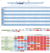
Figure 1.
Figure 1.
Various RBD mutations contribute to ACE2 binding and antibody evasion. (A) The most common mutational profiles for each variant of concern (VOC) within the receptor binding domain (RBD) of SARS-CoV-2 spike (S) protein. Data was obtained from Outbreak.info genomic reports. The amino acid position and the original amino acid of the S protein are shown at the top row of the table. NTD, amino-terminal domain; PANGO, Phylogenetic Assignment of Named Global Outbreak; RBD, receptor- binding domain; RBM, receptor binding motif; S1/S2, junction between the exposed S1 attachment domain and the partially buried S2 fusion domain; WHO, World Health Organization. The R346T and F486P in mutation table indicate the specific mutation of BQ.1.1 and XBB.1.5 relative to their parental strains, BQ.1 and XBB.1, respectively. Created with Biorender. (B) Enhancement of ACE2 affinity for VOCs [Δ−log(KD) values] with the wildtype as a reference. Means are calculated from published studies reporting ACE2 affinity data of VOCs. Colors represents the level of ACE2 binding enhancement: strong [Δ−log(KD) > 0.8]; moderate [Δ−log(KD) = 0.5–0.8]; mild [Δ−log(KD) = 0.3–0.5]; slightly increased affinity [Δ−log(KD) < 0.3]. (C) Geometric mean fold reduction in neutralization (mFRN) values of monoclonal antibodies (mAbs) against VOCs relative to the wildtype reference. Means are calculated from published studies reporting neutralization data for the indicated mAbs. Full datasets are available in Supplementary Data File 1, and statistics in Supplementary Data File 2. Colors represent the strength of mAbs resistance: strong (mFRN > 100); moderate (mFRN = 10–100); mild (mFRN = 3–10); no resistance (mFRN = 1–3); increased sensitivity (mFRN < 1). Bam + Ete, bamlanivimab + etesevimab; REGEN-COV (Cas + Imd), casirivimab + imdevimab; Evusheld (Tix + Cil), cilgavimab + tixagevimab; NA, no neutralization data reported for the antibody-variant pair at the time of writing.
Figure 2.
Figure 2.
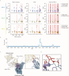
Epitope mutations drive resistance to antibody neutralization. (A) Geometric mean fold reduction in neutralization (mFRN) data for each class of monoclonal antibody (mAb). The mFRN values (y-axis) was determined using the wildtype pseudoviruses containing only one single mutation in the RBD of S protein (positions 305–534, x-axis) (Shang et al., 2020), with the wildtype pseudovirus as the reference control. Each dot represents the mFRN value of one mutation-mAb pair (mutation sites refer to Fig. 1A). The colors of dots represent the corresponding testing mAbs. Horizonal dashed line shows the mFRN = 3 threshold. Mutation sites of representative VOCs Beta, Gamma, Delta and Omicron XBB.1 (see Fig. 1A) are indicated by vertical red lines. The full dataset of mFRN values for each mAb in the presence of single RBD mutations are summarized in the Supplementary Data File 3. (B) Total escape scores of bebtelovimab (LY-CoV1404) determined by a full spike deep mutational scanning system at each site in the BA.1 RBD. A detailed explanation of escape score could be found in the reference (Yu et al., 2022). An interactive data set is available at Github. (C) X-ray crystal structure of the bebtelovimab Fab bound to the S protein RBD (PDB 7MMO). The rectangular region indicates the Wuhan-Hu-1 RBD epitope recognized by bebtelovimab and is showed as ribbons in the zoomed view. The key escape sites, such as N450 and P499, correspond to the region (site 444-450 and 499) with escape score > 10 (see Fig. 2B). (D) Key escape sites in (C) are showed as atoms.
Figure 3.
Figure 3.
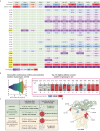
Potent epistatic mutations reverse the deleterious summed effect on ACE2 affinity. (A) Heat map depicting relative enhancement in ACE2 affinity for variants of concern (VOCs) (top row) and individual mutations (other rows). Each column shows data for each variant, including Alpha, Beta, Gamma, Delta, BA.1, BA.2, BA.4/5, BA.2.75, BQ.1.1, XBB.1, and XBB.1.5. The data in each row are the calculated logarithmic negative KD [Δ−log(KD)] value, to present the effect of mutations on ACE2 binding affinity. These values are calculated by comparing the KD value of a mutated S protein with that of a wildtype S protein under the same experimental condition. The effect for each constituent VOC mutation individually on a wild type background, together with their summed effect is shown. At the left-most column of the table, the original amino acids of the S protein and their corresponding positions are shown; and the amino acids (K417, F486, Q493, G496, Q498, N501, and Y505) directly contacting ACE2 are highlighted. Different colors represent the changed levels on ACE2 affinity: strong enhancement [Δ−log(KD) > 0.8]; moderate enhancement [Δ−log(KD) = 0.5–0.8]; mild enhancement [Δ−log(KD) = 0.3–0.5]; slightly enhancement [Δ−log(KD) < 0.3]; slightly decreased affinity [Δ−log(KD) = −0.5–0]; moderate decreased affinity [Δ−log(KD) = −0.5–−0.8]; strongly decreased affinity [Δ−log(KD) < −0.8]. “−” indicates no mutation at this position for the corresponding variants/column.(B) Systematic analysis of ACE2 binding affinity for RBD proteins containing all possible combinations of 15 mutations in the RBD of BA.1 variant. Left: distribution of ACE2 binding affinity using all possible mutational intermediates of BA.1 (N = 215 = 32,768 RBD genotypes tested). Binding affinity is shown as −log(KD). The vertical lines indicate the −log(KD) for wildtype Wuhan-Hu-1 strain and Omicron BA.1 variant, respectively. An interactive data browser is available at Github. Right: among the top 1% intermediates with a superior ACE2 affinity (a total of 321 genotypes), relative proportions (%) of each amino acid are shown at all 15 mutation sites. On these RBD positions, the amino acids for BA.1 variant and the corresponding amino acid in wildtype Wuhan-Hu-1 strain are also shown. Colors depict the genotypes preference at each position: preferred (genotype proportion > 60%); no apparent preference (genotype proportion = 40%–60%); unappreciated (genotype proportion < 40%). (C) Two scenarios when comparing the effect of individual BA.1 constituent mutations in the wildtype backbone [Δ−log(KD) values from Fig. 3A] with their proportions among the top 1% variants with highest ACE2 affinity (genotype proportions from Fig. 3B). (D) Co-crystal structure of Omicron BA.1 RBD and ACE2 receptor (PDB ID 7WPB). Mutated residues are shown, and their surfaces are colored as the corresponding residues in Fig. 3B.
Figure 4.
Figure 4.
Maintaining the balance between ACE2 affinity and mAb evasion for the viral fitness. (A) To inhibit infection, the neutralizing antibody competitively binds to SARS-CoV-2 RBD, while the interaction between the viral S protein and the host receptor ACE2 is abolished. Created with Biorender. (B) Upper left: In the presence of certain escape mutation, a neutralizing antibody might lose its neutralizing capacity. However, some escape mutations could also decrease the ACE2-S binding affinity, reducing the viral infectivity and fitness. Upper right: some RBD mutations might exert epistatic effects to enhance ACE2 binding. Bottom: For each SARS-CoV-2 variant, a delicate balance needs to be kept between its immune evasion capacity (achieved through escape mutations) and sufficient ACE2 binding affinity (maintained by epistatic mutations). Created with Biorender.

Similar articles

Cited by

References

    1. Addetia A, Piccoli L, Case JB. et al.. Neutralization, effector function and immune imprinting of Omicron variants. Nature 2023;621:592–601. - PMC - PubMed
    1. Alcantara MC, Higuchi Y, Kirita Y. et al.. Deep mutational scanning to predict escape from bebtelovimab in SARS-CoV-2 Omicron subvariants. Vaccines (Basel) 2023;11:711. - PMC - PubMed
    1. Andreano E, Piccini G, Licastro D. et al.. SARS-CoV-2 escape from a highly neutralizing COVID-19 convalescent plasma. Proc Natl Acad Sci USA 2021;118:e2103154118. - PMC - PubMed
    1. Barton MI, MacGowan SA, Kutuzov MA. et al.. Effects of common mutations in the SARS-CoV-2 Spike RBD and its ligand, the human ACE2 receptor on binding affinity and kinetics. Elife 2021;10:e70658. - PMC - PubMed
    1. Bouhaddou M, Reuschl AK, Polacco BJ. et al.. SARS-CoV-2 variants evolve convergent strategies to remodel the host response. Cell 2023;186:4597–4614.e26. - PMC - PubMed

MeSH terms

Supplementary concepts