

A chronic pro-inflammatory environment contributes to the physiopathology of actinic lentigines
- PMID: 38438410
- PMCID: PMC10912228
- DOI: 10.1038/s41598-024-53990-5
A chronic pro-inflammatory environment contributes to the physiopathology of actinic lentigines
Abstract
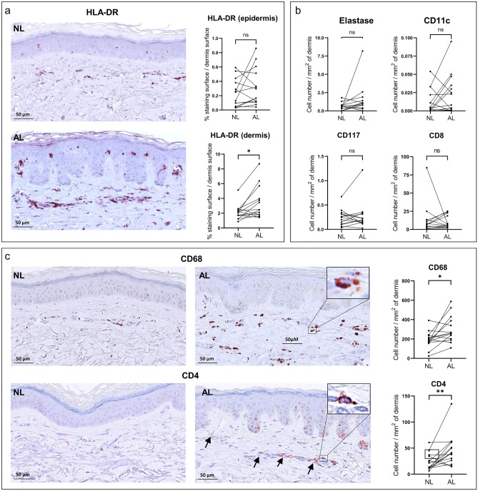
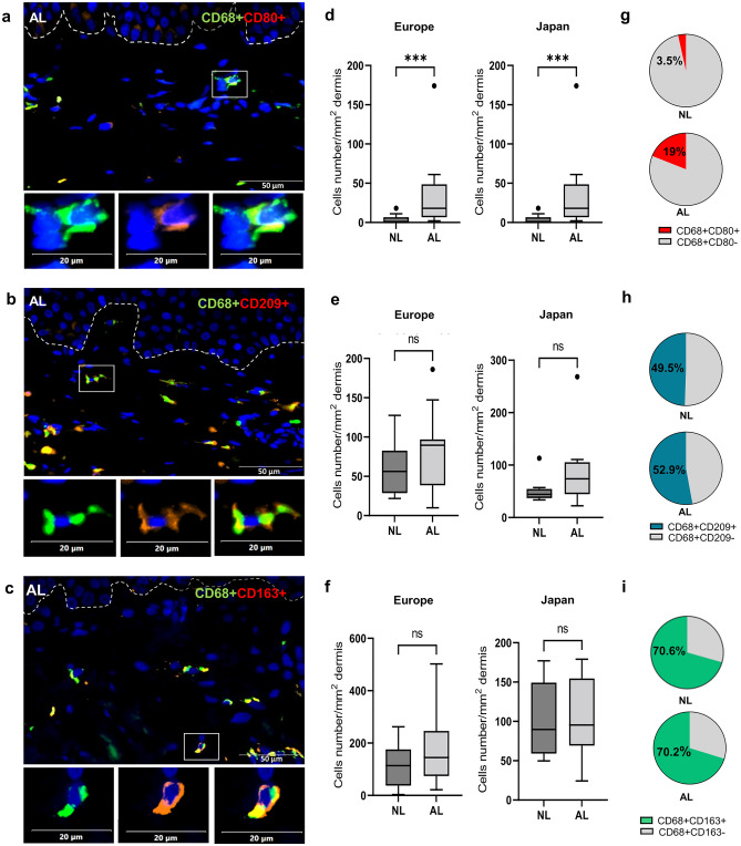
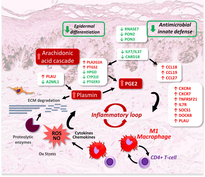
Actinic lentigines (AL) or age spots, are skin hyperpigmented lesions associated with age and chronic sun exposure. To better understand the physiopathology of AL, we have characterized the inflammation response in AL of European and Japanese volunteers. Gene expression profile showed that in both populations, 10% of the modulated genes in AL versus adjacent non lesional skin (NL), i.e. 31 genes, are associated with inflammation/immune process. A pro-inflammatory environment in AL is strongly suggested by the activation of the arachidonic acid cascade and the plasmin pathway leading to prostaglandin production, along with the decrease of anti-inflammatory cytokines and the identification of inflammatory upstream regulators. Furthermore, in line with the over-expression of genes associated with the recruitment and activation of immune cells, immunostaining on skin sections revealed a significant infiltration of CD68+ macrophages and CD4+ T-cells in the dermis of AL. Strikingly, investigation of infiltrated macrophage subsets evidenced a significant increase of pro-inflammatory CD80+/CD68+ M1 macrophages in AL compared to NL. In conclusion, a chronic inflammation, sustained by pro-inflammatory mediators and infiltration of immune cells, particularly pro-inflammatory M1 macrophages, takes place in AL. This pro-inflammatory loop should be thus broken to normalize skin and improve the efficacy of age spot treatment.
© 2024. The Author(s).
Conflict of interest statement
All authors are employees of L’Oréal.
Figures
Similar articles
-
Actinic lentigines from Japanese and European volunteers share similar impaired biological functions.J Dermatol Sci. 2022 Jul;107(1):8-16. doi: 10.1016/j.jdermsci.2022.07.001. Epub 2022 Jul 2. J Dermatol Sci. 2022. PMID: 35817661
-
Morphological and molecular characterization of actinic lentigos reveals alterations of the dermal extracellular matrix.Br J Dermatol. 2017 Dec;177(6):1619-1632. doi: 10.1111/bjd.15697. Epub 2017 Nov 16. Br J Dermatol. 2017. PMID: 28570000
-
Development of actinic lentigines due to multiple sub-erythemal exposure to UVA1 radiation in Asian skin.Exp Dermatol. 2023 Nov;32(11):2034-2037. doi: 10.1111/exd.14937. Epub 2023 Sep 16. Exp Dermatol. 2023. PMID: 37715544
-
Role of In Vivo Reflectance Confocal Microscopy in the Analysis of Melanocytic Lesions.Acta Dermatovenerol Croat. 2018 Apr;26(1):64-67. Acta Dermatovenerol Croat. 2018. PMID: 29782304 Review.
-
Macrophages: The Good, the Bad, and the Gluttony.Front Immunol. 2021 Aug 12;12:708186. doi: 10.3389/fimmu.2021.708186. eCollection 2021. Front Immunol. 2021. PMID: 34456917 Free PMC article. Review.
References
-
- Monestier S, Gaudy C, Gouvernet J, Richard MA, Grob JJ. Multiple senile lentigos of the face, a skin ageing pattern resulting from a life excess of intermittent sun exposure in dark-skinned caucasians: A case-control study. Br. J. Dermatol. 2006;154:438–444. doi: 10.1111/j.1365-2133.2005.06996.x. - DOI - PubMed
MeSH terms
Substances
LinkOut - more resources
Full Text Sources
Molecular Biology Databases
Research Materials
