. 2024 Mar 2;15(1):1939.
doi: 10.1038/s41467-024-46239-2.

Structure-guided engineering of biased-agonism in the human niacin receptor via single amino acid substitution

Affiliations

Structure-guided engineering of biased-agonism in the human niacin receptor via single amino acid substitution

Manish K Yadav et al. Nat Commun. .

Abstract

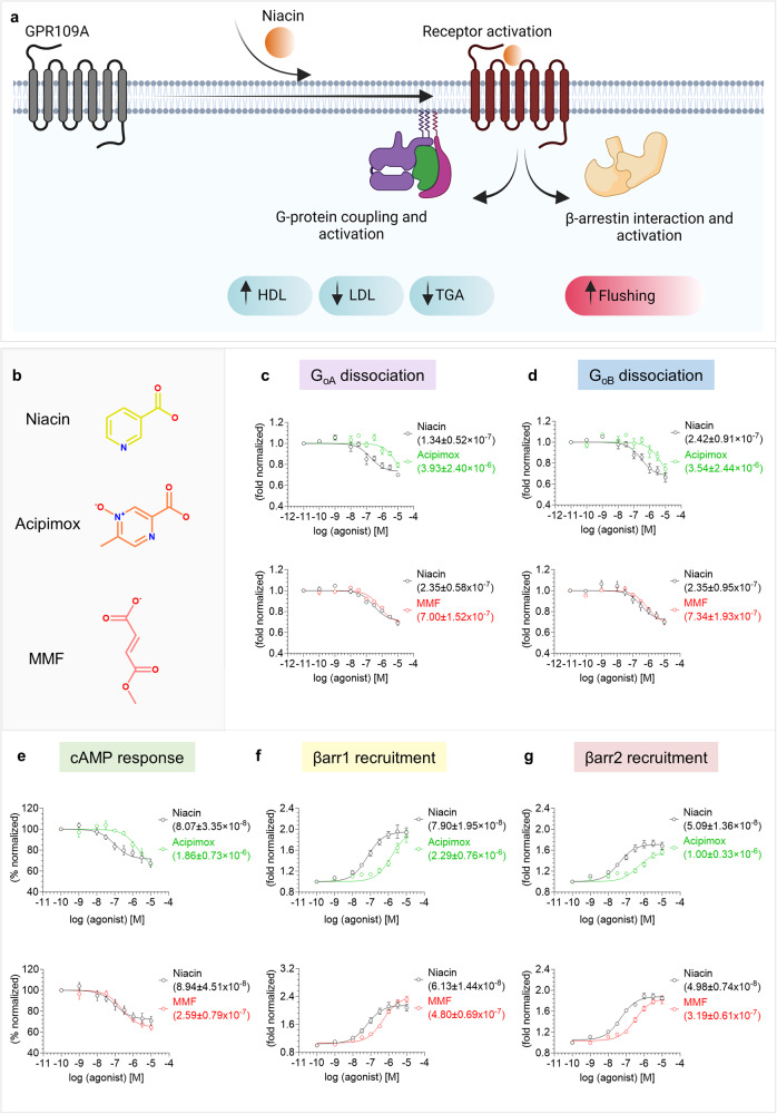
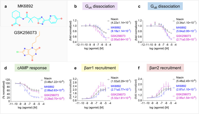
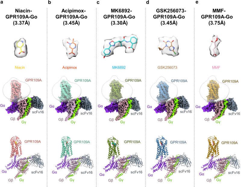
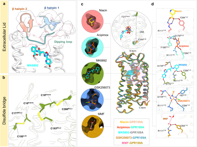
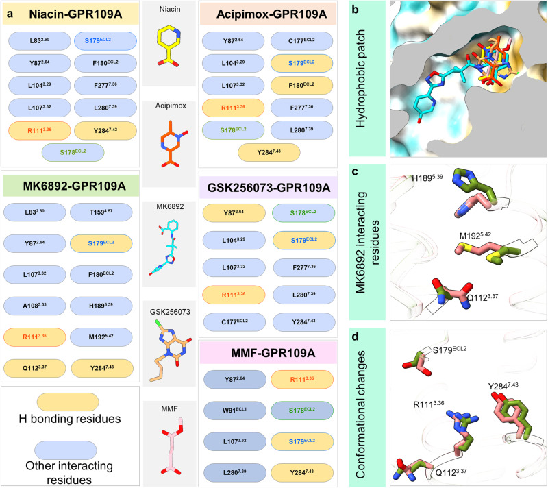
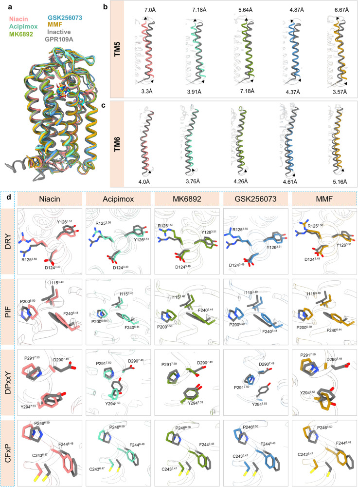
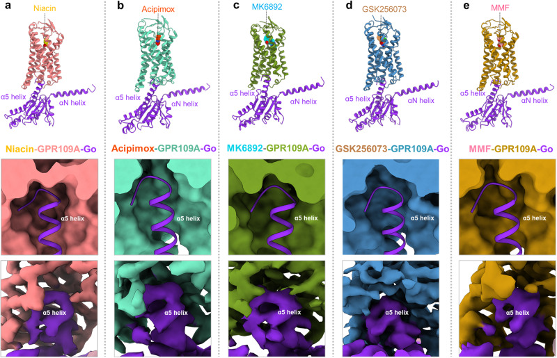
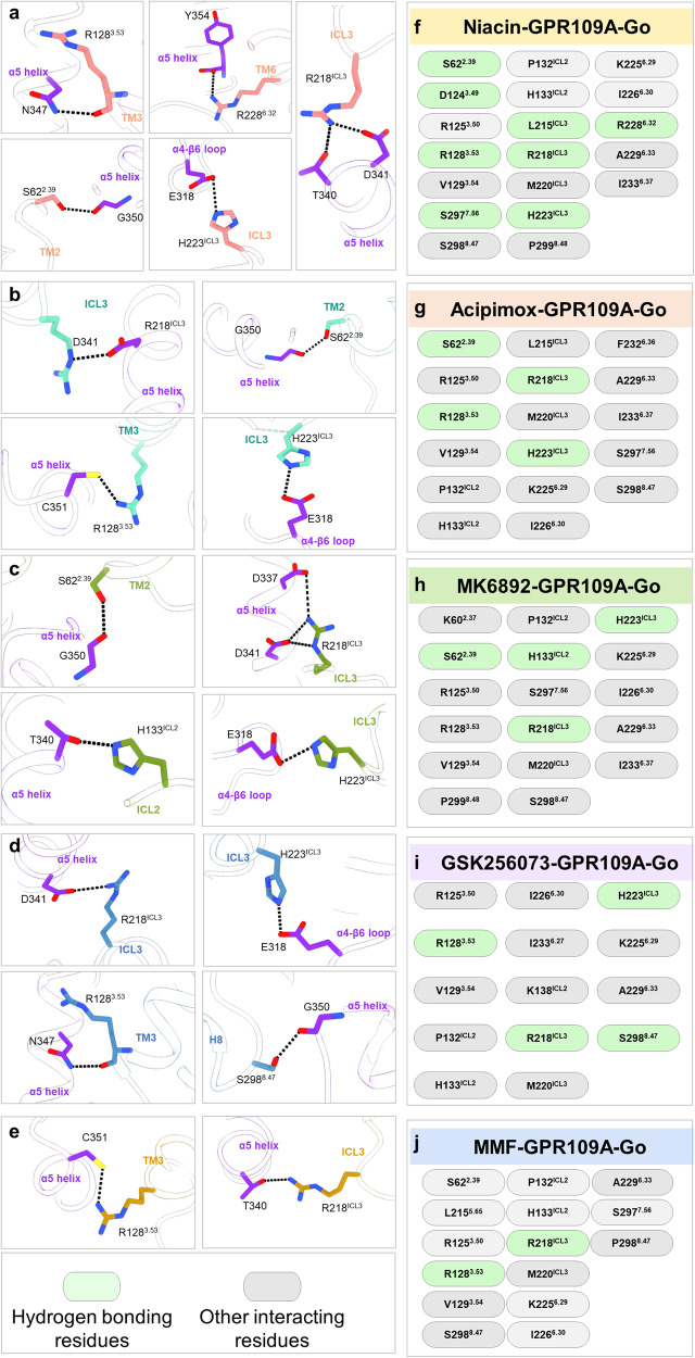
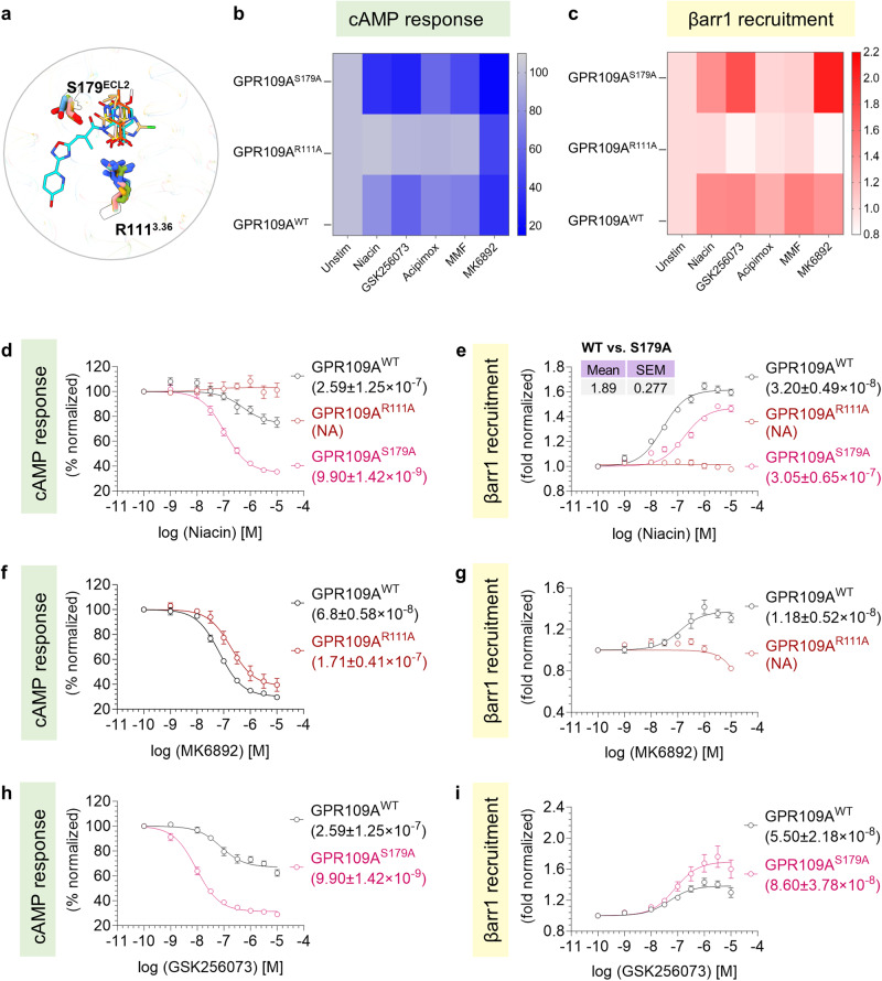
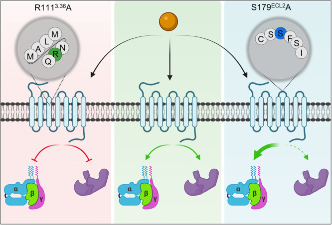
The Hydroxycarboxylic acid receptor 2 (HCA2), also known as the niacin receptor or GPR109A, is a prototypical GPCR that plays a central role in the inhibition of lipolytic and atherogenic activities. Its activation also results in vasodilation that is linked to the side-effect of flushing associated with dyslipidemia drugs such as niacin. GPR109A continues to be a target for developing potential therapeutics in dyslipidemia with minimized flushing response. Here, we present cryo-EM structures of the GPR109A in complex with dyslipidemia drugs, niacin or acipimox, non-flushing agonists, MK6892 or GSK256073, and recently approved psoriasis drug, monomethyl fumarate (MMF). These structures elucidate the binding mechanism of agonists, molecular basis of receptor activation, and insights into biased signaling elicited by some of the agonists. The structural framework also allows us to engineer receptor mutants that exhibit G-protein signaling bias, and therefore, our study may help in structure-guided drug discovery efforts targeting this receptor.


Conflict of interest statement

The authors declare no competing interests.

Figures

Fig. 1
Fig. 1. Pharmacological profiling of niacin, acipimox, and MMF on GPR109A.
a Diagrammatic illustration of GPR109A activation and downstream signaling outcomes (created with BioRender.com). b Chemical structures of niacin, acipimox, and MMF. c, d NanoBiT-based heterotrimeric GoA/GoB dissociation assay in response to acipimox and MMF with niacin as a reference ligand, (mean ± SEM; n = 3–4 independent experiments, i.e., for GoA dissociation with acipimox: n = 4, for GoA dissociation with MMF: n = 3, for GoB dissociation in response to acipimox and MMF: n = 4; fold normalized with the minimum concentration for each ligand as 1). e Acipimox and MMF stimulated decrease in forskolin-induced cAMP level measured by GloSensor assay (mean ± SEM; n = 3–4 independent experiments, i.e, for acipimox induced response: n = 3 and for MMF induced response: n = 4; % normalized with the minimum concentration for each ligand as 100). f, g NanoBiT-based βarr1/2 recruitment in response to acipimox and MMF (mean ± SEM; n = 4–5 independent experiments, i.e., for βarr1 recruitment: n = 4, for βarr2 recruitment in response to acipimox: n = 5 and in response to MMF: n = 4; fold normalized with the minimum concentration for each ligand as 1). Source data is provided as Source Data file.
Fig. 2
Fig. 2. Pharmacological profiling of niacin, MK6892 and GSK256073 on GPR109A.
a Chemical structure of MK6892 and GSK256073. NanoBiT-based heterotrimeric GoA/GoB dissociation assay in response to MK6892 and GSK256073 with niacin as a reference ligand. b, c showing GoA and GoB dissociation respectively (mean ± SEM; n = 4; fold normalized with the minimum concentration for each ligand as 1). d Agonist stimulated forskolin-induced cAMP decrease measured by GloSensor assay (mean ± SEM; n = 4; % normalized with the minimum concentration for each ligand as 100). e, f βarr1/2 recruitment downstream of GPR109A in response to MK6892 and GSK256073 was studied by NanoBiT-based assay (mean ± SEM; n = 6; fold normalized with the minimum concentration for each ligand as 1). Source data is provided as Source Data file.
Fig. 3
Fig. 3. Overall architecture of niacin, acipimox, MK6892, GSK256073 and MMF bound GPR109A-G protein complexes.
Map and ribbon diagram of the ligand-bound GPR109A-Go complexes (front view) and the cryo-EM densities of the ligands (sticks) are depicted as transparent surface representations. a niacin-GPR109A-Go: Light coral: GPR109A, blue violet: miniGαo, rosy brown: Gβ1, chartreuse: Gɣ2, gray: ScFv16, b acipimox-GPR109A-Go: medium aquamarine: GPR109A, blue violet: miniGαo, rosy brown: Gβ1, chartreuse: Gɣ2, gray: ScFv16, c MK6892-GPR109A-Go: olive drab: GPR109A, blue violet: miniGαo, rosy brown: Gβ1, chartreuse: Gɣ2, gray: ScFv16, d GSK256073-GPR109A-Go: Steel blue: GPR109A, blue violet: miniGαo, rosy brown: Gβ1, chartreuse: Gɣ2, gray: ScFv16, e MMF-GPR109A-Go: Dark golden rod: GPR109A, blue violet: miniGαo, rosy brown: Gβ1, chartreuse: Gɣ2, gray: ScFv16.
Fig. 4
Fig. 4. Structural features of ligand binding pocket of GPR109A.
a, b Structural features of ligand (MK6892) bound-GPR109A, N-terminal β-hairpin, close-up view of ECL2 dipping into the orthosteric pocket and ribbon diagram of disulfide bridges formed at the ligand binding pocket. c Superposed niacin, acipimox, MK6892, GSK256073 and MMF bound GPR109A structures highlighting the orthosteric binding pocket (cross-sections of GPR109A bound to the individual ligand). d GPR109A ligand binding pocket highlighting the major interactions of the individual ligand (black dotted line represents H-bond and ionic interactions).
Fig. 5
Fig. 5. Ligand binding interface of GPR109A.
a List of GPR109A residues interacting with ligands, with key interacting residues highlighted (R1113.36 – orange, S178ECL2 – green, S179ECL2 – blue). b Cross-section of GPR109A orthosteric pocket depicting the hydrophobic patch surrounding the individual ligand. c Interacting residues of GPR109A with the extended part of MK6892. d Conformational changes of GPR109A residues interacting with MK6892 with respect to niacin.
Fig. 6
Fig. 6. Major conformational changes on GPR109A activation.
a Superimposition of inactive GPR109A with receptor bound to niacin, acipimox, MK6892, GSK256073, and MMF. b, c Displacements of TM5, TM6 upon GPR109A activation in the structures of niacin, acipimox, MK6892, GSK256073, and MMF bound GPR109A respectively. d Conformational changes in the conserved microswitches (DRY, PIF, NPxxY, CW/FxP) in the active structure of GPR109A.
Fig. 7
Fig. 7. GPR109A-G-protein interacting interface.
ae Representation of α5 helix of Gαo docking into the cytoplasmic core of GPR109A bound to niacin, acipimox, MK6892, GSK256073 and MMF, respectively.
Fig. 8
Fig. 8. GPR109A-G-protein interacting residues.
ae Key interactions between Gαo residues and residues of the cytoplasmic core of GPR109A. Black dotted line represents the H-bond. fj List of GPR109A residues interacting with Gαo in niacin, acipimox, MK6892, GSK256073, and MMF bound structures.
Fig. 9
Fig. 9. Contribution of orthosteric pocket residues on GPR109A mediated signaling.
a Cartoon representation of residues interacting via H-bond with niacin (yellow), acipimox (orange red), MK6892 (cyan), GSK256073 (sandy brown), and MMF (light pink). b-c cAMP response downstream of GPR109AWT, GPR109AR111A, and GPR109AS179A in response to the indicated ligands was studied by GloSensor assay (b) (mean ± SEM; n = 3 independent experiments; % normalized with the minimum concentration for each ligand as 100) and NanoBiT-based βarr1 recruitment assay (c) (mean ± SEM; n = 3 independent experiments; fold normalized with the minimum concentration for each ligand as 1). d, f, h cAMP decrease studied by GloSensor assay downstream of GPR109AWT, and mutants in response to niacin, MK6892, and GSK256073 (mean ± SEM; n = 3–5 independent experiments; n = 3 in response to niacin and MK6892 and n = 5 in response to GSK256073; % normalized with the minimum concentration for each ligand as 100). e, g, i NanoBiT-based βarr1 recruitment downstream of GPR109AWT, and mutants in response to niacin, MK6892, and GSK256073 (mean ± SEM; n = 3–4 independent experiments; n = 3 in response to MK6892, n = 4 in response to niacin, and GSK256073; fold normalized with the minimum concentration for each ligand as 1). Bias factor for the mutant GPR109AS179A (shown in the inset of e) was calculated using the software https://biasedcalculator.shinyapps.io/calc/ (Detailed formula is provided in methods section). The compiled data of G-protein activation and βarr1 recruitment assays (mean ± SEM, n = 3–4 independent experiments; n = 3 for cAMP response and n = 4 for βarr1 recruitment) was used for the calculation. During bias factor calculation GPR109AWT was considered as reference and observed G-protein bias with GPR109AS179A upon stimulation with niacin. Source data is provided as Source Data file.
Fig. 10
Fig. 10. Structure guided bias signaling.
Schematic depicting the effect of two mutants GPR109AS179A and GPR109AR111A in G-protein activation and βarr1 recruitment in response to niacin. Source data is provided as source data file. (Created with BioRender.com).

Similar articles

Cited by

References

    1. Soga T, et al. Molecular identification of nicotinic acid receptor. Biochem. Biophys. Res. Commun. 2003;303:364–369. doi: 10.1016/S0006-291X(03)00342-5. - DOI - PubMed
    1. Tunaru S, Lattig J, Kero J, Krause G, Offermanns S. Characterization of determinants of ligand binding to the nicotinic acid receptor GPR109A (HM74A/PUMA-G) Mol. Pharmacol. 2005;68:1271–1280. doi: 10.1124/mol.105.015750. - DOI - PubMed
    1. Wise A, et al. Molecular identification of high and low affinity receptors for nicotinic acid. J. Biol. Chem. 2003;278:9869–9874. doi: 10.1074/jbc.M210695200. - DOI - PubMed
    1. Hanson J, et al. Nicotinic acid- and monomethyl fumarate-induced flushing involves GPR109A expressed by keratinocytes and COX-2-dependent prostanoid formation in mice. J. Clin. Investig. 2010;120:2910–2919. doi: 10.1172/JCI42273. - DOI - PMC - PubMed
    1. Maciejewski-Lenoir D, et al. Langerhans cells release prostaglandin D-2 in response to nicotinic acid. J. Investig. Dermatol. 2006;126:2637–2646. doi: 10.1038/sj.jid.5700586. - DOI - PubMed