. 2024 Mar 2;15(1):1924.
doi: 10.1038/s41467-024-46119-9.

Polycomb-mediated silencing of miR-8 is required for maintenance of intestinal stemness in Drosophila melanogaster

Affiliations

Polycomb-mediated silencing of miR-8 is required for maintenance of intestinal stemness in Drosophila melanogaster

Zoe Veneti et al. Nat Commun. .

Abstract

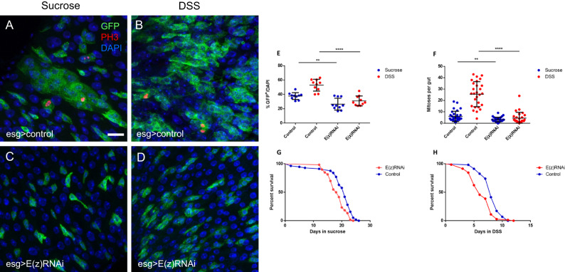
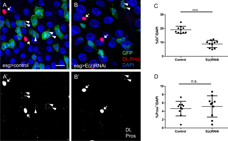
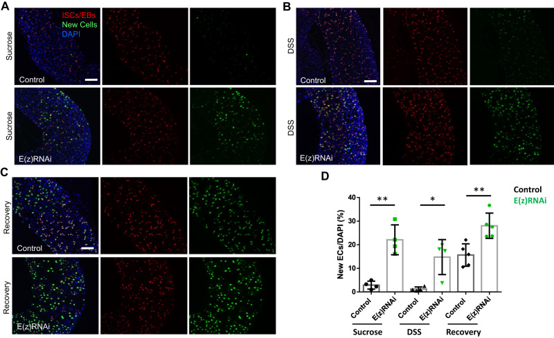
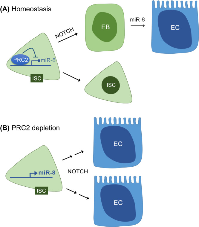
Balancing maintenance of self-renewal and differentiation is a key property of adult stem cells. The epigenetic mechanisms controlling this balance remain largely unknown. Herein, we report that the Polycomb Repressive Complex 2 (PRC2) is required for maintenance of the intestinal stem cell (ISC) pool in the adult female Drosophila melanogaster. We show that loss of PRC2 activity in ISCs by RNAi-mediated knockdown or genetic ablation of the enzymatic subunit Enhancer of zeste, E(z), results in loss of stemness and precocious differentiation of enteroblasts to enterocytes. Mechanistically, we have identified the microRNA miR-8 as a critical target of E(z)/PRC2-mediated tri-methylation of histone H3 at Lys27 (H3K27me3) and uncovered a dynamic relationship between E(z), miR-8 and Notch signaling in controlling stemness versus differentiation of ISCs. Collectively, these findings uncover a hitherto unrecognized epigenetic layer in the regulation of stem cell specification that safeguards intestinal homeostasis.


Conflict of interest statement

The authors declare no competing interests.

Figures

Fig. 1
Fig. 1. E(z) is required for ISC proliferation and midgut regeneration in Drosophila.
Immunofluorescence phospho-Histone 3 (pH3) staining of posterior midguts from 7 day old w1118 control adult female flies expressing esg-Gal4, tubGal80ts, UAS-GFP (esgts > GFP) fed with 5% sucrose solution (A) or 3% DSS (B) for 2 days prior to dissection; ISCs/EBs (GFP, green), mitotic cells (pH3, red), nuclei (DAPI, blue). Note the increase in the percentage of GFP+ cells after DSS treatment in (B) vs. (A). Young (3 days old) mated females were shifted to 29 °C for 2 days to express E(z)-RNAi and then fed with sucrose (C) or DSS (D) for two additional days at 29 °C before being processed as described in (A, B). Note the absence of the DSS-induced increase in the percentage of GFP+ cells in (D) vs. (B). Scale bar; 25 μm, applicable to (AD) images. E Quantification of the GFP+ cells as a percentage of total DAPI-stained cells. Data are represented as mean ± SD. Two-tailed, unpaired t-test: p = 0.0014 for sucrose and p < 0.0001 for DSS fed flies. Zoom-in images from ten posterior midguts were randomly selected for counting. F Quantification of pH3+ positive cells representing mitotic ISCs in control and E(z)-RNAi midguts. Data represented as mean ± SD. A decreased number of pH3+ cells was observed upon induction of E(z)-RNAi after both sucrose (Two-tailed, unpaired t-test, p = 0.0029) and DSS (p < 0.0001) treatment. Cells were counted from whole guts, n = 32 for control flies fed with sucrose, n = 30 for E(z)-RNAi flies fed with sucrose, n = 27 for control flies fed with DSS and n = 27 for E(z)-RNAi flies fed with DSS, pooled from three independent experiments. G, H Survival curves for control and E(z)-RNAi flies fed with sucrose (p = 0.0004) or DSS (p < 0.0001; log-rank (Mantel–Cox) test). The number of flies (n) assessed is n = 57 for control flies fed with sucrose, n = 60 for E(z)-RNAi flies fed with sucrose, n = 58 for control flies fed with DSS and n = 72 for E(z)-RNAi flies fed with DSS, pooled from three independent experiments.
Fig. 2
Fig. 2. E(z) depletion leads to loss of ISCs.
Flies with esgts > GFP were crossed to either UAS-E(z)-RNAi to knockdown E(z) in intestinal progenitor cells or to w1118 to generate the respective control genotype (control). A, B Control and E(z)-RNAi posterior midguts were immunostained at 7 days post induction for the ISC marker Delta (Dl, cytoplasmic puncta in GFP+ cells, arrowheads) and the EE marker Prospero (Pros, solid nuclear staining in GFP-negative cells, arrows). Scale bar; 20 μm, applies to all images. C, D Quantification of Dl+ and Pros+ cells as percentages of total DAPI-stained cells (Two-tailed unpaired t-test: p < 0.0001 for Dl+ cells). Randomly selected posterior midgut zoom-in images from each of n = 10 flies were analyzed per marker and genotype. Data are represented as mean ± SD.
Fig. 3
Fig. 3. Loss of E(z) function induces precocious differentiation into ECs.
AB’ Midguts from adult flies containing nucleus-localized GFP-labeled control MARCM clones (AA’) or clones of the catalytically inactive allele E(z)731 (BB’), stained for DAPI (blue). Guts were dissected from adult flies 10 days after clone induction. Scale bar; 50 μm. A higher magnification for each of the squared areas in (A) and (B) is depicted in (A’) and (B’) respectively. C Quantification of the cell numbers of the control or inactive E(z)731 allele-carrying clones of the indicated genotypes. Results shown are the means ± SD of n = 31 for control and n = 44 for E(z)731 clones (p < 0.0001, two-tailed unpaired t-test). DE’' Immunofluorescence staining of control (DD'') and E(z)731 (EE'') MARCM clones for Delta (Dl) and Prospero (Pros). Control clones have at least one Dl+ cell per clone and roughly 25% of the clones contain one Pros+ cell, whereas E(z)731 clones rarely contain a Dl+ cell and never a Pros+ cell. Scale bar; 25 μm. F Quantification of Dl and Pros-positive cells from n = 237 cells/15 clones for control and n = 87 cells/20 E(z)731 clones.
Fig. 4
Fig. 4. Knock-down of E(z) promotes EB to EC transition.
The G-TRACE lineage tracing reporter is driven by esg-Gal4-tubGal80ts to label progenitor cells with UAS-RFP and newly produced progeny cells with nuclear EGFP, via the expression of UAS-FLP and Ubi-p63EFRT-stop-FRT-EGFP. Flies carrying these transgenes were crossed either to w1118 (control) or to UAS-E(z)-RNAi. Collected progeny was aged for 4–7 days after eclosion and expression of transgenes was induced by 29 °C temperature shift. Newly differentiated ECs are identified as large cells expressing nuclear EGFP and low/no RFP. In control flies fed with sucrose (A) or DSS (B) for 36 h at 29 °C, newly produced ECs are scarcely detected in the R4 region of posterior midgut, whereas flies expressing E(z)-RNAi displayed a marked appearance of new ECs. Scale bars; 50 μm. C Flies were fed with DSS for 36 h to induce ISC proliferation and then left to recover under normal feeding conditions for 4 days. Control flies resume production of new ECs after 4 days of recovery from DSS to normal feeding conditions which is further increased upon E(z) knock-down. Scale bar; 50 μm. D Quantification of newly produced ECs per genotype and treatment. The number of large size cells with nuclear EGFP were counted from R4 posterior midguts and normalized to 100 cells stained with DAPI (n = 4 for control and E(z)-RNAi flies fed with sucrose or DSS, n = 5 for flies left to recover after DSS treatment). Values are presented as mean ± S.D. Two-tailed, unpaired t-test was used for statistical analysis: **p = 0.0011 (sucrose), *p = 0.01 (DSS), **p = 0.0047 (recovery after DSS).
Fig. 5
Fig. 5. E(z) depletion reduces chromatin-bound H3K27me3 and leads to the up-regulation of miR-8 expression.
Α Immunofluorescence staining for H3K27me3 (red) in control and E(z)-RNAi midguts after 7 days of RNAi induction and 2 days of feeding with sucrose or DSS. Scale bar; 25 μm. B Genome browser snapshot of a region in chromosome 2R containing the miR-8 locus, where H3K27me3 was found reduced in E(z)-RNAi vs. control sorted GFP-expressing progenitor cells. C Quantification of l(2)gl, ph-p and Psc mRNA levels in ISCs/EBs depleted of E(z) relative to E(z)-proficient cells. Total RNA was extracted from two biological replicates of FACS-sorted GFP+ progenitor cells from 100 control and 100 E(z)-RNAi fly midguts for each replicate and expression levels were assessed by reverse transcription qPCR normalized to rp49, as indicated. D miR-8-3p and miR-8-5p levels increase upon E(z)-RNAi induction. Expression was normalized to 2S rRNA. The origin of 3p and 5p is shown in the upper panel of (D).
Fig. 6
Fig. 6. The microRNA miR-8 is a functional target of E(z)/PRC2.
Α In control flies fed with sucrose, miR-8-EGFP-sensor expression is detected mainly in cells with small nuclei (arrowheads) but is absent from cells with large nuclei (arrows), suggesting low miR-8 levels in ISCs/EBs and EEs and higher expression in committed EBs and ECs. Flies induced to express E(z)-RNAi for 7 days by an esg-Gal4 driver which does not express GFP (esg-Gal4, tubGal80ts) have decreased number of miR-8-sensor positive cells. Note the high levels of sensor GFP in the visceral muscle layer surrounding the gut, suggesting absence of mir-8 expression in those cells. Scale bar; 25 μm. B Dual immunostaining of midguts from control or E(z)-RNAi flies with Dl (arrowheads) and Pros (arrows) antibodies. Lower expression of the mir-8-sensor was seen in ISCs of flies induced to express E(z)-RNAi by an esg-Gal4 driver which does not express GFP (esg-Gal4, tubGal80ts). In addition to the loss of miR-8-sensor-GFP, note the increased diameter and lower levels of Delta in a remaining ISC upon 14 days of E(z)-RNAi induction (arrowhead in lower panel). Scale bar; 25 μm. C The growth defects in E(z)731 mutant clones are partly alleviated by depletion of miR-8. Depletion of miR-8 by UAS-miR-8-sponge leads to expansion of GFP+ E(z)731 MARCM clones visualized 10 days after heat shock induction. Scale bar; 50 μm. D Quantification of the cell numbers per clone of the indicated genotypes. Results shown are the means ± SD of n = 10 clones for each genotype. E(z)731 mutant clones with depletion of miR-8 have a greater number of cells than E(z)731 mutant clones alone. Two-tailed, unpaired t-test: p = 0.0059.
Fig. 7
Fig. 7. Notch is required for the loss of stemness induced by E(z) depletion.
AH The RNAi-mediated knock-down of Notch leads to accumulation of Dl+ and Pros+ cells in MARCM control clones 10 days after heat-shock (AD). E(z)731 mutant clones depleted of Notch (EH) proliferate to a lesser extent than Notch-RNAi alone, and also accumulate Dl+ and Pros+ cells. Compared to E(z)731 null MARCM clones that hardly proliferate (Figs. 3 and 6), the simultaneous depletion of Notch displays a dramatically elevated number of cells per clone (E). Scale bar; 25 μm; arrowheads show representative Dl+ cells; light blue dotted arrows show representative Pros+ cells and yellow arrows indicate large Dl and Pros cells, most likely representing enterocytes. I, J Quantification of the average cell numbers per clone of the indicated genotypes. Results shown are the means ± SD of n = 10 clones for each genotype. E(z)731 mutant clones depleted of Notch (EH) grow to a lesser extent than those of Notch-RNAi alone (Two-tailed, unpaired t-test: p = 0.0059) (I), but significantly more than E(z)731 null clones (J, see also Figs. 3 and 6). (Two-tailed, unpaired t-test: p < 0.0001). K The reduced survival of flies bearing Notch RNAi in ISCs/EBs is partly alleviated upon simultaneous E(z) knock-down. Survival curves of the indicated lines crossed to esg-Gal4 driver are shown for the indicated genotypes (p = 0.0053, log-rank (Mantel–Cox) test, n = 55 for N-RNAi, n = 51 for E(z)-RNAi, n = 47 for Notch plus E(z)-RNAi and n = 62 for w1118 flies, pooled from three independent experiments).
Fig. 8
Fig. 8. Proposed model of PRC2-mediated regulation of intestinal stem cell (ISC) fate in Drosophila.
Based on the results reported herein, we propose a dynamic relationship between E(z), miR-8 and Notch signaling in controlling stemness vs. differentiation of ISCs. Our data suggest that E(z)/PRC2 is required in ISCs to maintain Notch target genes, such as miR-8, in a repressive chromatin state, functioning as a brake to precocious differentiation caused by basal Notch signaling (A). Disruption of E(z)/PRC2 function specifically in ISCs leads to loss of stemness and differentiation toward the EC lineage associated with Notch-mediated upregulation of miR-8 and, consequently, reduced levels of the miR-8 target Escargot (B).

Similar articles

References

    1. Kassis JA, Kennison JA, Tamkun JW. Polycomb and trithorax group genes in Drosophila. Genetics. 2017;206:1699–1725. - PMC - PubMed
    1. Piunti A, Shilatifard A. The roles of Polycomb repressive complexes in mammalian development and cancer. Nat. Rev. Mol. Cell Biol. 2021;22:326–345. - PubMed
    1. Czermin B, et al. Drosophila enhancer of Zeste/ESC complexes have a histone H3 methyltransferase activity that marks chromosomal Polycomb sites. Cell. 2002;111:185–196. - PubMed
    1. Cao R, et al. Role of histone H3 lysine 27 methylation in Polycomb-group silencing. Science. 2002;298:1039–1043. - PubMed
    1. Muller J, et al. Histone methyltransferase activity of a Drosophila Polycomb group repressor complex. Cell. 2002;111:197–208. - PubMed

LinkOut - more resources