. 2024 Mar 19;98(3):e0181523.
doi: 10.1128/jvi.01815-23. Epub 2024 Feb 29.

Interaction between the SFTSV envelope glycoprotein Gn and STING inhibits the formation of the STING-TBK1 complex and suppresses the NF-κB signaling pathway

Affiliations

Interaction between the SFTSV envelope glycoprotein Gn and STING inhibits the formation of the STING-TBK1 complex and suppresses the NF-κB signaling pathway

Yupei Jia et al. J Virol. .

Abstract

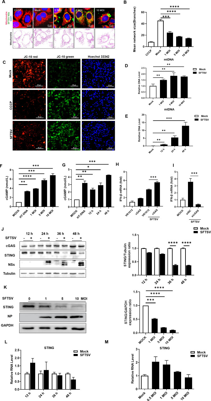
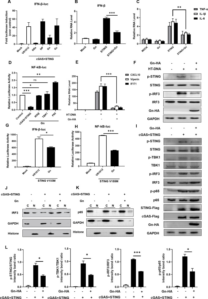
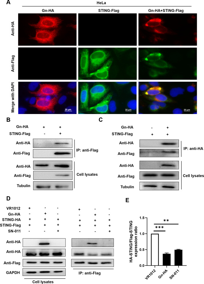
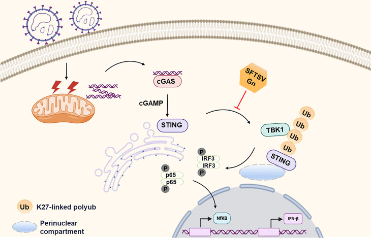
Severe fever with thrombocytopenia syndrome virus (SFTSV) is an emerging tick-borne bunyavirus with high pathogenicity. There has been a gradual increase in the number of reported cases in recent years, with high morbidity and mortality rates. The cyclic GMP-AMP synthase (cGAS)-stimulator of interferon genes (STING) signaling pathway plays an important role in the innate immune defense activated by viral infection; however, the role of the cGAS-STING signaling pathway during SFTSV infection is still unclear. In this study, we investigated the relationship between SFTSV infection and cGAS-STING signaling. We found that SFTSV infection caused the release of mitochondrial DNA into the cytoplasm and inhibits downstream innate immune signaling pathways by activating the cytoplasmic DNA receptor cGAS. We found that the SFTSV envelope glycoprotein Gn was a potent inhibitor of the cGAS-STING pathway and blocked the nuclear accumulation of interferon regulatory factor 3 and p65 to inhibit downstream innate immune signaling. Gn of SFTSV interacted with STING to inhibit STING dimerization and inhibited K27-ubiquitin modification of STING to disrupt the assembly of the STING-TANK-binding kinase 1 complex and downstream signaling. In addition, Gn was found to be involved in inducing STING degradation, further inhibiting the downstream immune response. In conclusion, this study identified the important role of the glycoprotein Gn in the antiviral innate immune response and revealed a novel mechanism of immune escape for SFTSV. Moreover, this study increases the understanding of the pathogenic mechanism of SFTSV and provides new insights for further treatment of SFTS.

Importance: Severe fever with thrombocytopenia syndrome virus (SFTSV) is a newly discovered virus associated with severe hemorrhagic fever in humans. However, the role of the cyclic GMP-AMP synthase (cGAS)-stimulator of interferon genes (STING) signaling pathway during SFTSV infection is still unclear. We found that SFTSV infection inhibits downstream innate immune signaling pathways by activating the cytoplasmic DNA receptor cGAS. In addition, SFTSV Gn blocked the nuclear accumulation of interferon regulatory factor 3 and p65 to inhibit downstream innate immune signaling. Moreover, we determined that Gn of SFTSV inhibited K27-ubiquitin modification of STING to disrupt the assembly of the STING-TANK-binding kinase 1 complex and downstream signaling. We found that the SFTSV envelope glycoprotein Gn is a potent inhibitor of the cGAS-STING pathway. In conclusion, this study highlights the crucial function of the glycoprotein Gn in the antiviral innate immune response and reveals a new method of immune escape of SFTSV.

Keywords: Gn; cyclic GMP-AMP synthase (cGAS); infection; innate immunity; mitochondrial DNA; severe fever with thrombocytopenia syndrome virus (SFTSV); stimulator of interferon genes (STING).


Conflict of interest statement

The authors declare no conflict of interest.

Figures

Fig 1
Fig 1
mtDNA released into the cytosol upon SFTSV infection is captured by cGAS and inhibits the cGAS-STING signaling pathway in the late stages of infection. SFTSV infection induces mitochondrial damage to release mtDNA into the cytosol, where it is captured by cGAS, resulting in the inhibition of the cGAS-STING signaling pathway in the late stages of infection. (A, B) Confocal micrographs of mitochondria in HeLa cells infected with SFTSV after 24 h. The results were analyzed via the MiNA workflow, and ImageJ was used to calculate the ratio of cells that contained fragmented mitochondria to total cells. Treatment with CCCP (5 µM) was performed as the positive control. (C) Mitochondrial membrane potential values after SFTSV infection were measured using the JC-10 mitochondrial membrane potential fluorescent probe. CCCP (5 µM) was used as the positive control. (D) Abundance of mtDNA in the cytosol of HeLa cells infected with SFTSV at various MOIs (MOI = 0, 1, 5, and 10). The abundances of specific DNA fragments were quantified by qPCR. (E) Abundance of mtDNA in the cytosol of SFTSV-infected HeLa cells over time (12, 24, and 48 h). The abundances of specific DNA fragments were quantified by qPCR. (F, G) A reagent kit was used to measure the amount of cGAMP released after infection with SFTSV at different MOIs and for different durations. (H) cGAS (200 ng of plasmid) was overexpressed in HeLa cells. The cells were infected with SFTSV, and RNA isolated 48 h postinfection (hpi) was analyzed for interferon (IFN)-β expression by RT-qPCR. A t-test was used for statistical analysis. (I) cGAS was knocked down in HeLa cells. The cells were infected with SFTSV, and RNA isolated 6 hpi was analyzed for IFN-β expression by RT-qPCR. A t-test was used for statistical analysis. (J) HeLa cells were infected with SFTSV at an MOI of 1. The cells were collected at 12, 24, 36, and 48 hpi, and the expression of cGAS and STING was confirmed by Western blotting. The gray values were analyzed by ImageJ and Prism software. (K) HeLa cells were infected with SFTSV (MOI = 0, 1, 5, and 10) and collected 36 h postinfection, and the expression of cGAS and STING was analyzed by Western blotting. The gray values were analyzed by ImageJ and Prism software. (L) HeLa cells were infected with SFTSV at an MOI of 1. The cells were collected 12, 24, 36, and 48 hpi, and the mRNA level of STING was measured via RT-qPCR. (M) HeLa cells were infected with SFTSV (MOI = 0, 0.5, 1, 5, or 10), the cells were collected 48 h postinfection, and the mRNA level of STING was measured via RT-qPCR.
Fig 2
Fig 2
The SFTSV viral protein Gn inhibits the cGAS-STING-mediated innate immune response. SFTSV Gn inhibited the cGAS-STING-mediated downstream innate immune response. (A) HEK293T cells were cotransfected with IFN-β Luc and the cGAS-STING, NSs, NP, Gn, or Gc expression vector. A luciferase assay and Western blot analysis were performed 36 h after transfection. (B) HeLa cells were transfected with the VR1012, Gn (200 ng), STING (200 ng), or STING and Gn plasmids. Cellular RNA was extracted 36 h after transfection, and the RNA level of IFN-β was measured by RT-qPCR. (C) HeLa cells were transfected with VR1012, Gn (200 ng), STING (200 ng), or STING and Gn plasmids. Cellular RNA was extracted 36 h after transfection, and the RNA levels of TNF-α, IL-1β, and IL-6 were measured by RT-qPCR. (D) Comparison of Gn-mediated inhibition of NF-κB signaling induced by cGAS (200 ng), STING (200 ng), IKKβ (200 ng), TBK1 (200 ng), and p65 (200 ng). HEK293T cells were cotransfected with the NF-κB-Luc (200 ng) and cGAS (200 ng), STING (200 ng), IKKβ (200 ng), TBK1 (200 ng), or p65 (200 ng) expression vectors in the presence or absence of the Gn expression vector. The values in cells transfected with cGAS (200 ng), STING (200 ng), IKKβ (200 ng), TBK1 (200 ng), or p65 (200 ng) alone were set to 1, as appropriate. (E) HeLa cells were transfected with VR1012, HT-DNA, or HT-DNA and Gn (200 ng) for 36 h, after which the RNA levels of CXCL10, viperin, and IFIT1 were measured via RT-qPCR. A t-test was used for statistical analysis. (F) The degree of phosphorylation of STING and IRF3 was evaluated. (G, H) HEK293T cells were transfected with the IFN-β Luc/NF-κB Luc reporter plasmid and pRL-SV40, VR1012, STING V155M, or STING V155M and Gn. Transactivation of the luciferase reporter was evaluated 36 h after transfection. (I) HeLa cells were cotransfected with STING-Flag (200 ng), cGAS-Flag (200 ng), Gn-HA (200 ng), or the control vector. After 36 h, the cells were harvested, and the protein expression levels were analyzed by immunoblotting. (J, K) Gn inhibits the nuclear translocation of IRF3 and p65. HEK293T cells were transfected with the control vector, STING-Flag (200 ng), cGAS-Flag (200 ng), or STING-Flag and cGAS-Flag plus Gn-HA (200 ng), as indicated. The cells were harvested, total cell lysates were prepared, and the nuclear (N) and cytoplasmic (C) fractions were separated 36 h after transfection. The indicated proteins were analyzed by immunoblotting using anti-p65 and anti-IRF3 antibodies. Glyceraldehyde-3-phosphate dehydrogenase (GAPDH and histone were used as controls and detected using anti-GAPDH and anti-histone antibodies, respectively. (L) Degradation levels of p-STING, p-TBK1, p-IRF3, and p-p65 in (I) were quantified using ImageJ and Prism, respectively.
Fig 3
Fig 3
Gn interacts with STING and affects STING dimerization. Gn interacts with STING and affects STING dimerization. (A) Colocalization of intracellular Gn and STING. HeLa cells were transfected with STING-Flag alone, Gn-HA alone, or STING-Flag plus Gn-HA, as shown. 4',6-diamidino-2-phenylindole (DAPI) staining was performed to visualize nuclei. (B, C) Identification of the Gn–STING interaction by coimmunoprecipitation. HEK293T cells were transfected with Gn and the STING-Flag or control vector as indicated. Cell lysates were prepared and subjected to immunoprecipitation using anti-Flag/anti-HA beads 48 h after transfection. Proteins in the precipitate samples were separated by SDS-PAGE; an anti-HA antibody was used to detect Gn-HA, and an anti-Flag antibody was used to detect STING-Flag. (D) Gn affects STING dimerization. HEK293T cells were cotransfected with STING-Flag, STING-HA, and Gn-HA or the control vector. SN-011 was used as the positive control. HeLa cells were pretreated with SN-011 for 5 h (2 µM). After 48 h, the cells were harvested, and protein expression levels were measured by immunoblotting with anti-Flag, anti-HA, or anti-GAPDH antibodies. (E) STING dimerization was decreased in the presence of Gn as described in (D); binding was quantified using ImageJ software. The means and standard deviations are presented.
Fig 4
Fig 4
Gn inhibits the K27-linked ubiquitination of STING and suppresses its recruitment of TBK1. Gn inhibits the K27-linked ubiquitination of STING and its recruitment of TBK1. (A) Gn inhibits the modification of STING with WT-Ub. HEK293T cells were transfected with HA-Ub WT alone or cotransfected with STING-Flag, cGAS-Flag, and HA-Ub WT in the presence or absence of Gn. Cell lysates were prepared and subjected to immunoprecipitation with anti-Flag beads 48 h after transfection. Precipitated samples were prepared and reacted with an anti-Flag antibody to detect Flag-STING and an anti-HA antibody to detect HA-Ub WT and Gn-HA. GAPDH was used as the loading control. (B–E) Gn inhibits the K27-linked ubiquitination of STING. HEK293T cells were transfected with Myc-Ub K63/Myc-Ub K48/Myc-Ub K27/HA-Ub K63 alone or cotransfected with STING-Flag, cGAS-Flag, or Myc-Ub K63/Myc-Ub K48/Myc-Ub K27/HA-Ub K63 in the presence or absence of Gn. Cell lysates were prepared and subjected to immunoprecipitation with anti-Flag beads 48 h after transfection. Precipitated samples were prepared and reacted with an anti-Flag antibody to detect Flag-STING and an anti-Myc/anti-HA antibody to detect Myc-Ub K63/Myc-Ub K48/Myc-Ub K27/HA-Ub K63 and Gn-HA. GAPDH was used as the loading control. (F) Gn disrupts the interaction between STING and endogenous TBK1. HEK293T cells were transfected with cGAS-STING in the presence or absence of Gn. Cell lysates were prepared and subjected to immunoprecipitation using anti-Flag beads 36 h after transfection. Precipitated samples were prepared and reacted with an anti-Flag antibody to detect STING-Flag and an anti-TBK1 antibody to detect endogenous proteins. GAPDH was used as the loading control. SN-011 was used as the positive control. HeLa cells were pretreated with SN-011 for 5 h (2 µM). (G) STING-TBK1 binding was attenuated in the presence of Gn as described in (F); binding was quantified using ImageJ software. The means and standard deviations are presented.
Fig 5
Fig 5
SFTSV degrades STING via the autophagy pathway. (A) HeLa cells were transfected with VR1012 or Gn-HA (1,000 ng), and the cells were collected at 24, 36, and 48 h for immunoblotting to measure the expression of STING. (B) HeLa cells were transfected with 250, 500, or 1,000 ng of the Gn-HA plasmid. After 48 h, the cells were collected, and protein expression levels were measured by immunoblotting with anti-STING, anti-HA, or anti-GAPDH antibodies. (C) Quantitative analysis using ImageJ software. The averages and standard deviations are shown. (D) HeLa cells were transfected with VR1012 or Gn-HA. The cells were pretreated by adding Baf-A1 to the medium (final concentration, 20 nM) 12 h before transfection, and this medium was changed to a medium containing Baf-A1 at a final concentration of 20 nM after transfection. (E, F) With SFTSV-infected cells as a positive control group, the dose-dependent effect of Gn on endogenous STING expression was evaluated. (G) HeLa cells were infected with SFTSV, and Baf-A1 was added as described in (D). After 48 h, the cells were harvested, and protein expression levels were measured by immunoblotting. (H–J) HeLa cells were treated as described in (A). After 48 h, the cells were collected, and the mRNA levels of STING, IFN-α4, and IFN-β were measured by RT-qPCR.
Fig 6
Fig 6
The C-terminus of SFTSV Gn is required for the Gn-STING interaction. STING interacts with C-terminal fragments of Gn. (A) Various Gn truncation mutants. (B) Colocalization of the Gn truncation mutants and STING. HeLa cells were transfected with HA-Gn and STING-Flag alone or with HA-Gn-ΔN200/HA-Gn-ΔC200/HA-Gn-ΔM/HA-Gn 201–335 and STING-Flag, as shown. DAPI staining was performed to visualize nuclei. After 24 h, the cells were harvested, and colocalization was observed via immunofluorescence staining. (C) Identification of STING–Gn truncation mutant interactions by coimmunoprecipitation. HEK293T cells were transfected with STING-Flag and HA-Gn-ΔC200/HA-Gn-ΔM or the control vector as indicated. Cell lysates were prepared and subjected to immunoprecipitation using anti-HA beads 48 h after transfection. Representative immunoblot results are shown. (D) HEK293T cells were transfected with the IFN-β Luc reporter plasmid and pRL-SV40 plasmid simultaneously with the STING and Gn or Gn truncation plasmids. Cells were collected 36 h after transfection, and IFN-β promoter activity was evaluated via a luciferase reporter assay.
Fig 7
Fig 7
Amino acids 196–317 of STING are required for the STING–Gn interaction. Gn interacts with the CDN-binding domain (CBD) of STING. (A) Schematic of the STING functional domains and various STING truncation mutants. (B) Colocalization of Gn and STING truncation mutants. HeLa cells were sttransfected with STING-ΔCTT-Flag/STING 1–196-Flag/STING 196–340-Flag and Gn-HA as shown. DAPI staining was performed to visualize nuclei. After 24 h, the cells were harvested, and colocalization was observed via immunofluorescence staining. (C, D) Identification of Gn–STING truncation mutant interactions by coimmunoprecipitation. HEK293T cells were transfected with HA-Gn and STING-Flag/STING-ΔCTT-Flag/STING 1–317-Flag/STING 1–196-Flag/STING 196–340-Flag or the control vector as indicated. Cell lysates were prepared and subjected to immunoprecipitation using anti-Flag beads 48 h after transfection. Representative immunoblot results are shown.
Fig 8
Fig 8
SFTSV Gn enhances viral replication. (A, B) HeLa cells overexpressing Gn (250, 500, and 1,000 ng) were infected with SFTSV, and Gn was found to enhance viral replication. (C) Quantitative analysis using ImageJ software. The averages and standard deviations are shown. (D) Gn was overexpressed (500 ng) and cGAS was knocked down in virally infected cells; after 48 h, the impact on viral replication was evaluated.
Fig 9
Fig 9
Proposed model showing the mechanism by which SFTSV Gn inhibits the cGAS-STING signaling pathway. SFTSV infection causes the release of mitochondrial DNA into the cytoplasm and the activation of the cytoplasmic DNA receptor cGAS. SFTSV Gn is a potent inhibitor of the cGAS-STING pathway and blocks the nuclear accumulation of IRF3 and p65 to inhibit downstream signaling. Gn interacts with STING to inhibit STING dimerization, and Gn of SFTSV inhibits K27-linked ubiquitination of STING to disrupt the assembly of the STING-TBK1 complex and downstream signaling.

Similar articles

Cited by

References

    1. Yu X-J, Liang M-F, Zhang S-Y, Liu Y, Li J-D, Sun Y-L, Zhang L, Zhang Q-F, Popov VL, Li C, et al. . 2011. Fever with thrombocytopenia associated with a novel bunyavirus in China. N Engl J Med 364:1523–1532. doi:10.1056/NEJMoa1010095 - DOI - PMC - PubMed
    1. Kuhn J-O, Adkins S-O, Alioto D-O, Alkhovsky S-O, Amarasinghe G-O, Anthony SJ, Avšič-Županc T-O, Ayllón M-O, Bahl J-O, Balkema-Buschmann A-O, et al. . 2020. 2020 taxonomic update for phylum Negarnaviricota (Riboviria: Orthornavirae), including the large orders Bunyavirales and Mononegavirales - PMC - PubMed
    1. Zhang X, Liu Y, Zhao L, Li B, Yu H, Wen H, Yu X-J. 2013. An emerging hemorrhagic fever in China caused by a novel bunyavirus SFTSV. Sci China Life Sci 56:697–700. doi:10.1007/s11427-013-4518-9 - DOI - PubMed
    1. Zhan J, Wang Q, Cheng J, Hu B, Li J, Zhan F, Song Y, Guo D. 2017. Current status of severe fever with thrombocytopenia syndrome in China. Virol Sin 32:51–62. doi:10.1007/s12250-016-3931-1 - DOI - PMC - PubMed
    1. Takahashi T, Maeda K, Suzuki T, Ishido A, Shigeoka T, Tominaga T, Kamei T, Honda M, Ninomiya D, Sakai T, et al. . 2014. The first identification and retrospective study of severe fever with thrombocytopenia syndrome in Japan. J Infect Dis 209:816–827. doi:10.1093/infdis/jit603 - DOI - PMC - PubMed

MeSH terms