. 2024 Aug 1;147(8):2884-2896.
doi: 10.1093/brain/awae062.

Novel insight into atogepant mechanisms of action in migraine prevention

Affiliations

Novel insight into atogepant mechanisms of action in migraine prevention

Agustin Melo-Carrillo et al. Brain. .

Abstract

Recently, we showed that while atogepant-a small-molecule calcitonin gene-related peptide (CGRP) receptor antagonist-does not fully prevent activation of meningeal nociceptors, it significantly reduces a cortical spreading depression (CSD)-induced early response probability in C fibres and late response probability in Aδ fibres. The current study investigates atogepant effect on CSD-induced activation and sensitization of high threshold (HT) and wide dynamic range (WDR) central dura-sensitive trigeminovascular neurons. In anaesthetized male rats, single-unit recordings were used to assess effects of atogepant (5 mg/kg) versus vehicle on CSD-induced activation and sensitization of HT and WDR trigeminovascular neurons. Single cell analysis of atogepant pretreatment effects on CSD-induced activation and sensitization of central trigeminovascular neurons in the spinal trigeminal nucleus revealed the ability of this small molecule CGRP receptor antagonist to prevent activation and sensitization of nearly all HT neurons (8/10 versus 1/10 activated neurons in the control versus treated groups, P = 0.005). In contrast, atogepant pretreatment effects on CSD-induced activation and sensitization of WDR neurons revealed an overall inability to prevent their activation (7/10 versus 5/10 activated neurons in the control versus treated groups, P = 0.64). Unexpectedly however, in spite of atogepant's inability to prevent activation of WDR neurons, it prevented their sensitization (as reflected their responses to mechanical stimulation of the facial receptive field before and after the CSD). Atogepant' ability to prevent activation and sensitization of HT neurons is attributed to its preferential inhibitory effects on thinly myelinated Aδ fibres. Atogepant's inability to prevent activation of WDR neurons is attributed to its lesser inhibitory effects on the unmyelinated C fibres. Molecular and physiological processes that govern neuronal activation versus sensitization can explain how reduction in CGRP-mediated slow but not glutamate-mediated fast synaptic transmission between central branches of meningeal nociceptors and nociceptive neurons in the spinal trigeminal nucleus can prevent their sensitization but not activation.

Keywords: central sensitization; gepants; headache; migraine; pain; trigeminovascular.


Conflict of interest statement

All authors have completed the ICMJE uniform disclosure form at www.icmje.org/coi_disclosure.pdf (available on request from the corresponding author).

R.Bu. is the John Hedley-Whyte Professor of Anesthesia and Neuroscience at the Beth Israel Deaconess Medical Center and Harvard Medical School. He has received research support from the NIH: R01 NS094198-01A1, R37 NS079678, R01NS095655, R01 NS104296, R21 NS106345, Allergan, Teva, Dr. Ready, Eli Lilly, Trigemina and the Migraine Research Foundation. He is a reviewer for NINDS, holds stock options in AllayLamp and Percept; serves as consultant, advisory board member, or has received honoraria from: Alder, Allergan, Biohaven, Dr. Reddy's Laboratory, Eli Lilly, GlaxoSmithKline, Merck, Teva, and Trigemina. CME fees from Healthlogix, Medlogix, WebMD/Medscape, and Patents 9061025, 11732265.1, 10806890, US2021-0015908, WO21007165, US2021-0128724, WO210054. R.Br., B.D., A.M.A. and M.F.B. are employees of Allergan, an AbbVie Company.

Figures

Figure 1
Figure 1
Experimental design. Treatment group received intravenous atogepant (Ato). Control group received intravenous (IV) saline (vehicle). Stars depict times at which responses to mechanical stimulation of the dura and skin were determined. CSD = cortical spreading depression.
Figure 2
Figure 2
Recording sites (A and B), dural (C and D) and facial (E and F) receptive fields of all studied trigeminovascular neurons in the spinal trigeminal nucleus. Black and grey circles depict locations of lesions of high threshold (HT) and wide dynamic range (WDR) neurons in the different laminae of the upper cervical spinal cord segment. Red indicates locations and sizes of most sensitive areas of dural and cutaneous receptive fields. Inset in C depicts dural receptive field drawings. Created with Biorender.com.
Figure 3
Figure 3
Examples of CSD effects on activation and sensitization of individual WDR [A(i) and B(i)] and HT [A(ii) and B(ii)] neurons in untreated (vehicle) and treated (atogepant) animals. [A(i and ii)] Plots of firing rate before (blue) and 1 and 2 h after (red) cortical spreading depression (CSD) induction in an animal treated with saline (vehicle). Note that spontaneous firing and responses to mechanical stimulation of the dura and skin increased after the CSD. [B(i and ii)] Plots of firing rate before (blue) and 1 and 2 h after (green) CSD induction in an animal treated with atogepant. Note that spontaneous activity and responses to stimulation of the dura and skin did not increase after the CSD. Recording sites and locations of dural and cutaneous receptive fields of each of the four neurons are shown. HT = high threshold; VFH = von Frey hair; WDR = wide dynamic range.
Figure 4
Figure 4
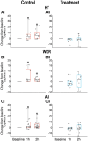
Percentage of HT, WDR and all neurons activated by CSD in the 10 animals treated with saline [Control, A(i), B(i) and C(i)] and 10 animals treated with atogepant [Treatment, A(ii), B(ii) and C(ii)]. Fisher exact was used to calculate the level of significance of the percentage differences between the groups. Plots of changes in spontaneous firing rate recorded in individual high threshold (HT) [A(iii and iv)], wide dynamic range (WDR) [B(iii and iv)] and all [C(iii and iv)] neurons before and 2 h after occurrence of cortical spreading depression (CSD). Red and blue lines depict neurons classified as activated and not activated, respectively.
Figure 5
Figure 5
Spontaneous activity changes (from baseline) recorded in HT [A(i and ii)], WDR [B(i and ii)] and all [C(i and ii)] neurons at 1 and 2 h after CSD induction. Box and whisker plots depict median and IQR (A and B, 10 neurons per group; C, 20 neurons per group). Scatter plots depict changes from baseline of individual neurons. *P < 0.05 Friedman test; post hoc/Tukey HSD. CSD = cortical spreading depression; HT = high threshold; WDR = wide dynamic range.
Figure 6
Figure 6
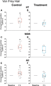
Changes from baseline in response to dural indentation 2 h after CSD induction. (A, C and E) Animals treated with saline. (B, D and F) Animals treated with atogepant. Box and whisker plots depict median and IQR (AD, 10 neurons per group; E and F, 20 neurons per group). Scatter plots depict changes from baseline of individual neurons. *P < 0.05 Wilcoxon signed rank test. CSD = cortical spreading depression; HT = high threshold; WDR = wide dynamic range.
Figure 7
Figure 7
Changes from baseline in responses to mechanical stimulation of the skin. (A) Brush, (B) pressure and (C) pinch 2 h after cortical spreading depression (CSD) induction in animals treated with saline (control) and atogepant (treatment). Box and whisker plots depict median and IQR (iiv, 10 neurons per group; v and vi, 20 neurons per group). Scatter plots depict changes from baseline of individual neurons. *P < 0.05 Wilcoxon signed rank test.
Figure 8
Figure 8
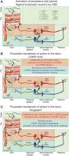
Hypothesized differences between mechanisms of action of atogepant (a small molecule CGRP-receptor antagonist and Amy1-receptor antagonist) and fremanezumab (an anti-CGRP-mAb). (A) Proposed sequence of events leading to cortical spreading depression (CSD)-induced calcitonin gene-related peptide (CGRP)-independent early activation of C-fibres and wide dynamic range (WDR) neurons, and CGRP-dependent delayed activation of Aδ-fibres and high threshold (HT) neurons. (B) Proposed mechanism of action of CGRP-mAb (monoclonal antibody) in the dura. This proposal integrates results from our previous studies,, showing that fremanezumab prevents the delayed activation and sensitization of Aδ-fibres and HT neurons but not the early activation and sensitization of C-fibres and WDR neurons. (C) Proposed mechanisms of action of Atogepant (Ato) in the dura. This proposal integrates results from the current and our previous study, showing that atogepant attenuates the early activation of C-fibres and delayed activation of the Aδ-fibres. The scientific framework presented in A is based on current evidence for (i) presence of CGRP in peptidergic C but not Aδ-fibres,; (ii) presence of CGRP receptors in Aδ but not C-fibres and in WDR and HT neurons,; (iii) CGRP release from central branches of C-fibres in the medullary and upper cervical dorsal horn,; (iv) a distinction between physiological and molecular processes that govern activation versus sensitization of central nociceptive neurons; (v) CGRP predominant role in neuronal sensitization,; (vi) predominant contribution of C-fibre nociceptors to the development of central sensitization; (vii) presence of AMY 1 receptors in both Aδ and C-fibres,; (viii) amylin analogue ability to provoke migraine-like headache; (ix) high level of amylin in the plasma of chronic migraine patients; and (x) speculation about pancreatic (b-cells) origin of amylin in the dura., Based on the above, we are proposing that by neutralizing the CGRP peptide, anti-CGRP monoclonal antibodies can prevent activation of both the CLR-RAMP1(CGRP receptor) and CTR-RAMP1 (Amy1 receptor) by CGRP, but not the activation of the CTR-RAMP1 by amylin (illustrated in B). In contrast, atogepant’s ability to block the CLR-RAMP1 as well as the CTR-RAMP1 can prevent their activation by CGRP as well as amylin (illustrated in C). In the context of migraine, this figure illustrates the potential importance of reducing C-fibre input and consequential CGRP release in nociceptive laminae of the spinal trigeminal nucleus as such interference can prevent the development and establishment of central sensitization—a physiological state tightly correlated with disease progression., (Please note that for simplicity, we did not mark presence of CGRP receptors in presynaptic terminals and consequently, did not discuss the possibility that activation of these receptors can contribute to the development of central sensitization by enhancing presynaptic glutamate release in the dorsal horn). Numbers in bold are explained at the bottom. 1. CSD; 2a. Vascular response; 2b. activation of C-fibers. 3. Activated C-fiber release of CGRP(and possibly adrenomedullin) in the dura, leading to activation of Aδ-fibers; 4a. Activated C-fibers release glutamate and CGRP in the dorsal horn, leading to activation and sensitization of WDR neurons and sensitization of HT neurons; 4b. Activated Aδ-fibers release glutamate that activates HT neurons; 5. Activated WDR and HT neurons send signals to the thalamus. AMPA = ionotropic glutamate receptors; CLR-RAMP1 = CGRP receptor; CTR-RAMP1 = amylin (Amy1) receptor; mGluR1 = metabotropic glutamate receptor 1; NK1 = neurokinin-1 (substance P) receptor; NMDA = N-methyl-D-aspartate (glutamate) receptor.

Similar articles

Cited by

References

    1. Messlinger K. The big CGRP flood—Sources, sinks and signalling sites in the trigeminovascular system. J Headache Pain. 2018;19:22. - PMC - PubMed
    1. Ebersberger A, Averbeck B, Messlinger K, Reeh PW. Release of substance P, calcitonin gene-related peptide and prostaglandin E2 from rat dura mater encephali following electrical and chemical stimulation in vitro. Neuroscience. 1999;89:901–907. - PubMed
    1. Messlinger K, Hanesch U, Baumgartel M, Trost B, Schmidt RF. Innervation of the dura mater encephali of cat and rat: ultrastructure and calcitonin gene-related peptide-like and substance P-like immunoreactivity. Anat Embryol (Berl). 1993;188:219–237. - PubMed
    1. Edvinsson L, Ekman R, Jansen I, McCulloch J, Uddman R. Calcitonin gene-related peptide and cerebral blood vessels: distribution and vasomotor effects. J Cereb Blood Flow Metab. 1987;7:720–728. - PubMed
    1. Kageneck C, Nixdorf-Bergweiler BE, Messlinger K, Fischer MJ. Release of CGRP from mouse brainstem slices indicates central inhibitory effect of triptans and kynurenate. J Headache Pain. 2014;15:7. - PMC - PubMed

MeSH terms

Substances