. 2024 Feb 8;25(4):2073.
doi: 10.3390/ijms25042073.

The Immunoregulatory and Regenerative Potential of Activated Human Stem Cell Secretome Mitigates Acute-on-Chronic Liver Failure in a Rat Model

Affiliations

The Immunoregulatory and Regenerative Potential of Activated Human Stem Cell Secretome Mitigates Acute-on-Chronic Liver Failure in a Rat Model

Barbara Cuadra et al. Int J Mol Sci. .

Abstract

Acute-on-chronic liver failure (ACLF) is a syndrome marked by sudden liver function decline and multiorgan failure, predominantly acute kidney injury (AKY), in patients with chronic liver disease. Unregulated inflammation is a hallmark of ACLF; however, the key drivers of ACLF are not fully understood. This study explores the therapeutic properties of human mesenchymal stem cell (MSC) secretome, particularly focusing on its enhanced anti-inflammatory and pro-regenerative properties after the in vitro preconditioning of the cells. We evaluated the efficacy of the systemic administration of MSC secretome in preventing liver failure and AKI in a rat ACLF model where chronic liver disease was induced using by the administration of porcine serum, followed by D-galN/LPS administration to induce acute failure. After ACLF induction, animals were treated with saline (ACLF group) or MSC-derived secretome (ACLF-secretome group). The study revealed that MSC-secretome administration strongly reduced liver histological damage in the ACLF group, which was correlated with higher hepatocyte proliferation, increased hepatic and systemic anti-inflammatory molecule levels, and reduced neutrophil and macrophage infiltration. Additionally, renal examination revealed that MSC-secretome treatment mitigated tubular injuries, reduced apoptosis, and downregulated injury markers. These improvements were linked to increased survival rates in the ACLF-secretome group, endorsing MSC secretomes as a promising therapy for multiorgan failure in ACLF.

Keywords: acute-on-chronic liver failure; in vitro preconditioning; mesenchymal stem cells; multiorgan failure; secretome.


Conflict of interest statement

The authors declare no conflicts of interest.

Figures
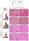
Figure 1
Figure 1
MSC-Secretome administration enhances survival rate and attenuates ACLF hepatic injury. To establish the ACLF model, rats were exposed to porcine serum administration for 11 weeks (cirrhosis development), followed by the administration of LPS/DGalN as an acute challenge to induce ACLF. Survival rates and hepatic damage were assessed up to 7 days post-acute challenge: (A) Kaplan–Meier survival analysis was carried out for control, ACLF, and ACLF + Sec groups; n = 10 in the control group, n = 35 in ACLF and ACLF + Sec groups. (B) Masson’s trichrome and H&E staining were performed to evaluate MSC-secretome effects in the ACLF model. Representative micrographs show liver histology of ACLF rats treated with the vehicle (ACLF group) and ACLF rats treated with the MSC secretome derived from 1 × 10⁶ in vitro preconditioned MSCs (ACLF + Sec), 16 and 24 h after ACLF induction (scale bars represent 250 µm). (C) Scoring of necrosis, periportal inflammation (solid bar), pericentral inflammation (lined bar), and congestion 24 h post-acute injury. N/O = not observed. Data are presented as mean ± SEM, whereas dots represet individual values, n = 5; * p < 0.05 vs. control group; & p < 0.05 vs. cirrhosis group; # p < 0.05 vs. ACLF group.
Figure 2
Figure 2
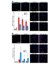
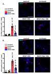
MSC-secretome administration reduces apoptotic rate and promotes hepatocyte proliferation after ACLF induction. Apoptosis and cellular proliferation were analyzed at 8, 16, and 24 h post-ACLF induction in all experimental groups. MSC secretome’s effect on apoptosis was assessed via TUNEL staining (FITC—green), and proliferation was assessed via PCNA immunoreactivity (Alexa Fluor 555—red); in both cases, the nuclei were counterstained with DAPI (blue): (A) Representative micrographs of cell apoptosis determined by TUNEL (white arrowheads). (B) Representative micrographs of hepatocyte proliferation determined by PCNA labeling (white arrowheads). Quantification of positive nuclei per 100 hepatic cells was carried out using digital imaging. Data represent mean ± SEM for 30 fields/animal, six animals/group. Scale bars represent 50 µm. Complementary semiquantitative analysis of apoptosis and proliferation was performed via Western blotting for (C) cleaved caspase 3 and (D) PCNA in liver tissue 8, 12, 16, and 24 h post-ACLF induction. Protein levels were normalized against GAPDH. Data are presented as mean ± SEM, whereas dots represet individual values, n = 5; * p < 0.05 vs. control group; & p < 0.05 vs. cirrhosis group; # p < 0.05 vs. ACLF group.
Figure 2
Figure 2
MSC-secretome administration reduces apoptotic rate and promotes hepatocyte proliferation after ACLF induction. Apoptosis and cellular proliferation were analyzed at 8, 16, and 24 h post-ACLF induction in all experimental groups. MSC secretome’s effect on apoptosis was assessed via TUNEL staining (FITC—green), and proliferation was assessed via PCNA immunoreactivity (Alexa Fluor 555—red); in both cases, the nuclei were counterstained with DAPI (blue): (A) Representative micrographs of cell apoptosis determined by TUNEL (white arrowheads). (B) Representative micrographs of hepatocyte proliferation determined by PCNA labeling (white arrowheads). Quantification of positive nuclei per 100 hepatic cells was carried out using digital imaging. Data represent mean ± SEM for 30 fields/animal, six animals/group. Scale bars represent 50 µm. Complementary semiquantitative analysis of apoptosis and proliferation was performed via Western blotting for (C) cleaved caspase 3 and (D) PCNA in liver tissue 8, 12, 16, and 24 h post-ACLF induction. Protein levels were normalized against GAPDH. Data are presented as mean ± SEM, whereas dots represet individual values, n = 5; * p < 0.05 vs. control group; & p < 0.05 vs. cirrhosis group; # p < 0.05 vs. ACLF group.
Figure 3
Figure 3
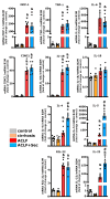
MSC-secretome administration increases hepatic anti-inflammatory cytokine expression after ACLF induction. The hepatic mRNA levels of the cytokines MCP-1, TNF-α, IL-6, CINC1, IL-1β, IL-18, IL-4, IL-5, IL-22, and IL-22 were quantified by RT-qPCR 16 h post-ACLF induction (LPS/DGalN administration), normalized against β2 microglobulin (B2M) and expressed as fold change vs. control group. Data are presented as mean ± SEM, whereas dots represet individual values (n = 5) (* p < 0.05 vs. control group; & p < 0.05 vs. cirrhosis group; # p < 0.05 vs. ACLF group).
Figure 4
Figure 4
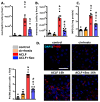
MSC-secretome administration decreases hepatic neutrophil and macrophage infiltration after ACLF induction. Hepatic infiltration of (A) neutrophils (labeled with RP1) and (B) monocytes (labeled with His48) were evaluated by flow cytometry 16 h post-ACLF induction, n = 4. (C) Myeloperoxidase (MPO) levels in hepatic tissues reflect neutrophil infiltration; data were normalized to protein amount and expressed as fold change vs. control group, n = 6. (D) Macrophage infiltration (F4/80 Alexa 555, red) in hepatic tissue 16 h post-ACLF induction was assessed by confocal microscopy. Nuclei were counterstained with DAPI (blue). Scale bars represent 50 µm. Quantification of positive cells was carried out by digital imaging analysis. Data are presented as mean ± SEM for 30 fields/animal, six animals/group, whereas dots represet individual values; * p < 0.05 vs. control group; & p < 0.05 vs. cirrhosis group; # p < 0.05 vs. ACLF group.
Figure 5
Figure 5
MSC-secretome administration increases hepatic Nrf2 and HO-1 levels, decreasing oxidative damage after ACLF induction: (A) Nrf2 levels were quantified in the nuclear fraction of hepatic tissue 16 h post-ACLF induction. Values were normalized against histone H4. (B) Heme oxygenase 1 (HO-1) levels were determined in total liver tissue fraction at the same time point, and data were normalized against GAPDH. Results for both markers are presented as the mean ± SEM, n = 5, and expressed as fold change vs. control group. (C) Given the role of Nrf2 and HO-1 in oxidative stress management, levels of 8-hydroxy-2′-deoxyguanosine (8-OHdG) in plasma (left panel) and hepatic tissue (right panel) were assessed 24 h post-ACLF induction by ELISA. Data are presented as mean ± SEM, whereas dots represet individual values, n = 8; * p < 0.05 vs. control group; & p < 0.05 vs. cirrhosis group; # p < 0.05 vs. ACLF group.
Figure 6
Figure 6
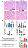
MSC-secretome administration mitigates acute kidney injury associated with ACLF. Histological evaluation of renal effects of MSC-secretome administration in ACLF murine model: (A) Representative renal tissue micrographs 24 h post-ACLF induction, stained with PAS. ACLF group exhibited histological alterations like brush border loss (black arrows), tubular dilatation (asterisks), and cell detachment (blue arrows). Scale bars represent 100 µm. (B) mRNA levels of tubular damage markers HMGB1, IL-18, Kim-1, and NGAL were quantified by RT-qPCR at 8, 16, 24 h, and 7 days post-ACLF induction (LPS/NGalN administration) and normalized against GAPDH expression. Data are presented as mean ± SEM (n = 6) and expressed as fold change vs. control group. (C) Changes in NGAL and Kim-1 levels were confirmed by Western blotting 16 h post-ACLF induction. Data are presented as mean ± SEM (n = 5), whereas dots represet individual values; * p < 0.05 vs. control group; & p < 0.05 vs. cirrhosis group; # p < 0.05 vs. ACLF group.
Figure 7
Figure 7
MSC-Secretome Administration Reduces Apoptotic Rate of Renal Cells Post-ACLF Induction. MSCs-secretome´s effects on renal apoptosis rate was analyzed at 8, 16 and 24 h post-ACLF induction by TUNEL staining (FITC––green) and confocal microscopy. The nuclei were counterstained with DAPI (blue). (A) Representative micrographs of TUNEL-stained apoptosis in tubular sections (white arrowshead). Quantification of positive nuclei per 100 cells was done using digital imaging analysis. Data are presented as mean ± SEM for 30 fields/animal, six animals/group. Scale bars represent 50 µm. (B) Complementary apoptosis analysis via Western blot quantifying cleaved caspase-3 levels 16 h post-ACLF induction. Levels were normalized against GAPDH, data are presented as mean ± SEM n = 5; whereas dots represents individual values. Expression undetected (N/D) in control group * p < 0.05; & p < 0.05 vs. cirrhosis group; # p < 0.05 vs. ACLF group.
Figure 8
Figure 8
MSC-Secretome Administration Reduces Renal Macrophage and Lymphocyte Infiltration in the ACLF animal model. The inflammatory response was assessed by evaluating the infiltration of (A) macrophages (F4/80 Alexa 555, red) and (B) T lymphocytes (CD3 Alexa 555, red) by confocal microscopy. Nuclei counterstained with DAPI (blue). Scale bars = 50 µm. Quantification of positive cells was carried out using digital imaging analysis. Data are presented as mean ± SEM of 30 fields/animal, six animals/group, whereas dots represet individual values; * p < 0.05 vs. control group; & p < 0.05 vs. cirrhosis group; # p < 0.05 vs. ACLF group.

Similar articles

References

    1. Luo J., Li J., Li P., Liang X., Hassan H.M., Moreau R., Li J. Acute-on-chronic liver failure: Far to go—A review. Crit. Care. 2023;27:259. doi: 10.1186/s13054-023-04540-4. - DOI - PMC - PubMed
    1. Moreau R., Jalan R., Gines P., Pavesi M., Angeli P., Cordoba J., Durand F., Gustot T., Saliba F., Domenicali M., et al. Acute-on-chronic liver failure is a distinct syndrome that develops in patients with acute decompensation of cirrhosis. Gastroenterology. 2013;144:1426–1437. doi: 10.1053/j.gastro.2013.02.042. - DOI - PubMed
    1. Sarin S.K., Choudhury A., Sharma M.K., Maiwall R., Al Mahtab M., Rahman S., Saigal S., Saraf N., Soin A.S., Devarbhavi H., et al. Acute-on-chronic liver failure: Consensus recommendations of the Asian Pacific association for the study of the liver (APASL): An update. Hepatol. Int. 2019;13:353–390. doi: 10.1007/s12072-019-09946-3. - DOI - PMC - PubMed
    1. Fernández J., Acevedo J., Wiest R., Gustot T., Amoros A., Deulofeu C., Reverter E., Martínez J., Saliba F., Jalan R., et al. Bacterial and fungal infections in acute-on-chronic liver failure: Prevalence, characteristics and impact on prognosis. Gut. 2018;67:1870–1880. doi: 10.1136/gutjnl-2017-314240. - DOI - PubMed
    1. Yang L., Wu T., Li J., Li J. Bacterial Infections in Acute-on-Chronic Liver Failure. Semin. Liver Dis. 2018;38:121–133. doi: 10.1055/s-0038-1657751. - DOI - PubMed

Substances

LinkOut - more resources