. 2024 Jan 23;46(2):1047-1063.
doi: 10.3390/cimb46020066.

An Expeditious Neutralization Assay for Porcine Reproductive and Respiratory Syndrome Virus Based on a Recombinant Virus Expressing Green Fluorescent Protein

Affiliations

An Expeditious Neutralization Assay for Porcine Reproductive and Respiratory Syndrome Virus Based on a Recombinant Virus Expressing Green Fluorescent Protein

Juan Wang et al. Curr Issues Mol Biol. .

Abstract

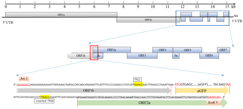
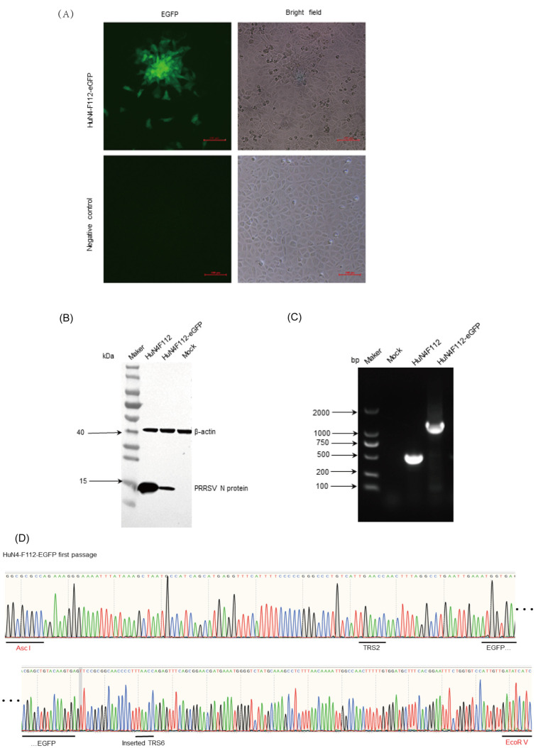
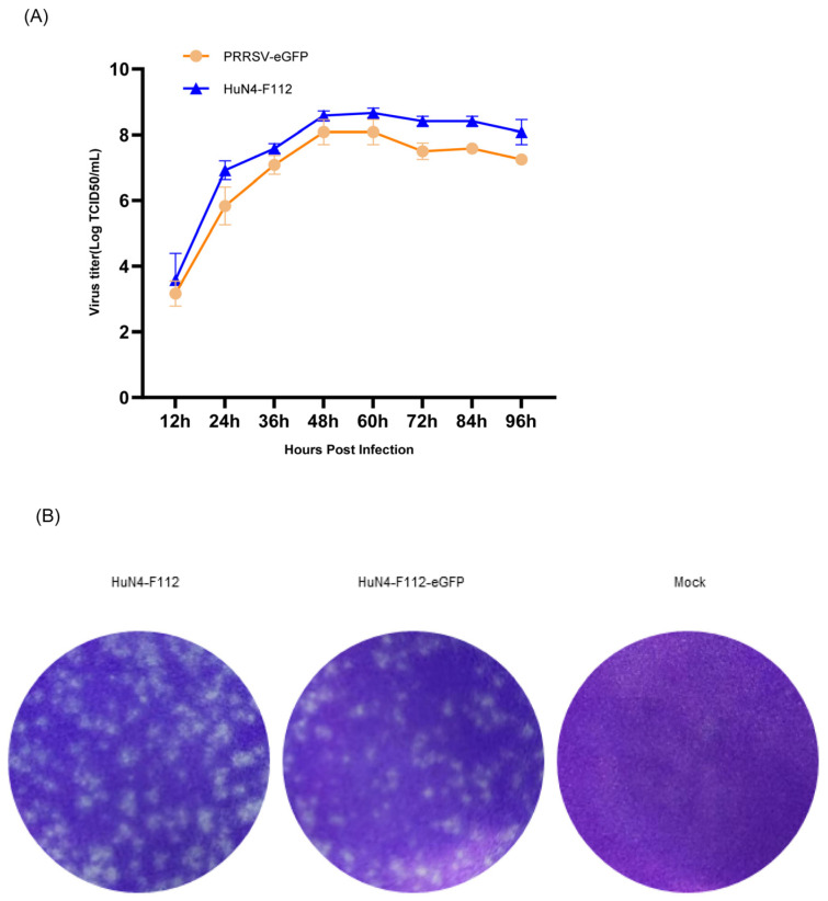
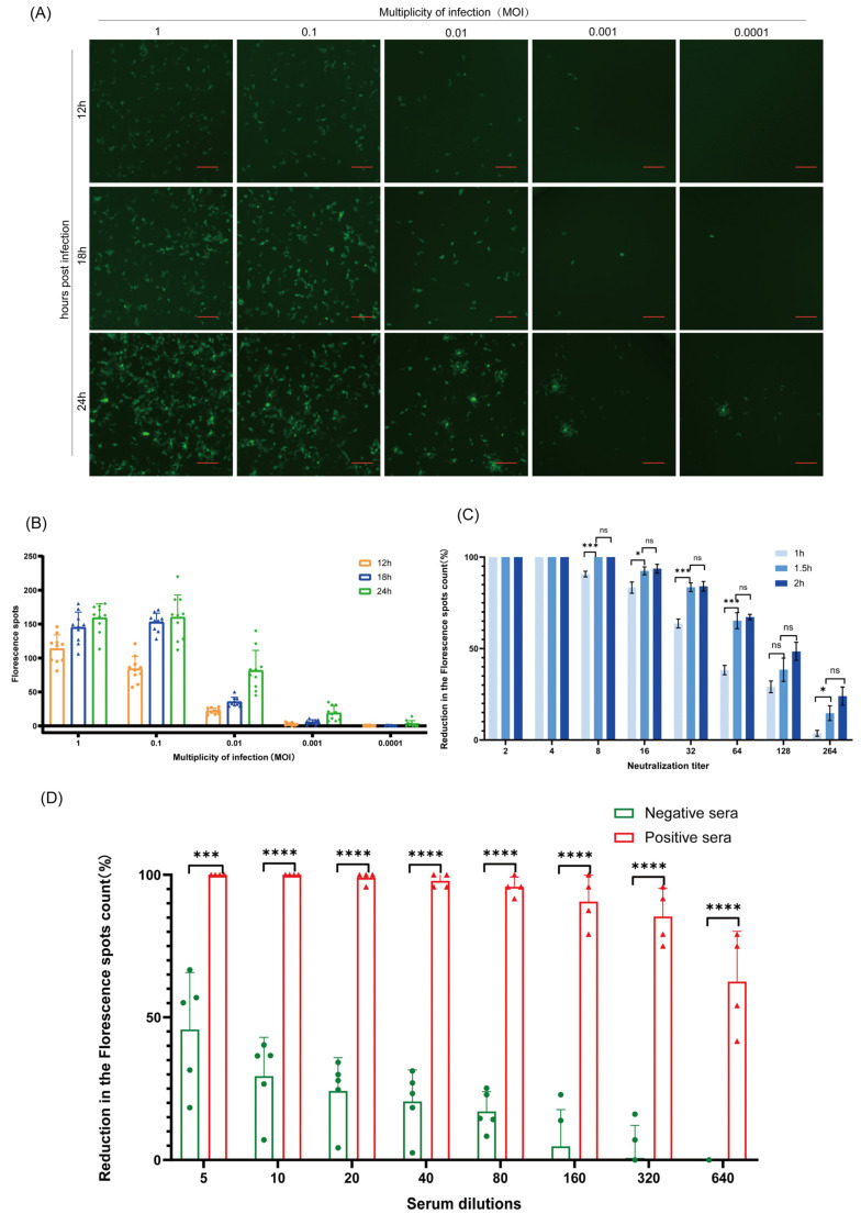
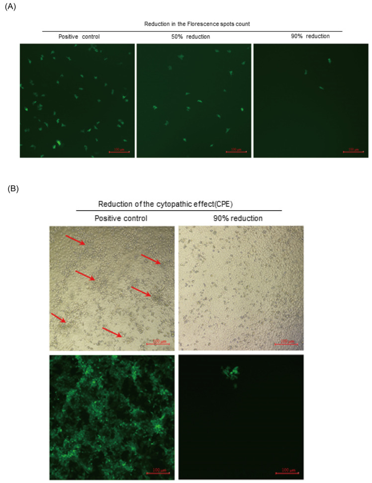
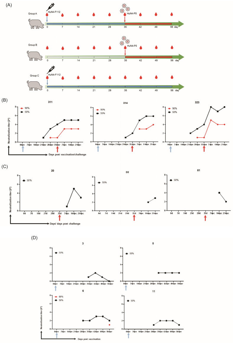
Due to the extensive genetic and antigenic variation in Porcine Reproductive and Respiratory Syndrome Virus (PRRSV), as well as its rapid mutability and evolution, PRRS prevention and control can be challenging. An expeditious and sensitive neutralization assay for PRRSV is presented to monitor neutralizing antibodies (NAbs) in serum during vaccine research. Here, a PRRSV expressing eGFP was successfully rescued with reverse genetics based on the infectious clone HuN4-F112-eGFP which we constructed. The fluorescent protein expressions of the reporter viruses remained stable for at least five passages. Based on this reporter virus, the neutralization assay can be easily used to evaluate the level of NAbs by counting cells with green fluorescence. Compared with the classical CPE assay, the newly developed assay increases sensitivity by one- to four-fold at the early antibody response stage, thus saving 2 days of assay waiting time. By using this assay to unveil the dynamics of neutralizing antibodies against PRRSV, priming immunity through either a single virulent challenge or only vaccination could produce limited NAbs, but re-infection with PRRSV would induce a faster and stronger NAb response. Overall, the novel HuN4-F112-eGFP-based neutralization assay holds the potential to provide a highly efficient platform for evaluating the next generation of PRRS vaccines.

Keywords: PRRSV; enhanced GFP; infectious clones; neutralization assay; neutralizing antibodies; reporter virus.


Conflict of interest statement

The authors declare no conflicts of interest. The funders had no role in the design of the study; in the collection, analyses, or interpretation of data; in the writing of the manuscript; or in the decision to publish the results.

Figures

Figure 1
Figure 1
Schematic diagram of the HuN4-F112 infectious clone with the eGFP gene inserted between open reading frame 1b (ORF1b) and ORF2a. The eGFP gene was flanked by the Asc I and EcoR V sites for insertion. The red letters is DNA sequences of the two restriction sites (Asc I and EcoR V). After digestion with Asc I and EcoR V, the eGFP gene, fused with PRRSV TRS2, was inserted at the C terminus of the ORF1b gene in the HuN4-F112 infectious clone. The recombinant PRRSV full-length cDNA clone harboring eGFP was named HuN4-F112-eGFP.
Figure 2
Figure 2
Recovery and identification of the recombinant virus HuN4-F112-eGFP. (A) The cytopathic effect (CPE), characterized by cellular rounding and clumping, was observed 4 days post-transfection. At the same position, the fluorescence expression was monitored under an inverted fluorescence microscope. Scale bar: 100 µm. (B) MARC-145 cells were analyzed using Western blotting to detect PRRSV N proteins at 24 h post-infection. (C) PCR confirmation of recombinant plasmids HuN4-F112-eGFP with F1 and R1. The HuN4-F112-eGFP and parental HuN4-F112 were, respectively, used as the cDNA to amplify the insert fragments. (D) Sequencing result of the eGFP gene in HuN4-F112-eGFP for the first passage. The underlinings marked the DNA sequences of the special fragments in the sequencing result. The Asc I and EcoR V were the restriction sites.
Figure 3
Figure 3
Biological characteristics of HuN4-F112-eGFP. (A) Comparison of virus multistep growth curves of HuN4-F112-eGFP and parental HuN4-F112. MARC-145 cells were infected with the recombinant viruses (P5) and the parental viruses at an MOI of 0.01. Virus supernatants, collected at 12 h intervals from 0 to 96 hpi, were measured using the TCID50 assay. Each data point represents the mean ± deviation of duplicates. (B) Viral plaque morphology assays comparing HuN4-F112-eGFP to parental HuN4-F112. MARC-145 cells were infected with serial diluted recombinant viruses and covered with 1% low-melting-point agarose supplemented with 2% FBS. At 3 days post-infection, the plaques formed by virus infection were visualized using crystal violet staining.
Figure 4
Figure 4
Genetic stability of the HuN4-F112-eGFP reporter virus. (A) The expression of eGFP in the MARC-145 cells infected with each virus passage. Scale bars represent 200 μm. (B) RNA extracted from cells infected with the P1–P5 viruses was analyzed using RT-PCR with F1 and R1 primers. The corresponding regions in the parental HuN4-F112 viruses were amplified as a control. The PCR products underwent visualization through 1% agarose gel electrophoresis. (C) The titers of the P0–P5 viruses were measured using the TCID50 assay. The number of black dots on the bars represents the number of replicates per data. (D) The growth kinetics of HuN4-F112-eGFP at the second and fifth passages were determined with viral titer tests. Results are means ± SD (triplicate).
Figure 5
Figure 5
Development of a sensitive HuN4-F112-eGFP-based neutralization assay. (A) Fluorescent images of MARC-145 cells infected with HuN4-F112-eGFP over time. MARC-145 cells were cultured in 96-well plates and infected with HuN4-F112-eGFP at MOIs of 1, 0.1, 0.01, 0.001, and 0.0001. The cells were monitored for fluorescence expression under an inverted fluorescence microscope (Nikon, Japan) at 12, 18, and 24 h post-infection. Scale bars represent 200 μm. (B) GFP fluorescence dot count for images in (A). We counted the MARC-145 cells with GFP fluorescence in 10 wells for each viral infection dose at 12, 18, and 24 h post-infection using the counting method in Section 3.10. The number of dots on the bars represents the number of counting images. (C) Incubation time optimization. Each diluted serum was mixed with an equal volume of HuN4-F112-eGFP for periods of 1h, 1.5 h, and 2 h at 37 °C. The experiment was performed according to the procedure described in Section 3.7. The fluorescence inhibition ratio was compared for the different incubation times. ns p > 0.05, * p < 0.05, *** p < 0.001. (D) The effect of serum dilution on the neutralization assays. Four PRRSV-vaccinated pig sera and five negative pig sera were first pre-diluted, 1:5, in 0% DMEM, and then serially diluted two-fold with 2% DMEM. Then, virus neutralization assays were conducted, as previously described in Section 3.7. The fluorescence inhibition ratio was compared at different serum dilutions. *** p < 0.001, **** p < 0.0001. The number of dots on the bars represents the number of sera samples. All pig sera were heat-inactivated at 56 °C for 30 min. Error bars indicate the standard deviations.
Figure 6
Figure 6
A comparison of the HuN4-F112-eGFP-based neutralization assay with the traditional CPE reduction assay. (A) Fluorescent images were captured to record the number of cells with green fluorescence in the field of view at 18 hpi using the HuN4-F112-eGFP-based neutralization assay. Scale bars represent 100 μm. (B) Images were captured to record the cytopathic effect (CPE) at 72 hpi using the CPE-based neutralization assay under bright and GFP fields. The red arrows sign the appearance of CPEs. Scale bars represent 100 μm.
Figure 7
Figure 7
HP-PRRSV neutralizing antibody response patterns detected using a GFP neutralization assay. (A) All pig sera were divided into three groups. Group A included sera from three pigs that were injected with the HuN4-F112 virus on day 0 and challenged with HuN4-P5 at 35 days post-vaccination. Group B included sera from three pigs that were only challenged with HuN4-P5 on the 35th day. Group C consisted of sera from four pigs that were injected with the HuN4-F112 virus on day 0. (BD) The neutralization titers of serum samples from each piglet in Group A (B), Group B (C), and Group C (D), collected at 0, 7, 14, 21, 28, 35, 42, 49, and 56 days post-vaccination. Immunized piglets showed faster and stronger NAb responses after a virulent challenge. Blue arrows suggest that pigs were injected with the HuN4-F112 virus on 0 dpv, and red arrows suggest that pigs were challenged with HuN4-P5 on 35 dpv.

Similar articles

References

    1. Neumann E.J., Kliebenstein J.B., Johnson C.D., Mabry J.W., Bush E.J., Seitzinger A.H., Green A.L., Zimmerman J.J. Assessment of the economic impact of porcine reproductive and respiratory syndrome on swine production in the United States. J. Am. Vet. Med. Assoc. 2005;227:385–392. doi: 10.2460/javma.2005.227.385. - DOI - PubMed
    1. Mengeling W.L., Lager K.M., Vorwald A.C. Clinical consequences of exposing pregnant gilts to strains of porcine reproductive and respiratory syndrome (PRRS) virus isolated from field cases of “atypical” PRRS. Am. J. Vet. Res. 1998;59:1540–1544. - PubMed
    1. Wensvoort G., Terpstra C., Pol J.M., ter Laak E.A., Bloemraad M., de Kluyver E.P., Kragten C., van Buiten L., den Besten A., Wagenaar F., et al. Mystery swine disease in The Netherlands: The isolation of Lelystad virus. Vet. Q. 1991;13:121–130. doi: 10.1080/01652176.1991.9694296. - DOI - PubMed
    1. Allende R., Lewis T.L., Lu Z., Rock D.L., Kutish G.F., Ali A., Doster A.R., Osorio F.A. North American and European porcine reproductive and respiratory syndrome viruses differ in non-structural protein coding regions. Pt 2J. Gen. Virol. 1999;80:307–315. doi: 10.1099/0022-1317-80-2-307. - DOI - PubMed
    1. Nelsen C.J., Murtaugh M.P., Faaberg K.S. Porcine reproductive and respiratory syndrome virus comparison: Divergent evolution on two continents. J. Virol. 1999;73:270–280. doi: 10.1128/JVI.73.1.270-280.1999. - DOI - PMC - PubMed