. 2024 Oct;61(10):7466-7480.
doi: 10.1007/s12035-024-04059-x. Epub 2024 Feb 23.

The Long Non-Coding RNA NR3C2-8:1 Promotes p53-Mediated Apoptosis through the miR-129-5p/USP10 Axis in Amyotrophic Lateral Sclerosis

Affiliations

The Long Non-Coding RNA NR3C2-8:1 Promotes p53-Mediated Apoptosis through the miR-129-5p/USP10 Axis in Amyotrophic Lateral Sclerosis

Dejiang Pang et al. Mol Neurobiol. 2024 Oct.

Abstract

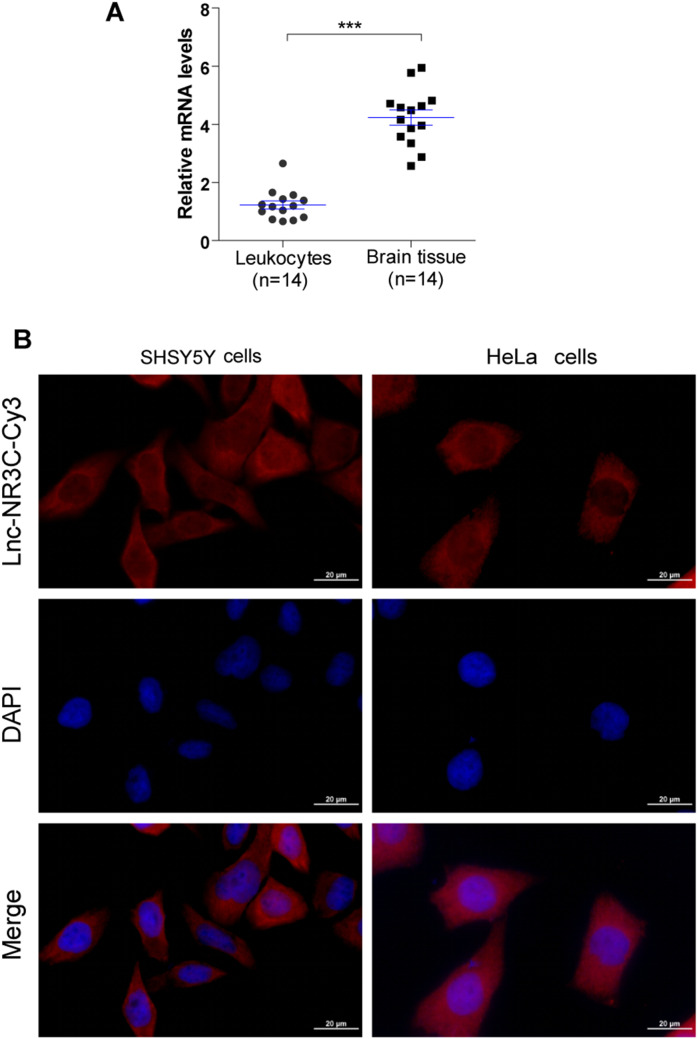
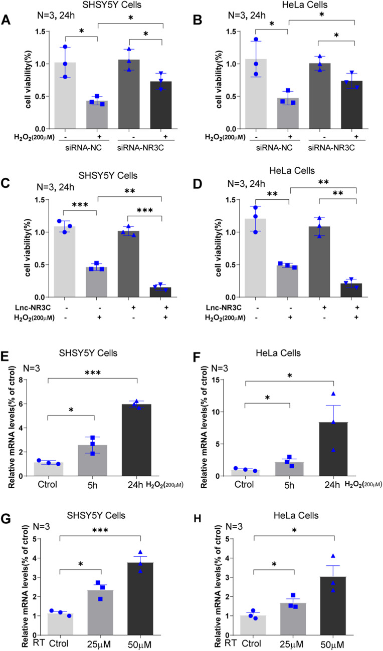
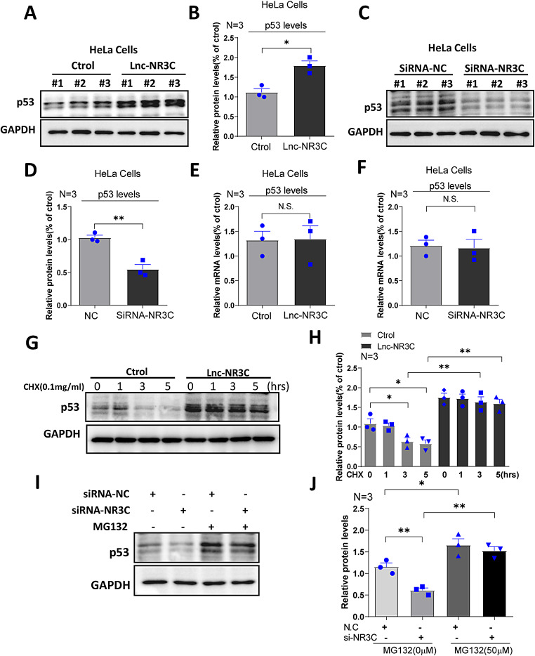
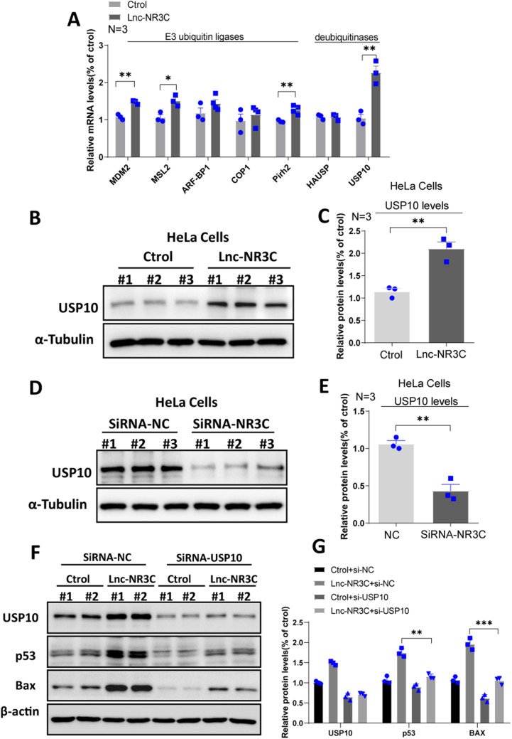
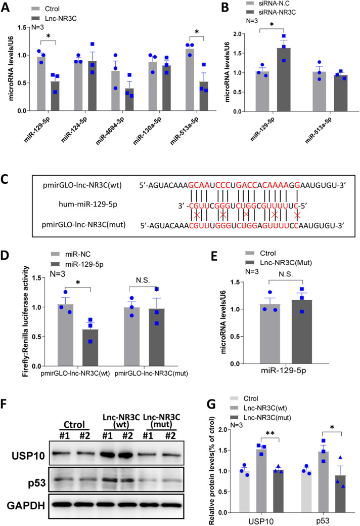
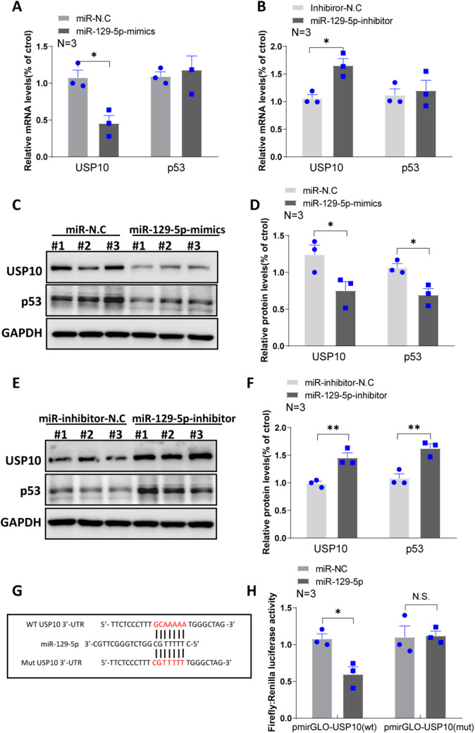
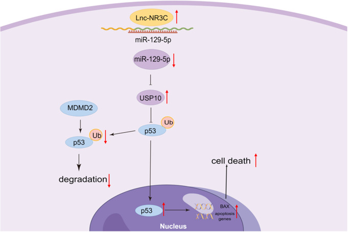
Motor neuron degeneration in amyotrophic lateral sclerosis (ALS) is a form of apoptosis, but the mechanisms underlying this neuronal cell death remain unclear. Numerous studies demonstrate abnormally elevated and active p53 in the central nervous system of ALS patients. Activation of p53-regulated pro-apoptotic signaling pathways may trigger motor neuron death. We previously reported decreased expression of the long non-coding RNA NR3C2-8:1 (Lnc-NR3C) in leukocytes of ALS patients. Here, we show lnc-NR3C promotes p53-mediated cell death in ALS by upregulating USP10 and promoting lnc-NR3C-triggered p53 activation, resulting in cell death. Conversely, lnc-NR3C knockdown inhibited USP10-triggered p53 activation, thereby protecting cells against oxidative stress. As a competitive endogenous RNA, lnc-NR3C competitively binds miR-129-5p, regulating the usp10/p53 axis. Elucidating the link between Lnc-NR3C and the USP10/p53 axis in an ALS cell model reveals a role for long non-coding RNAs in activating apoptosis. This provides new therapeutic opportunities in ALS.


Conflict of interest statement

The authors confirm that there are no conflicts of interest.

Figures

Fig. 1
Fig. 1
LncRNA-NR3C is expressed in the CNS and mainly localized in the cell cytoplasm. (A). lncRNA-NR3C expression in human peripheral blood leukocytes and brain tissues (peritumor tissue of glioma patients) was analysed via qRT–PCR. (***p < 0.001). (B). Localization of lncRNA-NR3C by RNA-FISH in SHSY5Y and HeLa cells. Nuclei are stained blue (DAPI), and lncRNA-NR3C is stained red. Scale bars represent 20 μm
Fig. 2
Fig. 2
LncRNA-NR3C is involved in cell survival and induced by oxidative stress. (A-B). The cell viability of SHSY5Y cells (A) and HeLa cells (B) with lnc-NR3C knockdown were examined under H2O2 (200µM,24 h) treatment by the CCK-8 assay.(*p < 0.05). (C-D).The cell viability of SHSY5Y cells (C) and HeLa cells (D) with lnc-NR3C overexpression were examined under H2O2 (200µM,24 h) treatment by the CCK-8 assay. (**p < 0.01,***p < 0.001). (E-F).Lnc-NR3C is induced by oxidative stress evoked by H2O2 (200µM,5 and 24 h) in SHSY5Y (E) and HeLa cells (F). (*p < 0.05,***p < 0.001). (G-H).Lnc-NR3C is induced by oxidative stress evoked by rotenone (RT,25µM and 50µM,24 h) in SHSY5Y (G) and HeLa cells (H). (*p < 0.05,***p < 0.001)
Fig. 3
Fig. 3
Lnc-NR3C promotes apoptosis. (A-B) After overexpressing lnc-NR3C cell apoptosis were assessed by Hochest/PI staining in the presence of H2O2(500µM,6 h).Cells were costained with Hoechst (blue), and PI (red), and visualized by fluorescence microscope (A).The total cell death is statistically analysed(B). (*p < 0.05,**p < 0.01, N.S. no statistical significance). (C-F)Western blot analysis of apoptosis-related proteins Bcl-2, Bax and Cleaved -PARP1 in human HeLa(C-D) and SH-SY5Y cells(E-F) with overexpressing lnc-NR3C.Quantification and statistical analysis of three independent experiments in HeLa(D) and SHSY5Y cells(F).(*p < 0.05,**p < 0.01,***p < 0.001)
Fig. 4
Fig. 4
Overexpression of lnc-NR3C increases p53 protein stability. (A-B) Western blot analysis and quantification statistical analysis of the p53 proteins in human HeLa cells with overexpressing lnc-NR3C. (*p < 0.05). (C-D) Western blot analysis and quantification statistical analysis of the p53 proteins in human HeLa cells with knockdown lnc-NR3C. (**p < 0.01). (E-F) qRT–PCR analysis of the p53 in human HeLa cells with overexpressing and knockdown lnc-NR3C. (N.S. no statistical significance. (G-H) Western blot analysis and quantification statistical analysis of the p53 proteins in human HeLa cells treating with cycloheximide (CHX,0.1 mg/ml,1 h,3 and 5 h) after overexpressing lnc-NR3C. (*p < 0.05,**p < 0.01). (I-J) Western blot analysis and quantification statistical analysis of the p53 proteins in human HeLa cells treating with MG132 (50 µM,5 h) after knockdown lnc-NR3C. (*p < 0.05, **p < 0.01) (The concentration of MG132 was incorrectly labelled, and we should correct it to 50 µM in the figure and legends of data J. See the attachment, named Fig. 4)
Fig. 5
Fig. 5
Overexpression of lnc-NR3C increases USP10 protein level. (A) qRT–PCR analysis of E3 ubiquitin ligases and deubiquitinases,which have also been shown to regulate p53 stability, in human HeLa cells with overexpressing lnc-NR3C.(*p < 0.05, **p < 0.01). (B-C)Western blot analysis and quantification statistical analysis of the USP10 proteins in human HeLa cells with overexpressing lnc-NR3C. (**p < 0.01). (D-E)Western blot analysis and quantification statistical analysis of the USP10 proteins in human HeLa cells with knockdown lnc-NR3C. (**p < 0.01). (F-G)Western blot analysis and quantification statistical analysis of the p53 and Bax in human HeLa cells with knockdown USP10 after overexpressing lnc-NR3C. ( **p < 0.01,***p < 0.001)
Fig. 6
Fig. 6
Overexpression of lnc-NR3C increases p53 protein stability. (A) qRT–PCR analysis of potential target miRNAs for lnc-NR3C in human HeLa cells with overexpressing lnc-NR3C. (*p < 0.05). (B) qRT–PCR analysis of potential target miRNAs for lnc-NR3C in human HeLa cells with knockdown lnc-NR3C. (*p < 0.05). (C) Schematic representation of the predicted binding sites for miR-129-5p in lnc-NR3C. (D) Luciferase activity in 293T cells cotransfected with miR-129-5p and luciferase reporters that contained lnc-NR3C (left) or the indicated mutant transcript (right). (*p < 0.05, N.S. no statistical significance). (E) qRT–PCR analysis of miR-129-5p in human HeLa cells with overexpressing mutated lnc-NR3C.(N.S. no statistical significance). (F-G) Western blot analysis and quantification statistical analysis of the USP10 and p53 in human HeLa cells with overexpressing lnc-NR3C and mutated lnc-NR3C. (*p < 0.05,**p < 0.01)
Fig. 7
Fig. 7
miR-129-5p inhibits the expression of USP10. (A-B) qRT–PCR analysis of the USP10 and p53 in human HeLa cells with overexpressing and inhibiting miR-129-5p. (*p < 0.05). (C-D) Western blot analysis and quantification statistical analysis of the USP10 and p53 in human HeLa cells with overexpressing miR-129-5p. (*p < 0.05). (E-F) Western blot analysis and quantification statistical analysis of the USP10 and p53 in human HeLa cells with inhibiting miR-129-5p. (*p < 0.05,**p < 0.01). (G) Schematic representation of the predicted binding sites for miR-129-5p in USP10 3’-UTR. (H) Luciferase activity in 293T cells cotransfected with miR-129-5p and luciferase reporters that contained USP10 3’-UTR (left) or the indicated mutant transcript (right). (*p < 0.05, N.S. no statistical significance)
Fig. 8
Fig. 8
Model. The long non-coding RNA NR3C2-8:1(lnc-NR3C) promotes p53-mediated apoptosis through the miR-129-5p/USP10 Axis

Similar articles

Cited by

References

    1. Feldman EL et al (2022) Amyotrophic lateral sclerosis. Lancet 400(10360):1363–1380 - PMC - PubMed
    1. Goutman SA et al (2022) Recent advances in the diagnosis and prognosis of amyotrophic lateral sclerosis. Lancet Neurol 21(5):480–493 - PMC - PubMed
    1. Mead RJ et al (2023) Amyotrophic lateral sclerosis: a neurodegenerative disorder poised for successful therapeutic translation. Nat Rev Drug Discov 22(3):185–212 - PMC - PubMed
    1. Sathasivam S, Shaw PJ (2005) Apoptosis in amyotrophic lateral sclerosis–what is the evidence? Lancet Neurol 4(8):500–509 - PubMed
    1. Taylor JP, Brown RH Jr., Cleveland DW (2016) Decoding ALS: from genes to mechanism. Nature 539(7628):197–206 - PMC - PubMed

MeSH terms