. 2024 Nov 15;18(6):1014-1025.
doi: 10.5009/gnl230304. Epub 2024 Feb 22.

Tumor-Derived Exosomal Circular RNA Pinin Induces FGF13 Expression to Promote Colorectal Cancer Progression through miR-1225-5p

Affiliations

Tumor-Derived Exosomal Circular RNA Pinin Induces FGF13 Expression to Promote Colorectal Cancer Progression through miR-1225-5p

Xianghui Liao et al. Gut Liver. .

Abstract

Background/aims: : Colorectal cancer (CRC) is a common malignant tumor, and circular RNAs (circRNAs) are abnormally expressed in CRC. However, the function and underlying mechanism of circRNA pinin (circ-PNN; hsa_circ_0101802) in CRC remain unclear.

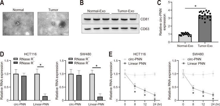
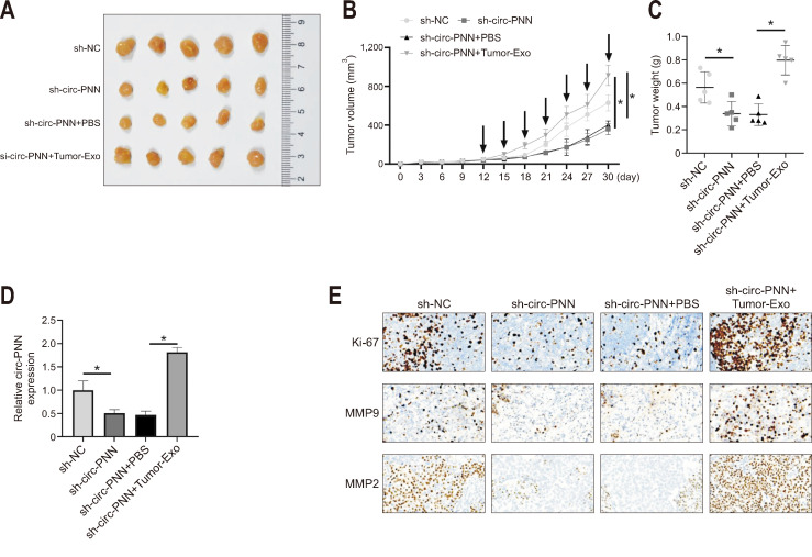
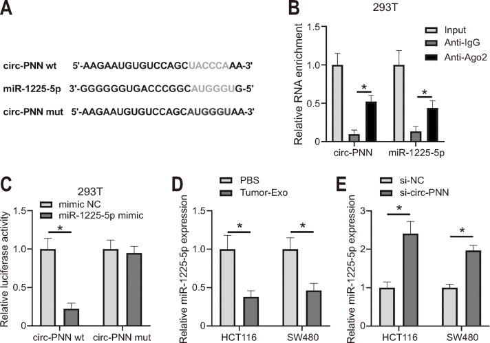
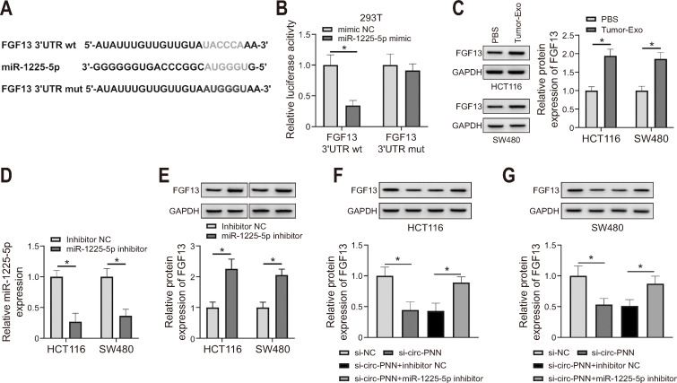
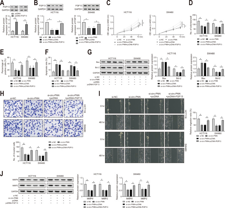
Methods: : Exosomes were isolated from the plasma of CRC patients and identified by transmission electron microscopy and Western blotting. The RNA expression levels of circ-PNN, miR-1225-5p, and fibroblast growth factor 13 (FGF13) were measured by quantitative real-time polymerase chain reaction. Cell proliferation was detected by Cell Counting K-8, colony formation, and 5-ethynyl-2'-deoxyuridine assays. Cell apoptosis was assessed by flow cytometry. The expression of apoptosis and metastasis-related proteins was evaluated by Western blotting. The associations among circ-PNN, miR-1225-5p, and FGF13 were confirmed by dual-luciferase report assay and RNA immunoprecipitation assay. A xenograft model was used to verify the function of circ-PNN in tumor formation in vivo.

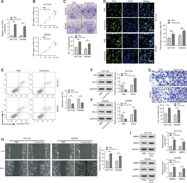
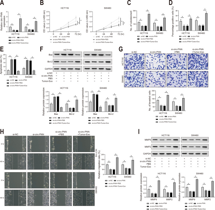
Results: : circ-PNN expression was upregulated in plasmic exosomes derived from CRC patients. The expression of circ-PNN and FGF13 was upregulated, while miR-1225-5p expression was downregulated in CRC cells incubated with plasmic exosomes derived from CRC patients. Tumor-derived exosomes promoted the proliferation, migration, and invasion but inhibited apoptosis of CRC cells. Moreover, the addition of tumor-derived exosomes partly reversed the inhibitory effect of circ-PNN knockdown on CRC tumor progression in vitro and in vivo. Thus, circ-PNN acts as a sponge for miR-1225-5p to regulate FGF13 expression.

Conclusions: : Tumor-derived exosomal circ-PNN promoted CRC progression through the regulation of the miR-1225-5p/FGF13 pathway, providing a potential therapeutic target for CRC.

Keywords: Circular RNA pinin; Colorectal neoplasms; Exosomes; Fibroblast growth factor 13; miR-1225-5p.


Conflict of interest statement

CONFLICTS OF INTEREST

No potential conflict of interest relevant to this article was reported.

Figures

Fig. 1
Fig. 1
Characterization of exosomes (Exos) derived from the plasma of colorectal cancer patients. (A) Transmission electron microscopy image of normal-Exos and tumor-Exos. (B) The protein levels of CD81 and CD63 were measured by Western blotting. (C) The expression of circ-PNN in normal-Exos and tumor-Exos was measured by qRT-PCR. (D) After RNase R treatment, the levels of circ-PNN and its linear transcript PNN were examined by qRT-PCR. (E) For actinomycin D assay, the levels of circ-PNN and its linear transcript PNN were measured by qRT-PCR. circ-PNN, circular RNA pinin; qRT-PCR, quantitative real-time polymerase chain reaction. *p<0.05.
Fig. 2
Fig. 2
Tumor-derived exosomes (Exos) enhance proliferation, invasion and migration, but promote apoptosis in colorectal cancer cells. Colorectal cancer cells were treated with tumor-Exos or PBS. (A) The level of circ-PNN was assessed by qRT-PCR. (B-D) Cell proliferation was examined by Cell Counting K-8, colony formation and EdU assays. (E) Cell apoptosis were monitored by flow cytometry. (F) The protein levels of Bax and Bcl-2 were detected by Western blotting. (G) Cell invasion was determined by transwell assay. (H) Cell migration was monitored by wound healing assay. (I) The protein levels of MMP9 and MMP2 were assessed by Western blotting. PBS, phosphate-buffered saline; circ-PNN, circular RNA pinin; qRT-PCR, quantitative real-time polymerase chain reaction; OD, optical density. *p<0.05.
Fig. 3
Fig. 3
Exosomal circ-PNN accelerates colorectal cancer progression in vitro. Colorectal cancer cells were transfected with si-NC or si-circ-PNN, and treated with tumor-Exos or PBS. (A) The level of circ-PNN was tested by qRT-PCR. (B-D) Cell proliferation was examined by Cell Counting K-8, colony formation and EdU assays. (E) Cell apoptosis was monitored by flow cytometry. (F) The protein levels of Bax and Bcl-2 were detected by Western blotting. (G) Cell invasion was determined by transwell assay. (H) Cell migration was monitored by wound healing assay. (I) The protein levels of MMP9 and MMP2 were assessed by Western blot analysis. circ-PNN, circular RNA pinin; si-NC, small hairpin RNA of negative control; si-circ-PNN, small hairpin RNA of circ-PNN; Exo, exosome; PBS, phosphate-buffered saline; qRT-PCR, quantitative real-time polymerase chain reaction. *p<0.05.
Fig. 4
Fig. 4
Exosomal circ-PNN regulates tumor growth in vivo. (A, B) The average tumor volume of xenograft tumor tissues was calculated based on measurements with a Vernier caliper. (C) Tumor weight of xenograft tumor tissues was tested. (D) The expression of circ-PNN was measured by qRT-PCR. (E) The expression levels of Ki-67, MMP9 and MMP2 in xenograft tumor tissues were detected by immunohistochemical. sh-NC, small hairpin RNA of negative control; circ-PNN, circular RNA pinin; sh-circ-PNN, small hairpin RNA of circ-PNN; Exo, exosome; qRT-PCR, quantitative real-time polymerase chain reaction; PBS, phosphate-buffered saline. *p<0.05.
Fig. 5
Fig. 5
circ-PNN interacts with miR-1225-5p. (A) The predicted binding sites between circ-PNN and miR-1225-5p are shown. (B, C) The correction between circ-PNN and miR-1225-5p was confirmed by RIP assay and Dual-Luciferase Reporter Assay. (D) The expression of miR-1225-5p was measured in colorectal cancer cells incubated with PBS or tumor exosome. (E) The expression of miR-1225-5p was measured in colorectal cancer cells with si-NC or si-circ-PNN transfection. circ-PNN, circular RNA pinin; PBS, phosphate-buffered saline; si-NC, small hairpin RNA of negative control; si-circ-PNN, small hairpin RNA of circ-PNN; NC, negative control; wt, wild type; mut, mutant. *p<0.05.
Fig. 6
Fig. 6
circ-PNN regulates FGF13 expression by modulating miR-1225-5p. (A) The predicted binding sites between miR-1225-5p and FGF13 were exhibited. (B) The correction between miR-1225-5p and FGF13 was confirmed by dual-luciferase report activity. (C) The level of FGF13 protein was measured in colorectal cancer (CRC) cells incubated with PBS or tumor exosome. (D) The expression of miR-1225-5p was measured by qRT-PCR in CRC cells with inhibitor NC or miR-1225-5p inhibitor transfection. (E) The expression of FGF13 in CRC cells with inhibitor NC or miR-1225-5p inhibitor transfection was measured. (F, G) The protein level of FGF13 in CRC cells with si-NC, si-circ-PNN, si-circ-PNN+inhibitor NC, or si-circ-PNN miR-1225-5p inhibitor transfection was detected by Western blot analysis. wt, wild type; mut, mutant; circ-PNN, circular RNA pinin; FGF13, fibroblast growth factor 13; PBS, phosphate-buffered saline; qRT-PCR, quantitative real-time polymerase chain reaction; Exo, exosome; NC, negative control; si-NC, small hairpin RNA of NC; si-circ-PNN, small hairpin RNA of circ-PNN. *p<0.05.
Fig. 7
Fig. 7
circ-PNN promotes colorectal cancer (CRC) proliferation, migration, and invasion by increasing FGF13 levels. (A) The protein level of FGF13 was tested by Western blot in CRC cells with pcDNA or pcDNA-FGF13 transfection. (B-J) CRC cells were transfected with si-NC, si-circ-PNN, si-circ-PNN+pcDNA or si-circ-PNN+pcDNA-FGF13, respectively. (B) The protein level of FGF13 in transfected cells was detected by Western blot analysis. (C-E) Cell proliferation was examined by Cell Counting K-8, colony formation and EdU assays. (F) Cell apoptosis was detected by flow cytometry. (G) The protein levels of Bax and Bcl-2 in transfected cells were detected by Western blotting. (H) Cells invasion was determined by transwell assay. (I) A wound healing assay was conducted to monitor cell migration. (J) The protein levels of MMP9 and MMP2 were assessed by Western blotting. circ-PNN, circular RNA pinin; FGF13, fibroblast growth factor 13; si-NC, small hairpin RNA of negative control; si-circ-PNN, small hairpin RNA of circ-PNN; pcDNA, pcDNA 3.1 (+) vector; OD, optical density. *p<0.05.

Similar articles

Cited by

References

    1. Kuipers EJ, Grady WM, Lieberman D, et al. Colorectal cancer. Nat Rev Dis Primers. 2015;1:15065. doi: 10.1038/nrdp.2015.65. - DOI - PMC - PubMed
    1. Brody H. Colorectal cancer. Nature. 2015;521:S1. doi: 10.1038/521S1a. - DOI - PubMed
    1. Goldstein DA, Zeichner SB, Bartnik CM, Neustadter E, Flowers CR. Metastatic colorectal cancer: a systematic review of the value of current therapies. Clin Colorectal Cancer. 2016;15:1–6. doi: 10.1016/j.clcc.2015.10.002. - DOI - PMC - PubMed
    1. Siegel RL, Miller KD, Goding Sauer A, et al. Colorectal cancer statistics, 2020. CA Cancer J Clin. 2020;70:145–164. doi: 10.3322/caac.21601. - DOI - PubMed
    1. Sun Z, Shi K, Yang S, et al. Effect of exosomal miRNA on cancer biology and clinical applications. Mol Cancer. 2018;17:147. doi: 10.1186/s12943-018-0897-7.ba9535a85a7f462385c41170a656b2bc - DOI - PMC - PubMed