. 2024 Mar 13;15(3):e0033524.
doi: 10.1128/mbio.00335-24. Epub 2024 Feb 21.

Diversity, origin, and evolution of the ESCRT systems

Affiliations

Diversity, origin, and evolution of the ESCRT systems

Kira S Makarova et al. mBio. .

Abstract

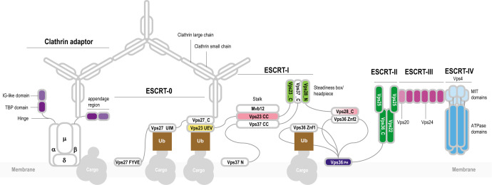
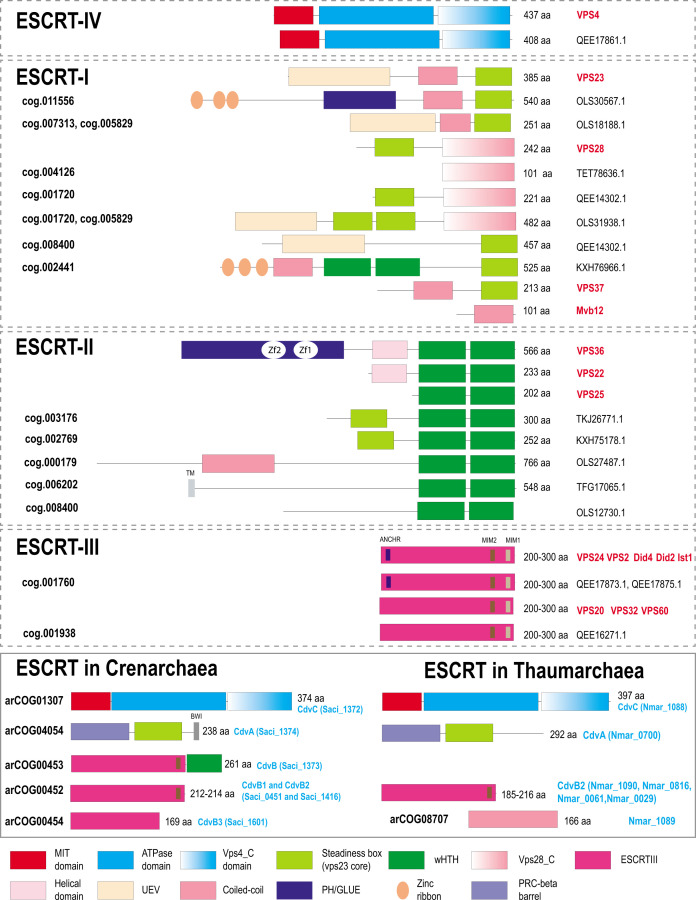
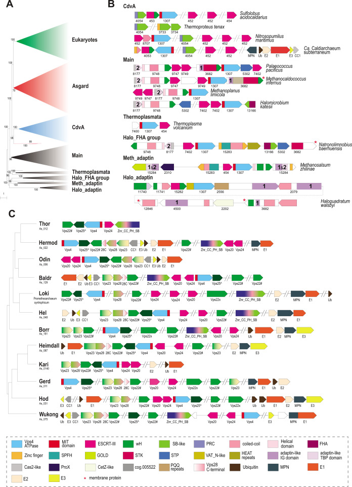
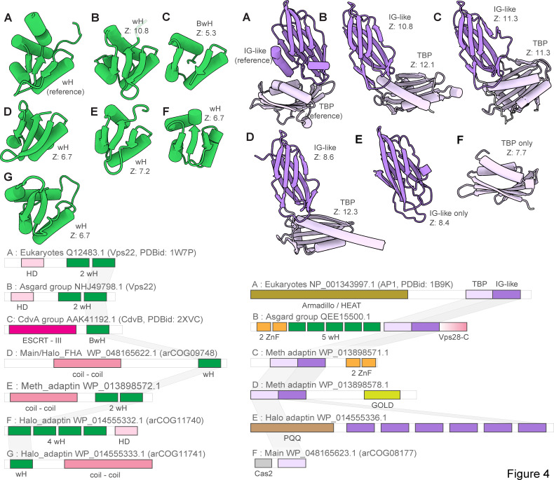
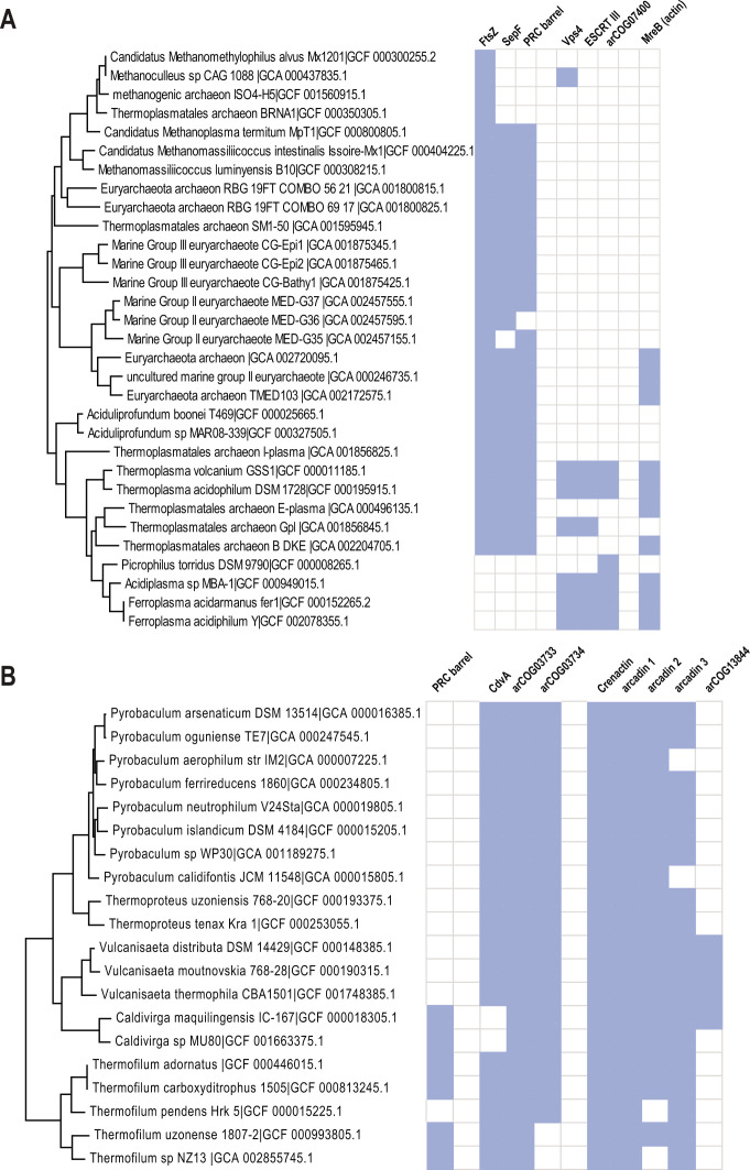
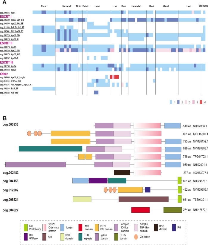
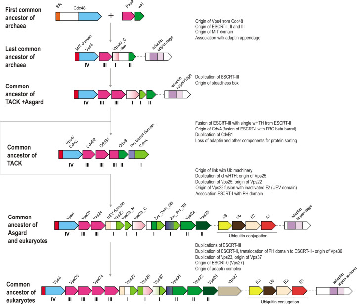
Endosomal sorting complexes required for transport (ESCRT) play key roles in protein sorting between membrane-bounded compartments of eukaryotic cells. Homologs of many ESCRT components are identifiable in various groups of archaea, especially in Asgardarchaeota, the archaeal phylum that is currently considered to include the closest relatives of eukaryotes, but not in bacteria. We performed a comprehensive search for ESCRT protein homologs in archaea and reconstructed ESCRT evolution using the phylogenetic tree of Vps4 ATPase (ESCRT IV) as a scaffold and using sensitive protein sequence analysis and comparison of structural models to identify previously unknown ESCRT proteins. Several distinct groups of ESCRT systems in archaea outside of Asgard were identified, including proteins structurally similar to ESCRT-I and ESCRT-II, and several other domains involved in protein sorting in eukaryotes, suggesting an early origin of these components. Additionally, distant homologs of CdvA proteins were identified in Thermoproteales which are likely components of the uncharacterized cell division system in these archaea. We propose an evolutionary scenario for the origin of eukaryotic and Asgard ESCRT complexes from ancestral building blocks, namely, the Vps4 ATPase, ESCRT-III components, wH (winged helix-turn-helix fold) and possibly also coiled-coil, and Vps28-like domains. The last archaeal common ancestor likely encompassed a complex ESCRT system that was involved in protein sorting. Subsequent evolution involved either simplification, as in the TACK superphylum, where ESCRT was co-opted for cell division, or complexification as in Asgardarchaeota. In Asgardarchaeota, the connection between ESCRT and the ubiquitin system that was previously considered a eukaryotic signature was already established.IMPORTANCEAll eukaryotic cells possess complex intracellular membrane organization. Endosomal sorting complexes required for transport (ESCRT) play a central role in membrane remodeling which is essential for cellular functionality in eukaryotes. Recently, it has been shown that Asgard archaea, the archaeal phylum that includes the closest known relatives of eukaryotes, encode homologs of many components of the ESCRT systems. We employed protein sequence and structure comparisons to reconstruct the evolution of ESCRT systems in archaea and identified several previously unknown homologs of ESCRT subunits, some of which can be predicted to participate in cell division. The results of this reconstruction indicate that the last archaeal common ancestor already encoded a complex ESCRT system that was involved in protein sorting. In Asgard archaea, ESCRT systems evolved toward greater complexity, and in particular, the connection between ESCRT and the ubiquitin system that was previously considered a eukaryotic signature was established.

Keywords: Asgard archaea; ESCRT; cell division; membrane remodeling.


Conflict of interest statement

The authors declare no conflict of interest.

Figures

Fig 1
Fig 1
General organization of the eukaryotic ESCRT system and its connection with other protein sorting complexes. The ESCRT machinery was thoroughly characterized in yeast and mammals (17, 19–21). The core of the system consists of four conserved, multisubunit complexes: ESCRT-I, ESCRT-II, ESCRT-III, and ESCRT-IV. ESCRT-0 subunits are not generally conserved and apparently differ among the eukaryotic phyla. ESCRT-0 subunits interact with protein cargo and recruit ESCRT-I complex. The best characterized subunit of ESCRT-0 is Vps27 which is a hub of protein-protein interactions (19). Vps27/HRS is a multidomain protein containing a VHS (Vps27/HRS/STAM) super-helix domain, an FYVE zinc finger, several coiled-coil regions, and a C-terminal P[ST]xP motif. Vps27 interacts with the ubiquitin E2 variant (UEV) domain of Vps23 (ESCRT-I) via the P[ST]xP motif and also with clathrin, ubiquitinated cargo, and lipids (19, 23, 24). ESCRT-I consists of four subunits: Vps28, Vps23, Vps37, and Mvb12 (Fig. 2). Three subunits, Vps23, Vps37, and Mvb12, form a stalk through the interaction between three long helices forming a coiled-coil structure. Vps23, Vps28, and Vps37 form a headpiece through the interaction between homologous helical hairpins known as the steadiness box (SB) (25). The C-terminal helical bundle domain of Vps28 interacts with ESCRT-II Vps36 via a Zn finger (Znf2) inserted into the split PH domain. ESCRT-II also consists of four subunits: Vps22, Vps36, and two copies of Vps25 (26). These proteins are paralogs that share a common region consisting of a pair of wH domains. In addition to the wH domains, Vps36 is fused to a PH domain and two zinc finger domains (the latter are absent in mammalian orthologs). Vps22 and Vps36 contain an additional helical domain upstream of the proximal wH domain, which likely promotes their interaction (Fig. 2). The N-terminal domain of VPS25 binds the C-terminal domain of VPS22, and the other VPS25 subunit contacts both VPS36 and VPS22, forming an asymmetric Y-shaped structure (26). Vps25 recruits the initial Vps20 subunit of ESCRT-III complex (26). ESCRT-III consists of several paralogous subunits, which belong to two groups, Vps2-like [MIM-1 (MIT-interacting motif) containing] and Vps20-like (MIM-2 containing), respectively (16, 27) (Fig. 2). Vps2 is targeted to the membrane where it nucleates Vps20 polymerization (16). Depolymerization of ESCRT-III is regulated by the Vps4 (16). Vps4 contains a diagnostic N-terminal three-helix bundle, the microtubule interacting and trafficking (MIT) domain which interacts with the C-terminal MIM-2 in ESCRT-III subunits (28). The ESCRT machinery is required for endosomal trafficking and is connected with other complexes involved in this process including clathrin, a large complex consisting of light and heavy subunits forming a triskelion (29). In eukaryotes, clathrin forms a cage-like scaffold around a vesicle and recruits Vps27. Clathrin is associated with clathrin adaptor complexes (AP) which mediate clathrin-dependent protein trafficking to and from endosomes (30, 31). AP complexes differ minimally in subunit composition. AP2, for example, consists of four subunits: alpha, beta, gamma, and mu (30, 31). Alpha and beta subunits are paralogs that contain an N-terminal HEAT repeats domain and a C-terminal appendage domain connected through an unstructured hinge interacting with clathrin. The appendage domain consists of two distinct subdomains, the proximal immunoglobulin-like (IG) beta sandwich fold and the distal TBP (TATA-box binding protein or helix-grip) fold (32). Appendage domains of alpha and beta subunits can recruit multiple additional proteins, whereas the N-terminal domains of alpha and beta subunits and the mu subunit interact with the membrane protein cargo (33, 34).
Fig 2
Fig 2
Domain organization and key motifs of ESCRT protein in eukaryotes, Asgard, and TACK archaea. Protein domains are shown as colored shapes according to the color code given beneath the schemes. Proteins are drawn roughly proportional to their size in amino acids which is indicated in the right. Domain boundaries for Saccharomyces cerevisiae are based on previously published structures and analyses (19, 24, 35). For archaeal proteins, domain boundaries are based either on previous analyses (22, 36–38) or on sequence analysis performed during this work as described under Materials and Methods. S. cerevisiae gene names are highlighted in red on the right. For Asgard and TACK archaea, protein size and accession are indicated on the right. asCOG numbers (39) are indicated for the Asgard proteins, and arCOG numbers (40) are indicated for the TACK proteins on the left. Signature sequence motifs are shown by small rectangles, and short names for the motifs are provided above the protein schematics (once for proteins with the same domain organization). Abbreviations: Zf, zinc finger; TM, transmembrane segment; UEV, ubiquitin E2 variant; MIM, MIT interaction motif; BWI, broken winged-helix interaction site. Not all proteins from cog.003176 are fused to SB, but most contain additional N-terminal subdomains distinct from those in eukaryotic homologs.
Fig 3
Fig 3
Phylogeny of Vps4 family ATPase and genomic neighborhoods of ESCRT systems in archaea. (A) Schematic representation of the phylogenetic tree of Vps4 protein family. The phylogenetic tree was built using IQ-Tree as described in Material and Methods. Major branches were collapsed and are designated on the right. Bootstrap values >70% calculated by IQ-Tree are shown. The complete tree in Newick format is available in the File S1. (B) Organization of selected ESCRT loci for each major branch of archaea (except Asgard). Genes are shown as arrows roughly proportional to gene size. Domains are indicated within the arrows for multidomain proteins, not to scale. Homologous genes and domains (or generic structural elements such as coiled-coil or helical domains) are color coded according to the code at the bottom of the figure. ArCOG numbers are indicated below the arrows. Numbers inside the arrows indicate IG domain (1) and TBP domain (2) in the adaptin appendage homology regions. (C) Components of ESCRT systems in selected genomes of 12 major Asgard lineages. The ESCRT related genes are shown for selected genomes of the 12 major lineages of Asgardarchaeota (39). The tree schematically shows the relationships among the lineages according to previously published phylogenetic analysis (39). Designations are the same as in B, except for multidomain proteins in which domains are not shown, but are explained in the color code schematics in the bottom of the figure. Protein names are indicated below the arrows. Designations: Vps22#, Asgard specific version of Vps22, cog.002769; Vps25^, Asgard specific version of Vps25, cog.002441; Znr_CC_PH_SB, Asgard specific protein family of cog.011556 (see Fig. 2). Abbreviations: wH, winged helix domain; Znr, zinc ribbon; SB, steadiness box; FHA, forkhead-associated domain; STK, serine/threonine protein kinase; STP, serine/threonine protein phosphatase; UEV, ubiquitin E2 variant; Ub, ubiquitin; CC, coiled-coil; PH, plextrin homology; SB, steadiness box; MIT, microtubule interacting and transport; HEPN, higher eukaryotes and prokaryotes nucleotide-binding domain; GOLD, Golgi dynamics domain; SPFH, stomatin, prohibitin, flotillin, and HflK; VAT_N, N-terminal domain of valosin-containing protein-like ATPase of Thermoplasma acidophilum; TBP, TATA-binding protein. Abbreviations for repetitive domains: HEAT, Huntington, Elongation Factor 3, PR65/A, TOR; PQQ, pyrrolo-quinoline quinone; PEGA, containing PEGA sequence motif. Abbreviations for ubiquitination pathway: E1, ubiquitin-activating enzyme; E2, ubiquitin-conjugating enzyme; E3, ubiquitin ligases; MPN, Mpr1/Pad1 N-terminal domain, deubiquitinating enzyme.
Fig 4
Fig 4
Winged helix and adaptin appendage domain homologs in eukaryotic and archaeal ESCRT systems. Left, comparison of winged helix domains (wH). Right, adaptin appendage-like regions. Multidomain proteins were trimmed to show only domains homologous to wH and adaptin appendage-like domain of Vps22 and alpha-adaptin appendage domain. Complete proteins are shown in Fig. S2, S7, and S8. Complete domain organizations of the respective proteins are shown underneath the structures. Letters denote the correspondence between structures and domain architectures. DALI Z-scores between the indicated domains and wH domain of Vps22 or IG-like and TBP domain of alpha-adaptin appendage domain are indicated. Abbreviations: IG, immunoglobulin; wH, winged helix domain; BwH, broken winged helix domain; TBP, TATA binding protein.
Fig 5
Fig 5
Phyletic patterns for putative cell division components of Thermoproteales and Thermoplasmata. The presence/absence patterns of cell division components for (A) Thermoplasmata and (B) Thermoproteales are shown. The patterns are overlayed on the respective species subtree. The tree built for all 524 arCOGs genomes using IQ-tree program (54) and concatenated alignment of ribosomal proteins. Blue indicates presence, and white indicates absence.
Fig 6
Fig 6
Phyletic patterns of ESCRT components and unique multidomain proteins in Asgardarchaeota. (A) Number of proteins in ESCRT asCOGs for 78 Agard phylum genomes. Major Asgard lineages are indicated above. asCOG numbers and a short description or gene or protein name are indicated on the left; the data were retrieved from asCOGs (41) with minimal corrections. The numbers of proteins are color coded as shown in the bottom right. Potentially ancestral (most widespread) asCOG are highlighted in green. (B) Domain architectures of Asgard proteins containing one or more domain related to ESCRT system. asCOG numbers are indicated on the left. Protein sizes and accession are indicated on the right. Homologous domains are shown by the same shape and color according to the color code provided in the bottom. Abbreviations are the same as in Fig. 3. Additional abbreviations: BAR, BIN, amphiphysin and Rvs161 and Rvs167 (yeast proteins) domain; TPR, tetratricopeptide repeats.
Fig 7
Fig 7
Origin and evolution of ESCRT systems. The inferred, hypothetical organizations of ESCRT systems in ancestral archaeal groups indicated on the left are shown. The inferred key events are described on the right. Designations of the genes and domains are the same as in Fig. 3. The Roman numerals under the genes correspond to (putative) components of the four ESCRT complexes. Abbreviations are the same as in Fig. 3, except for SR, substrate recognition domain.

Update of

Similar articles

Cited by

References

    1. Szathmáry E. 2015. Toward major evolutionary transitions theory 2.0. Proc Natl Acad Sci U S A 112:10104–10111. doi:10.1073/pnas.1421398112 - DOI - PMC - PubMed
    1. Koonin EV, Mulkidjanian AY. 2013. Evolution of cell division: from shear mechanics to complex molecular machineries. Cell 152:942–944. doi:10.1016/j.cell.2013.02.008 - DOI - PubMed
    1. Adams DW, Errington J. 2009. Bacterial cell division: assembly, maintenance and disassembly of the Z ring. Nat Rev Microbiol 7:642–653. doi:10.1038/nrmicro2198 - DOI - PubMed
    1. Caspi Y, Dekker C. 2018. Dividing the archaeal way: the ancient Cdv cell-division machinery. Front Microbiol 9:174. doi:10.3389/fmicb.2018.00174 - DOI - PMC - PubMed
    1. den Blaauwen T, Hamoen LW, Levin PA. 2017. The divisome at 25: the road ahead. Curr Opin Microbiol 36:85–94. doi:10.1016/j.mib.2017.01.007 - DOI - PMC - PubMed

MeSH terms

Substances

LinkOut - more resources