. 2024 Mar 13;15(3):e0282123.
doi: 10.1128/mbio.02821-23. Epub 2024 Feb 20.

Listeria adhesion protein orchestrates caveolae-mediated apical junctional remodeling of epithelial barrier for Listeria monocytogenes translocation

Affiliations

Listeria adhesion protein orchestrates caveolae-mediated apical junctional remodeling of epithelial barrier for Listeria monocytogenes translocation

Rishi Drolia et al. mBio. .

Abstract

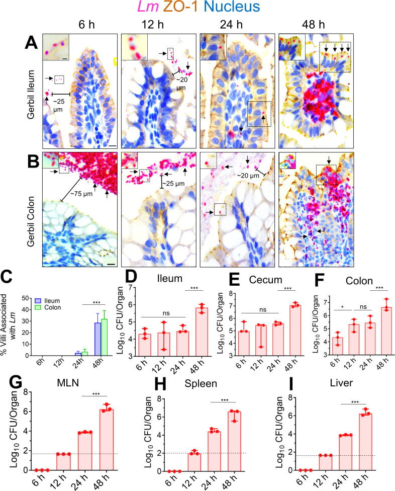
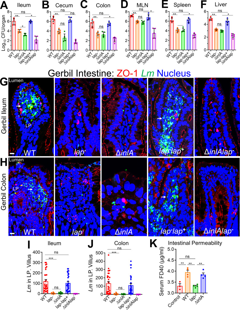
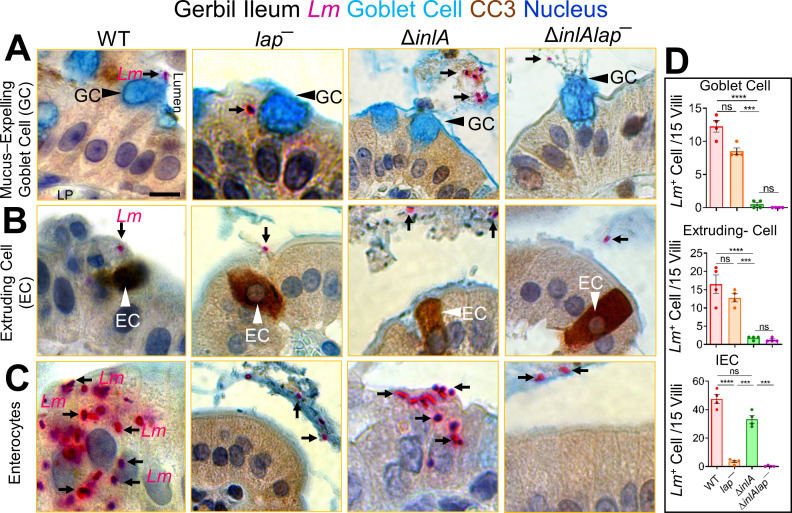
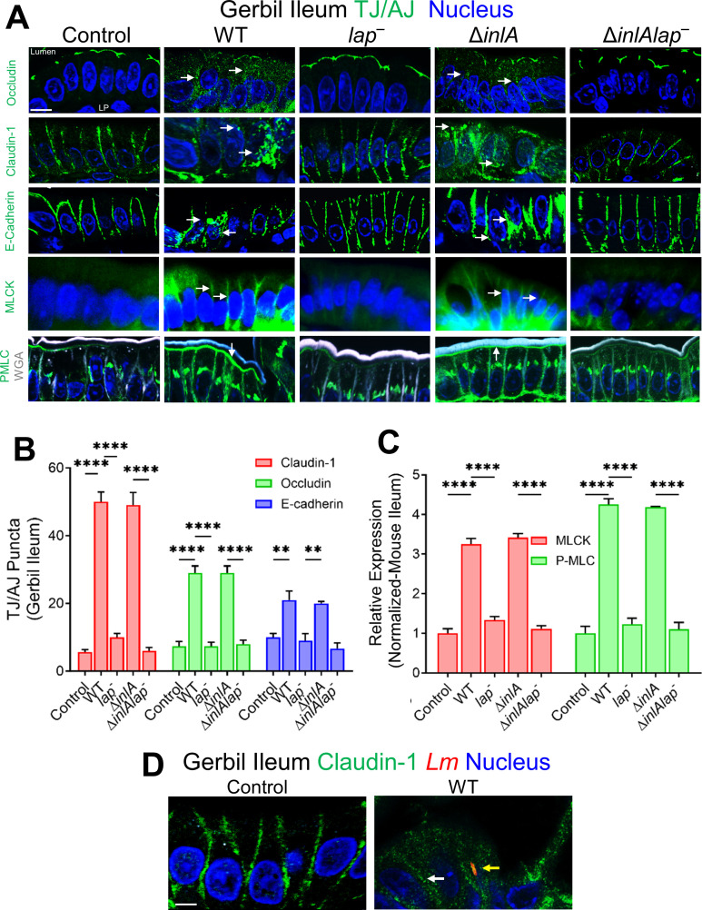
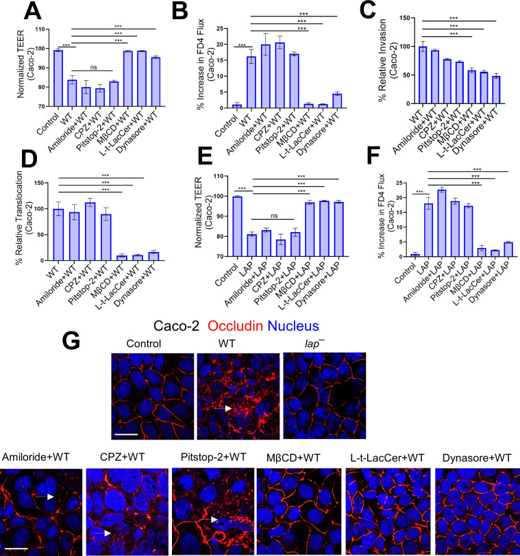
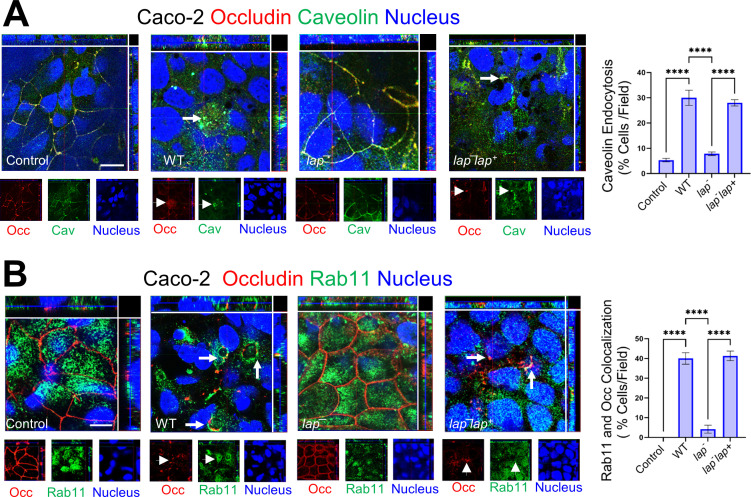
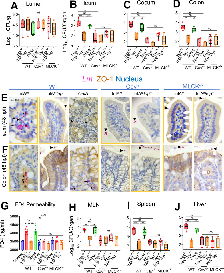
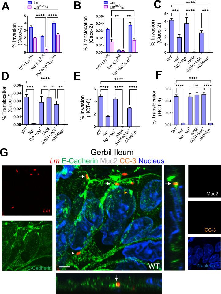
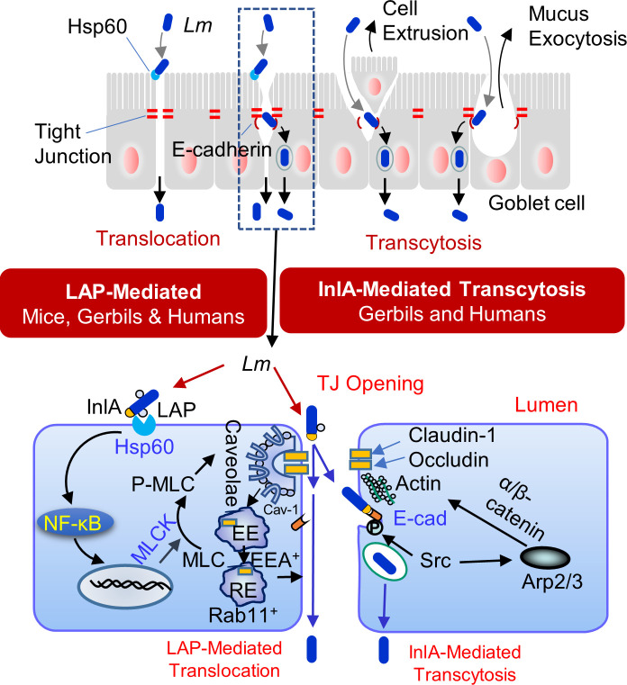
The cellular junctional architecture remodeling by Listeria adhesion protein-heat shock protein 60 (LAP-Hsp60) interaction for Listeria monocytogenes (Lm) passage through the epithelial barrier is incompletely understood. Here, using the gerbil model, permissive to internalin (Inl) A/B-mediated pathways like in humans, we demonstrate that Lm crosses the intestinal villi at 48 h post-infection. In contrast, the single isogenic (lap- or ΔinlA) or double (lap-ΔinlA) mutant strains show significant defects. LAP promotes Lm translocation via endocytosis of cell-cell junctional complex in enterocytes that do not display luminal E-cadherin. In comparison, InlA facilitates Lm translocation at cells displaying apical E-cadherin during cell extrusion and mucus expulsion from goblet cells. LAP hijacks caveolar endocytosis to traffic integral junctional proteins to the early and recycling endosomes. Pharmacological inhibition in a cell line and genetic knockout of caveolin-1 in mice prevents LAP-induced intestinal permeability, junctional endocytosis, and Lm translocation. Furthermore, LAP-Hsp60-dependent tight junction remodeling is also necessary for InlA access to E-cadherin for Lm intestinal barrier crossing in InlA-permissive hosts.

Importance: Listeria monocytogenes (Lm) is a foodborne pathogen with high mortality (20%-30%) and hospitalization rates (94%), particularly affecting vulnerable groups such as pregnant women, fetuses, newborns, seniors, and immunocompromised individuals. Invasive listeriosis involves Lm's internalin (InlA) protein binding to E-cadherin to breach the intestinal barrier. However, non-functional InlA variants have been identified in Lm isolates, suggesting InlA-independent pathways for translocation. Our study reveals that Listeria adhesion protein (LAP) and InlA cooperatively assist Lm entry into the gut lamina propria in a gerbil model, mimicking human listeriosis in early infection stages. LAP triggers caveolin-1-mediated endocytosis of critical junctional proteins, transporting them to early and recycling endosomes, facilitating Lm passage through enterocytes. Furthermore, LAP-Hsp60-mediated junctional protein endocytosis precedes InlA's interaction with basolateral E-cadherin, emphasizing LAP and InlA's cooperation in enhancing Lm intestinal translocation. This understanding is vital in combating the severe consequences of Lm infection, including sepsis, meningitis, encephalitis, and brain abscess.

Keywords: E-cadherin; Listeria adhesion protein (LAP); Listeria monocytogenes (Lm); caveolin; dynamin; gerbil; heat shock protein 60 (Hsp60); internalin A (InlA); intestinal barrier; tight junction (TJ); translocation.


Conflict of interest statement

A patent on LAP use as a tight junction modulator has been issued.

Figures

Fig 1
Fig 1
Kinetic analysis of the intestinal invasion of L. monocytogenes in orally infected gerbils.(A, B) Representative micrographs of ileal (A) or colonic villi (B) dual immunostained for ZO-1 (tight junction, brown) and Listeria (red, arrows) and counterstained with hematoxylin to stain the nucleus (blue) from gerbil orally challenged with ~3 × 10⁸ CFU of WT clinical strain (F4244, serovar 4b, CC6) at 6, 12, 24, and 48 hpi; bars, 10 µm. The boxed areas were enlarged; bars, 1 µm. Translocated Lm is observed in the lamina propria (arrows) at 48 hpi but confined in the lumen (arrows) at 6–24 hpi. (C) The graph representing quantitative measurements of infected ileal or colonic villi (%) ± SEM. Percentage of infected villi from 100 villi from a single gerbil, three gerbils per group, n = 300 villi. (D–I) Listeria counts (total CFU) in the intracellular location in the ileum (D) cecum (E) and colon (F); gentamicin-resistant CFU, and the mesenteric-lymph node (MLN) (G), spleen (H), and liver (I) at 6–48 hpi from two to three independent experiments. Dashed horizontal lines indicate the detection limit. Data (C–I) represent mean ± SEM of n = 3 gerbils per treatment from three independent experiments. Each point represents a single gerbil. ***P < 0.001; **P < 0.01; *P < 0.05; ns, no significance.
Fig 2
Fig 2
LAP and InlA promote L. monocytogenes translocation across the InlA-permissive gerbil intestinal barrier. (A–F) Female gerbils were orally challenged with ~3 × 10⁸ CFU of WT, lap ΔinlA, lap lap+, or the ΔinlA lap L. monocytogenes bacteria. The plots show the total CFU obtained from the ileum (intracellular) (A), cecum (intracellular) (B), colon (intracellular) (C), MLN (D), spleen (E), and liver (F) of gerbils (n = 4–6) at 48 hpi from three independent experiments. The bar and brackets represent the mean ± SEM for each group’s data points. All error bars represent mean ± SEM. **P < 0.01; *P < 0.05; ns, no significance. (G and H) Representative confocal immunofluorescence microscopic images of ileal (G) or colonic (H) tissue sections immunostained for ZO-1 (red), Listeria (green), and 4′,6-diamidino-2-phenylindole (blue; nucleus) from WT, lap ΔinlA, lap lap+, or the ΔinlA lap L. monocytogenes bacteria-challenged gerbils at 48 hpi, bars, 10 µm. Increased L. monocytogenes (green) was detected in the lamina propria of ileal (G) or colonic (H) tissue in WT and laplap+-challenged gerbils (arrows). (I and J) Graph representing quantitative measurements of L. monocytogenes counts in the lamina propria from ileal (I) or colonic (J) villi images (n = 30 villi) from three gerbils for each treatment. (K) Analysis of fluorescein isothiocyanate (FITC)-labeled 40 kDa dextran (FD40) permeability through the intestinal epithelium of uninfected (control) and L. monocytogenes-infected gerbils in serum at 48 hpi. FD40 was administered orally 4–5 h before sacrifice. Data represent mean ± SEM of n = 3–4 gerbils per treatment from two independent experiments. All error bars represent mean ± SEM. ***P < 0.001; **P < 0.01; *P < 0.05; ns, no significance.
Fig 3
Fig 3
L. monocytogenes translocation across absorptive enterocytes with luminally inaccessible E-cadherin is LAP-dependent. (A–C) Representative picto-micrographs of ileal tissue sections dual immunostained for Lm (pink) and cleaved caspase-3 (brown; extruding cells), and Alcian blue stained for delineating goblet cell (blue) from L.monocytogenes WT, lap, or ΔinlA-infected gerbils at 48 hpi. Lm (black arrows) association at and translocation across goblet cell (A, black arrowhead) and extruding cells in different phages of extrusion (B, white arrowhead) is InlA-dependent as Δinla stain shows no association at these sites. Lm (black arrows) association at and translocation across IECs (C) with luminally inaccessible E-cadherin is LAP-dependent as lap strain shows no association at these sites. The ΔinlAlap strain shows negligible Lm association at all the cellular sites. (D) Graph representing quantitative measurements of L. monocytogenes-infected cells (in each cell type or at each site) of villi images (n = 60 villi). Each point represents a single gerbil. All error bars represent SEM. ****P < 0.0001; ***P < 0.001; ns, no significance.
Fig 4
Fig 4
LAP promotes Lm-mediated junctional mislocalization and activates MLCK in gerbils. (A) Confocal immunofluorescence micrographs of the ileal tissue sections showing mislocalization (intracellular puncta, endocytosis) of claudin-1, occludin, and E-cadherin (green; arrows), and increased expression of MLCK and P-MLC (green; arrows) in WT, and ΔinlA-infected gerbils but intact localization of occludin, claudin-1, and E-cadherin and baseline expression of MLCK and P-MLC and in lap or the ΔinlAlap-challenged gerbils (arrows). Images are representative of five different fields from n = 3–4 gerbil per treatment. Scale bars, 10 µm. LP, lamina propria. (B and C) Quantitative analysis (mean ± SEM, n = 3 gerbil) of claudin-1, occludin, and E-cadherin puncta formation (B) and MLCK and P-MLC expression (C) from images of immunostained ileal tissues orally challenged with ~3 × 10⁸ CFU of WT, lap, ΔinlA, or the ΔinlAlap Lm 48 hpi. (D) Representative confocal immunofluorescence picto-micrographs of the gerbil ileal tissue sections immunostained for claudin-1 (green), Listeria (red; yellow arrow), and DAPI (blue; nucleus) from uninfected control or WT challenged gerbil at 48 hpi. Bars, 5 µm. Lm (yellow arrow) cells are seen in lamina propria in WT with mislocalization of claudin-1 (intracellular puncta, endocytosis with WT). Images are representative of five different fields from three gerbils. LP, lamina propria.
Fig 5
Fig 5
Inhibition of caveolin in cells prevents LAP-induced junctional endocytosis, barrier permeability, and Lm translocation. (A and B) TEER measurement of Caco-2 cell monolayer in transwell filter-insert treated pretreated with endocytic pathway inhibitors before Lm exposure (multiplicity of infection [MOI]; 50, 2 h) (A) and on the apical (AP)-to-basolateral (BL) flux of paracellular marker 4 kDa FITC-dextran (FD4) permeability (B). (C and D) Decreased Lm invasion (C) and translocation (D) at MOI of 50 through polarized Caco-2 cell monolayers following pre-treatment with methyl-β-cyclodextrin (MβCD) (10 µM, 30 min), Lt-Lacer (10 µM, 30 min), dynasore (10 µM, 30 min). (E and F) TEER measurement of Caco-2 cell monolayer in transwell filter-insert treated pretreated with endocytic pathway inhibitors before purified LAP exposure (2 µg/mL, 2 h) (E) and on the AP-to-BL flux of paracellular marker 4 kDa FITC-dextran (FD4) permeability (F). (G) Representative confocal immunofluorescence micrographs showing intact localization of occludin in Caco-2 cells pretreated with MβCD (10 µM, 30 min), Lt-Lacer (10 µM, 30 min), dynasore (10 µM, 30 min) before Lm exposure (MOI; 50, 45 min). Arrows depict the internalization of occludin.
Fig 6
Fig 6
Lm LAP reorganizes caveolin for junctional internalization and delivers it in early/recycling endosomal compartments. (A and B) Representative confocal immunofluorescence micrographs showing colocalization of internalized occludin in Caco-2 following Lm WT and laplap+ but not lap exposure (MOI; 50, 45 min) with caveolin (A, arrows) and Rab11 (B, arrows). Separated channels are shown individually at the bottom of the merged images for clarity. Quantitative analysis (mean ± SEM, n = 6) of caveolin (A) and Rab11 (B) colocalization with endocytosed occludin from images of immunostained Caco-2 cells infected before or after exposure with Lm (MOI; 50, 45 min) are presented in the right panels.
Fig 7
Fig 7
Listeria monocytogenes translocation and intestinal epithelial permeability are affected in caveolin-1 knockout mice. (A–D) Male and female wild-type C57BL/6 (Cav+/+, MLCK+/+) or the caveolin-1−/− knockout mice (Cav−/−) or the 210 kDa MLCK knockout mice (MLCK-/-) mice were orally gavaged with 5 × 10⁸ CFU of InlAm, InlAmlap, or ΔinlALm. The box plot shows the total CFU obtained from the lumen (A), ileum (intracellular) (B), cecum (intracellular) (C), and colon (intracellular) (D) of mice (n = 4–5) at 48 hpi from three independent experiments. The bar and brackets represent the median ± range for each group’s data points. Bar and brackets represent the median ± range. (E and F) Representative microscopic images of ileal (E) or colonic (F) tissue sections immunostained for ZO-1 (brown) Listeria (pink) from Lm-challenged mice at 48 hpi, bars, 10 µm. Arrows denote increased Lm detected in the lamina propria of ileal or colonic tissue in wild-type C57BL/6 mice challenged with InlAm or Δinla. Arrowheads denote bacteria restricted to the intestinal lumen. (G) Analysis of paracellular permeability of 4 kDa FITC-dextran (FD4) through the intestinal epithelium of uninfected (cont) or Lm-infected, WT (MLCK+/+ Cav+/+), the Cav−/−, or the MLCK−/− mice in serum at 48 hpi. FD4 was administered 4–5 h before sacrifice. Data represent mean ± SEM of three to six mice per treatment from three independent experiments. (H-J) The box plots show the total CFU obtained from the MLN (H), spleen (I), and liver (J) (n = 4–6) at 48 hpi of Lm–infected, WT (MLCK+/+ Cav+/+), the Cav−/−, or the MLCK−/− mice. Bar and brackets represent the median ± range, respectively, for the data points in each group. ***, P < 0.001; **, P < 0.01; *, P < 0.5; ns, no significance.
Fig 8
Fig 8
LAP-mediated endocytosis of epithelial junctions provides easier InlA access to the basolateral E-cadherin for Lm translocation. (A and B) Analysis of LinInlA invasion of Caco-2 and HCT-8 cells co-infected with a 1:1 (MOI; 50) mixture of LinInlA and Lm WT or LinInlA and lap. (C–F) Analysis of invasion (C and D) and translocation (transwell filter-inserts) (E and F) of Lm WT and isogenic strains in polarized Caco-2 and HCT-8 cell monolayers infected at an MOI of 50. Data represent mean ± SEM from three independent experiments, n = 6. (G) Representative confocal immunofluorescence picto-micrographs of the gerbil ileal tissue sections immunostained for E-cadherin (green), Listeria (red; arrows), Muc-2 (white, goblet cell), cleaved caspase-3 (orange), and DAPI (blue; nucleus) from WT-challenged gerbil at 48 hpi. Bars, 5 µm. Lm (arrows, yellow in merged images) was observed attached to the enterocytes in the absence of extruding or goblet cells and exiting into lamina propria in WT with co-localized E-cadherin at the adherens junctions. Separated channels are shown individually to the left or right of the merged images. The X-Z and Y-Z cross-sections were produced by orthogonal reconstructions from the z-stack scanning. Images are representative of five different fields from three gerbils. LP, lamina propria. ***, P < 0.001; **, P < 0.01; *, P < 0.5; ns, no significance.
Fig 9
Fig 9
Schematic showing the role of LAP and InlA in Listeria monocytogenes translocation across the InlA-permissive gut intestinal epithelial barrier. LAP-mediated L. monocytogenes translocation involves the interaction of LAP with epithelial Hsp60 for caveolin-1- and MLCK-mediated endocytosis of the tight junctions (claudin-1 and occludin) and the adherens junctions (E-cadherin), which is delivered in the EEA+ and Rab11+ early and late endosomes and subsequent epithelial barrier opening. The LAP-mediated apical junctional opening is also critical for providing InlA access to E-cadherin at the adherens junction. InlA/E-cadherin-mediated L. monocytogenes transcytosis occurs during epithelial cell extrusion or goblet cell mucus exocytosis.

Similar articles

Cited by

References

    1. Radoshevich L, Cossart P. 2018. Listeria monocytogenes: towards a complete picture of its physiology and pathogenesis. Nat Rev Microbiol 16:32–46. doi:10.1038/nrmicro.2017.126 - DOI - PubMed
    1. Pägelow D, Chhatbar C, Beineke A, Liu X, Nerlich A, van Vorst K, Rohde M, Kalinke U, Förster R, Halle S, Valentin-Weigand P, Hornef MW, Fulde M. 2018. The olfactory epithelium as a port of entry in neonatal neurolisteriosis. Nat Commun 9:4269. doi:10.1038/s41467-018-06668-2 - DOI - PMC - PubMed
    1. Drolia R, Bhunia AK. 2019. Crossing the intestinal barrier via Listeria adhesion protein and internalin A. Trends Microbiol 27:408–425. doi:10.1016/j.tim.2018.12.007 - DOI - PubMed
    1. Guttman JA, Finlay BB. 2009. Tight Junctions as targets of infectious agents. Biochim Biophys Acta 1788:832–841. doi:10.1016/j.bbamem.2008.10.028 - DOI - PubMed
    1. Zuo L, Kuo W-T, Turner JR. 2020. Tight Junctions as targets and effectors of mucosal immune homeostasis. Cell Mol Gastroenterol Hepatol 10:327–340. doi:10.1016/j.jcmgh.2020.04.001 - DOI - PMC - PubMed