. 2024 Feb 16;15(1):46.
doi: 10.1186/s13287-024-03659-7.

Gingival-derived mesenchymal stem cell therapy regenerated the radiated salivary glands: functional and histological evidence in murine model

Affiliations

Gingival-derived mesenchymal stem cell therapy regenerated the radiated salivary glands: functional and histological evidence in murine model

Hagar M Zayed et al. Stem Cell Res Ther. .

Abstract

Background: Radiotherapy in head and neck cancer management causes degeneration of the salivary glands (SG). This study was designed to determine the potential of gingival mesenchymal stem cells (GMSCs) as a cell-based therapy to regenerate irradiated parotid SG tissues and restore their function using a murine model.

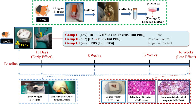
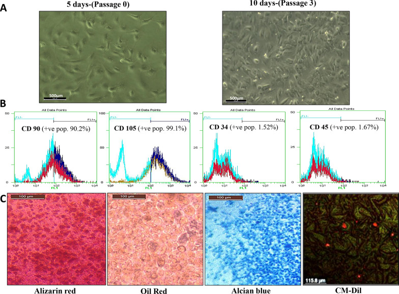
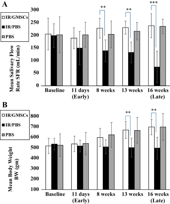
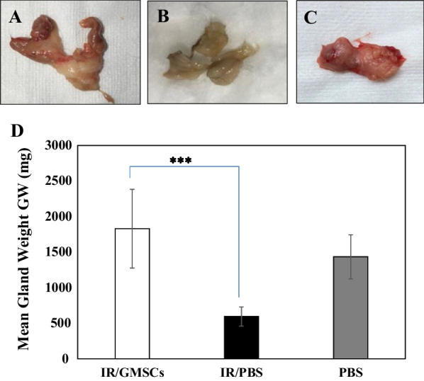
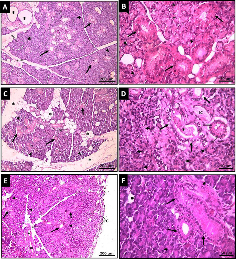
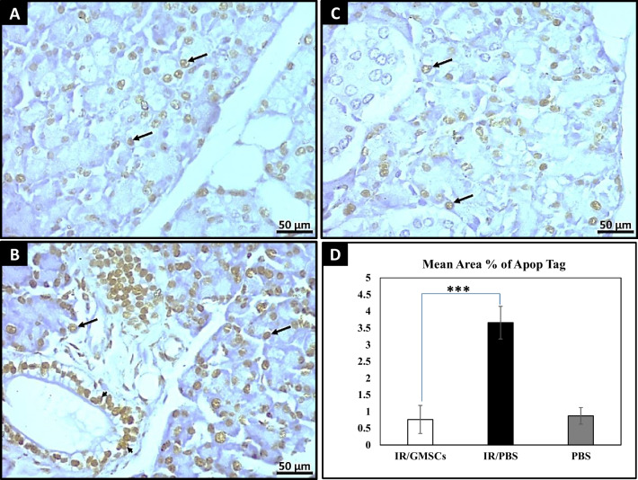
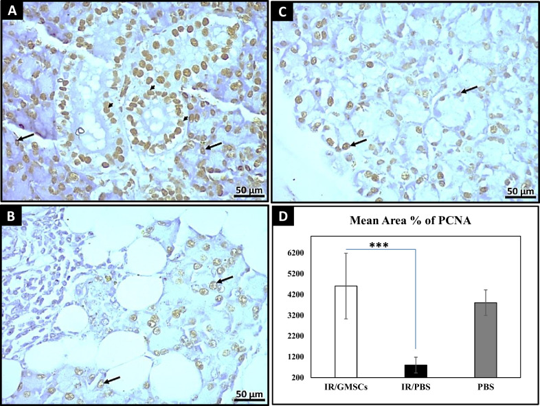
Methods: Cultured isolated cells from gingival tissues of 4 healthy guinea pigs at passage 3 were characterized as GMSCSs using flow cytometry for surface markers and multilineage differentiation capacity. Twenty-one Guinea pigs were equally divided into three groups: Group I/Test, received single local irradiation of 15 Gy to the head and neck field followed by intravenous injection of labeled GMSCs, Group II/Positive control, which received the same irradiation dose followed by injection of phosphate buffer solution (PBS), and Group III/Negative control, received (PBS) injection only. Body weight and salivary flow rate (SFR) were measured at baseline, 11 days, 8-, 13- and 16-weeks post-irradiation. At 16 weeks, parotid glands were harvested for assessment of gland weight and histological and immunohistochemical analysis.

Results: The injected GMSCs homed to degenerated glands, with subsequent restoration of the normal gland histological acinar and tubular structure associated with a significant increase in cell proliferation and reduction in apoptotic activity. Subsequently, a significant increase in body weight and SFR, as well as an increase in gland weight at 16 weeks in comparison with the irradiated non-treated group were observed.

Conclusion: The study provided a new potential therapeutic strategy for the treatment of xerostomia by re-engineering radiated SG using GMSCs.

Keywords: Anti-proliferating cell nuclear antigen; Apop-Tag Peroxidase; Guinea pigs; Oral derived stem cells; Xerostomia.


Conflict of interest statement

The authors declare that they have no known competing financial interests or personal relationships that could have appeared to influence the work reported in this paper.

Figures

Fig. 1
Fig. 1
Schematic representation of the experimental design and outcomes
Fig. 2
Fig. 2
Isolation, characterization, and tracing of GMSCs. A Inverted microscope micrographs of five days monolayer primary culture of GMSCs showing fibroblast-like spindle-shaped cells and other cells with star-shaped appearance. While GMSCs after 10 days (passage 3) of culturing showing 90% confluent cells of homogenous morphology of fibroblast-like spindle-shaped cells in monolayer culture. B Histogram analysis of cell surface markers of cultured GMSCs (passage 3) determined by flow cytometry showed positive expression of CD 90 and CD 105 surface markers and negative expression of CD 34 and CD 45 surface markers. C In vitro multilineage differentiation potential of GMSCs demonstrating osteogenic differentiation of cultured cells confirmed by mineralizing osteocytes stained with Alizarin red. Adipogenic differentiation was confirmed by the presence of lipid droplets stained red with Oil Red stain. Chondrogenic differentiation was confirmed by the presence of chondrocyte lineage cells stained with Alcian blue. The red fluorescence indicated that the CM-Dil-labeled cells were detected and evenly distributed within the differentiated gland tissues after 16 days
Fig. 3
Fig. 3
Graphical presentations of functional outcomes. A The mean salivary flow rate (SFR, mL/min) and B The mean body weight (BW, gm) at 11 days, 8 weeks, 13 weeks, and 16 weeks post-irradiation. **p ≤ 0.01 and *** p ≤ 0.001 [IR/GMSCSs] compared to the [IR/PBS] group. Data are expressed as the mean ± SD. n = 7 for each group
Fig. 4
Fig. 4
Macromorphological changes and weight of parotid gland. A GMSCs-treated group showing normal size and macroscopic structure, B Radiated group with smaller and degenerated appearance compared to C Control group. D Glandular weight (GW, gm) was significantly lower at 16 weeks post-irradiation. *** p ≤ 0.001 [IR/GMSCSs] compared to [IR/PBS] group
Fig. 5
Fig. 5
Photomicrographs of H&E-stained sections. A The GMSCs-treated parotid gland tissue section with restored normal structural architecture; the interlobular connective tissue is thin with some adipocytes (↑↑) and interlobular excretory ducts (*); each lobule is formed of nearly normal tightly packed serous acini (arrowheads) and numerous striated ducts (↑). (H&E ×100). B A higher magnification GMSCs-treated parotid gland tissue section showing closely packed serous acini with restoration of their basal basophilia and apical acidophilic granules (arrowheads); the intercalated ducts can be seen with narrow lumen and lined by simple squamous epithelium (*); the striated ducts have large lumina and lined by simple columnar cells that exhibit basal acidophilic striations (↑). (H&E X400). C The radiated parotid tissue section shows a remarkable increase in interlobular space with an apparent increase in adipocytes (*), denoting significant parenchymal atrophy with widely atrophied serous acini (arrowheads) and noticeable inter-acinar spaces (↑↑); the striated ducts exhibit noticeable dilatation (↑). (H&E ×100). D Higher magnification of the radiated parotid gland tissue section shows completely disorganized and atrophied serous acini with pyknotic nuclei (arrowheads) and noticeable inter-acinar spaces (↑↑); the striated duct exhibits vacuolated cytoplasm with pyknotic nuclei (↑) and dilated lumen (*). (H&E ×400). E The section of normal parotid tissue (control) showing normal structural architecture, surrounded by a capsule (C) arises from its loose interlobular connective tissue with some adipocytes (↑↑) and interlobular excretory ducts (*); each lobule is formed of tightly packed serous acini (arrowheads) and numerous striated ducts (↑). (H&E ×100). F A higher magnification of normal parotid tissue section (control) shows closely packed serous acini with basal basophilia and apical acidophilic granules (arrowheads), the striated ducts have large lumina and are lined by simple columnar cells with basal acidophilic striations (↑). (H&E ×400)
Fig. 6
Fig. 6
Apop-Tag peroxidase immunoreaction. A GMSCs-treated immune-stained section showing apparent low expression of positive apoptotic nuclei of the acinar cells (↑) after irradiation. (Apop Tag, ×400). B The immune-stained section of the radiated parotid gland shows high expression of positive apoptotic nuclei in both the acinar cells (↑) and the ductal cells (arrowheads). (Apop Tag, ×400). C The immune-stained section of normal control parotid salivary gland tissues shows the expression of positive apoptotic nuclei in the acinar cells (↑). (Apop Tag, ×400). D A bar graph showing a significantly higher mean area percentage of Apop-Tag immune positive reaction was recorded at 16 weeks post-irradiation, while there was no significant difference between the GMSCs-treated group and the control group. *** p ≤ 0.001 [IR/GMSCSs] compared to [IR/PBS] group
Fig. 7
Fig. 7
PCNA immunoreaction.A GMSCs-treated immune-stained section showing apparent high expression of positive PCNA nuclei of the ductal cells (arrowheads) as well as, acinar cells (↑) (PCNA, ×400). B The immune-stained section of the radiated parotid gland shows a low expression of PCNA-positive nuclei in the acinar cells (↑). (PCNA, ×400) C The immune-stained section of normal control parotid salivary gland tissues shows the high expression of PCNA-positive nuclei in the acinar cells (↑). D A bar graph showing that the GMSCs-treated group and Control group significantly recorded higher values of mean area percentage of PCNA immune positive reaction, while there was no significant difference between the stem cell group and the control group. *** p ≤ 0.001 [IR/GMSCSs] compared to [IR/PBS] group

Similar articles

References

    1. Vissink A, Jansma J, Spijkervet FKL, Burlage FR, Coppes RP. Oral sequelae of head and neck radiotherapy. Crit Rev Oral Biol Med. 2003;14:199–212. doi: 10.1177/154411130301400305. - DOI - PubMed
    1. Burlage FR, Coppes RP, Meertens H, Stokman MA, Vissink A. Parotid and submandibular/sublingual salivary flow during high dose radiotherapy. Radiother Oncol. 2001;61:271–274. doi: 10.1016/S0167-8140(01)00427-3. - DOI - PubMed
    1. Chitra S, Shyamala Devi CS. Effects of radiation and alpha-tocopherol on saliva flow rate, amylase activity, total protein and electrolyte levels in oral cavity cancer. Indian J Dent Res. 2008;19:213–218. doi: 10.4103/0970-9290.42953. - DOI - PubMed
    1. Jensen SB, Pedersen AM, Reibel J, Nauntofte B. Xerostomia and hypofunction of the salivary glands in cancer therapy. Support Care Cancer. 2003;11:207–225. doi: 10.1007/s00520-002-0407-7. - DOI - PubMed
    1. Fox PC. Salivary enhancement therapies. Caries Res. 2004;38:241–6. doi: 10.1159/000077761. - DOI - PubMed

LinkOut - more resources