. 2024 Feb 14:15:20417314241230633.
doi: 10.1177/20417314241230633. eCollection 2024 Jan-Dec.

Human induced pluripotent stem cell-derived planar neural organoids assembled on synthetic hydrogels

Affiliations

Human induced pluripotent stem cell-derived planar neural organoids assembled on synthetic hydrogels

Joydeb Majumder et al. J Tissue Eng. .

Abstract

The tailorable properties of synthetic polyethylene glycol (PEG) hydrogels make them an attractive substrate for human organoid assembly. Here, we formed human neural organoids from iPSC-derived progenitor cells in two distinct formats: (i) cells seeded on a Matrigel surface; and (ii) cells seeded on a synthetic PEG hydrogel surface. Tissue assembly on synthetic PEG hydrogels resulted in three dimensional (3D) planar neural organoids with greater neuronal diversity, greater expression of neurovascular and neuroinflammatory genes, and reduced variability when compared with tissues assembled upon Matrigel. Further, our 3D human tissue assembly approach occurred in an open cell culture format and created a tissue that was sufficiently translucent to allow for continuous imaging. Planar neural organoids formed on PEG hydrogels also showed higher expression of neural, vascular, and neuroinflammatory genes when compared to traditional brain organoids grown in Matrigel suspensions. Further, planar neural organoids contained functional microglia that responded to pro-inflammatory stimuli, and were responsive to anti-inflammatory drugs. These results demonstrate that the PEG hydrogel neural organoids can be used as a physiologically relevant in vitro model of neuro-inflammation.

Keywords: PEG hydrogels; disease modeling; neural tissue engineering; neuroinflammation; organoids.


Conflict of interest statement

The author(s) declared the following potential conflicts of interest with respect to the research, authorship, and/or publication of this article: W.L.M. and C.S.L. are co-founders and shareholders in Stem Pharm, Inc., which is focused on commercial applications of neural organoids. C.S.L., P.F.F., and W.D.R. are employees of Stem Pharm, Inc.

Figures

Graphical abstract
Graphical abstract
Figure 1.
Figure 1.
Schematic representation showing the culture of neural tissues on Matrigel or PEG hydrogels surfaces: (a) schematic protocol for the culture of neural tissues on the Matrigel or PEG hydrogels surfaces. PEG hydrogels were formed through photopolymerization to crosslink PEG molecules with matrix metalloproteinase (MMP)-degradable peptides and include a pendant cell adhesion peptide. Human induced pluripotent stem cells (iPSCs)-derived precursor cells were co-cultured on Matrigel and/or PEG hydrogel surface in angiogenesis 96 well plates. Endothelial cells (ECs) and pericytes (PCs) were seeded on day 0, followed by neural progenitor cells (NPCs) on day 7 and microglia (MGs) on day 12. Brightfield images of organoids cultured in a angiogenesis 96 well plate display planar morphology for tissues formed on PEG, and reticular morphology from tissues grown on Matrigel (b and c). 3D confocal images of neural tissues grown on Matrigel (top) or PEG hydrogel (bottom) surfaces (scale bar 500 and 100 µm). Tissues on PEG hydrogel surfaces displayed planar morphology while those on the Matrigel surface showed reticular morphology.
Figure 2.
Figure 2.
Characterization of vascular tissues in the neural organoids. Confocal images show CD31+ endothelial cells (green) in tissues formed on either Matrigel (a and b) or PEG hydrogels (c and d). (e) 3D reconstructions of CD31 vascular network within the planar neural organoid. Planar neural organoids on PEG hydrogels displayed denser and interconnected vascular network formation when compared to tissues formed on Matrigel at day 28. The vascular endothelial growth factor (VEGF) inhibitor pazopanib (PAZ) had a significant effect on CD31+ vascular networks (green) in neural tissues formed on Matrigel (e and f) or PEG hydrogels (g and h). (i) Measurement of VEGF protein from the culture samples of neural organoids formed on Matrigel (MTG) or PEG hydrogels (***p < 0.001; ****p < 0.0001). Scale bar 100 μm.
Figure 3.
Figure 3.
Characterization of neural marker proteins in the organoids. Immunofluorescence staining of neural tissues formed on Matrigel surface (a) and PEG hydrogel surface (f). Distribution of nuclei (b and g) with antibodies against neural marker proteins SOX2 (c and h), βIII-tubulin (d and i), and GFAP (e and j). Scale bar 100 μm. Immunofluorescence staining of neural organoids with antibodies against human brain cortical neural marker protein CUX1 (green; k and l); Scale bar 100 μm. (m) Measurement of brain-derived neurotrophic factor (BDNF) protein secreted by the neural tissues (*p < 0.05).
Figure 4.
Figure 4.
Characterization of synaptic marker proteins in the neural organoids. Immunofluorescence staining for the pre-synaptic marker protein (a and c) SNAP-25 (green) and post-synaptic marker protein (b and d) PSD-95 (red). Scale bar 50 μm. (e) Measurement of glutamate from the culture samples of neural tissues (****p < 0.0001).
Figure 5.
Figure 5.
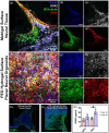
Characterization of microglia phenotypes in the neural organoids. Confocal images of the 3D neural tissues showing presence of the microglia marker protein IBA1 (red) and neural marker βIII-tubulin (green; a and c). A 3D reconstructions of segmented microglia show ramified morphology within the tissues (b and d). Scale bar 100 μm.
Figure 6.
Figure 6.
Gene expression and gene ontology analysis for planar neural organoids versus traditional brain organoids: (a) microscopic images displaying morphological features of the neural organoids assembled in traditional Matrigel suspension (top) or on a PEG hydrogel surface (bottom), (b) global patterns of gene expression in traditional brain organoids formed in Matrigel suspension versus planar neural organoids formed on PEG hydrogels, and (c) Gene ontology analysis for biological processes related to the growth of neural tissues.
Figure 7.
Figure 7.
Analysis of differentially expressed genes of the (a) forebrain, (b) midbrain, and (c) hindbrain in planar neural organoids formed on PEG hydrogels versus traditional brain organoids formed in Matrigel suspension. Fold change (Log2) plotted versus −Log10 of p-values of differential expression analysis. Green color points indicated positive fold change of those genes, red color points indicate negative fold change of the genes in the plots.
Figure 8.
Figure 8.
Analysis of differentially expressed genes. Fold change (Log2) plotted versus −Log10 of p-values of marker genes between planar neural organoids compared to traditional brain organoids: (a) brain micro-vascular markers, (b) cerebral cortex markers, (c and d) Synaptic transmission associated genes, (e) microglial cell marker genes, and (f) innate immune response marker genes. Green color points indicated positive fold change of those genes, red color points indicate negative fold change of the genes in the plots.
Figure 9.
Figure 9.
Lipopolysaccharide (LPS) induced inflammation in planar neural organoids: (a) LPS-induced TNF-α protein production in the media of planar neural organoid cultures, (b) LPS-induced IL-6 protein production in the media of the neural tissue culturesTreatment with the anti-inflammatory drugs Celecoxib (CEL) or Donepezil (DON) reduced the production of TNF-α and IL-6 in the culture medium. (C) Confocal microscopy images of the 3D neural organoids showing the ramified type morphology of microglia for the control treated group and ameboid type morphology of microglia for the LPS treated group. Scale bar 50 μm. MTG: Matrigel; PEG: polyethyleneglycol; LPS: lipopolysaccharide. **p < 0.01. ***p < 0.001. ****p < 0.0001).

Similar articles

Cited by

References

    1. Phillips JB. ‘EngNT’-engineering live neural tissue for nerve replacement. Emerg Top Life Sci 2021; 5: 699–703. - PMC - PubMed
    1. Heusinkveld HJ, Staal YCM, Baker NC, et al.. An ontology for developmental processes and toxicities of neural tube closure. Reprod Toxicol 2021; 99: 160–167. - PMC - PubMed
    1. Yan Y, Shin S, Jha B, et al.. Efficient and rapid derivation of primitive neural stem cells and generation of brain subtype neurons from human pluripotent stem cells. Stem Cells Transl Med 2013; 2: 862–870. - PMC - PubMed
    1. Yamada KM, Cukierman E, Modeling tissue morphogenesis and cancer in 3D. Cell 2007; 130: 601–610. - PubMed
    1. Pampaloni F, Reynaud EG, Stelzer EH. The third dimension bridges the gap between cell culture and live tissue. Nat Rev Mol Cell Biol 2007; 8: 839–845. - PubMed

LinkOut - more resources