. 2024 Feb 16;10(7):eadl4876.
doi: 10.1126/sciadv.adl4876. Epub 2024 Feb 14.

Skp1 proteins are structural components of the synaptonemal complex in C. elegans

Affiliations

Skp1 proteins are structural components of the synaptonemal complex in C. elegans

Joshua M Blundon et al. Sci Adv. .

Abstract

The synaptonemal complex (SC) is a zipper-like protein assembly that links homologous chromosomes to regulate recombination and segregation during meiosis. The SC has been notoriously refractory to in vitro reconstitution, thus leaving its molecular organization largely unknown. Here, we report a moonlighting function of two paralogous S-phase kinase-associated protein 1 (Skp1)-related proteins (SKR-1 and SKR-2), well-known adaptors of the Skp1-Cul1-F-box (SCF) ubiquitin ligase, as the key missing components of the SC in Caenorhabditis elegans. SKR proteins repurpose their SCF-forming interfaces to dimerize and interact with meiosis-specific SC proteins, thereby driving synapsis independent of SCF activity. SKR-1 enables the formation of the long-sought-after soluble complex with previously identified SC proteins in vitro, which we propose it to represent a complete SC building block. Our findings demonstrate how a conserved cell cycle regulator has been co-opted to interact with rapidly evolving meiotic proteins to construct the SC and provide a foundation for understanding its structure and assembly mechanisms.


Figures

Fig. 1.
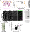
Fig. 1.. SKR proteins associate with SC proteins and localize to the SC central region.
(A) Schematic showing polyubiquitination of a substrate by an E2 ubiquitin-conjugating enzyme and the SCF ubiquitin ligase complex. RBX1, Ring-box protein 1. (B) A silver-stained SDS–polyacrylamide gel electrophoresis (PAGE) gel showing purified SKR-2–containing protein complexes using anti-Flag beads from a worm strain expressing SKR-2::3×Flag. N2 was used as a control. IP, immunoprecipitation. (C) A volcano plot showing proteins enriched in SKR-2 immunoprecipitates over N2 control. Normalized weighted spectra were transformed to logarithmic values (base 2) and analyzed by multiple unpaired t tests. The logarithm of fold change (base 2) is plotted on the x axis, and the negative logarithm of the P value (base 10) is plotted on the y axis. The P value of 0.05 is indicated by a horizontal dotted line. Proteins enriched in SKR-2 immunoprecipitates are found in the top right corner of the plot. F-box proteins (orange), SC proteins (green), CUL-1 (brown), SKR-1/2 (magenta), proteasome subunits (cyan), and other proteins (black) are highlighted and listed on the right. (D) SMLM localizations within the three boxed regions in fig. S2 (D and E) were straightened, aligned, and rendered to create averaged frontal (xy) and cross-sectional (xz) views. Scale bars, 100 nm. (E) Histogram counts (2-nm-wide bins) of straightened and aligned SMLM localizations along the x and z axes fitted with Gaussian distributions (black dashed lines). The respective SDs (σ) and peak-to-peak distance of HIM-3 (d) are indicated in the plot.
Fig. 2.
Fig. 2.. SKR-1 and SKR-2 are required for SC assembly.
(A) Schematic showing the skr alleles used in this study. (B) DAPI-stained oocyte nuclei at diakinesis from indicated genotypes. Scale bar, 3 μm. (C) Graph showing the number of DAPI bodies in diakinesis oocytes. The means ± SD are shown. ns, not significant (P > 0.9999); ****P < 0.0001 by Mann-Whitney U test. The numbers of oocytes scored are 30, 29, and 13 for wild-type, skr-2(kim66), and skr-2(ok1938), respectively. (D) Immunofluorescence images of pachytene nuclei from the indicated genotypes showing DNA, SYP-5 (green), and HIM-3 (blue) staining. Scale bar, 5 μm. (E) htp-3(tm3655) animals expressing SYP-2::GFP were treated with control (feeding) and skr-1/2 RNAi (microinjection), dissected, and stained for DNA and GFP. Scale bar, 5 μm.
Fig. 3.
Fig. 3.. Recombinant SKR exists as a dimer in solution.
(A) An SDS-PAGE gel showing the purification of recombinant 6×His-SKR-1WT and 6×His-SKR-1F115E. (B) SEC-MALS traces of 6×His-SKR-1WT (magenta) and 6×His-SKR-1F115E (light blue). Absorbance at 280 nm (A280) is shown on the left axis, and the measured molecular weight is shown on the right axis. mAU, milli–arbitrary units. (C) An AlphaFold model of a truncated C. elegans SKR-1 (amino acids 1 to 83, GGSG, and 96 to 143; magenta) superimposed onto the NMR structure of a truncated D. discoideum Skp1A dimer (gray; Protein Data Bank 6V88) (39). The inset shows the conserved phenylalanine (F97 in Dictyostelium Skp1A and F115 in C. elegans SKR-1) at the dimerization interface.
Fig. 4.
Fig. 4.. Dimerization of SKR proteins is essential for synapsis.
(A) Composite immunofluorescence images of a full-length gonad dissected from an skr-1F115E skr-2(kim66) animal are shown for DNA, HIM-3, and SYP-5 staining. Asterisk (*) indicates the distal tip. Scale bar, 50 μm. (B) Immunofluorescence images of pachytene nuclei from skr-2(kim66) and skr-1F115E skr-2(kim66) animals showing DNA, HTP-3, and SYP-2 staining. Scale bar, 5 μm. (C) DAPI-stained oocyte nuclei at diakinesis from skr-2(kim66) and skr-1F115E skr-2(kim66) animals. Scale bar, 3 μm. (D) Graph showing the number of DAPI bodies in diakinesis nuclei from indicated genotypes. The numbers of nuclei scored are shown below. The means ± SD is shown; ****P < 0.0001 by the Mann-Whitney U test.
Fig. 5.
Fig. 5.. SKR-2 interacts with the SYP proteins via the SCF-forming interfaces.
(A) An AlphaFold model of the C. elegans SKR-1 dimer with mutated residues highlighted. (B) An SEC-MALS trace of 6×His-SKR-1I155A,W173A. The absorbance at 280 nm is shown on the left axis, and the measured molecular weight is shown on the right axis. (C) Immunofluorescence images of pachytene nuclei from skr-2::3×flag worm strains harboring wild-type and the I153A and W171A mutation. DNA, SYP-5, and SKR-2::3×Flag staining are shown. Scale bar, 5 μm. (D) Immunoblots of worm lysates from the indicated genotypes probed against the 3×FLAG tag fused to the C terminus of SKR-2. N2 was used as a negative control, and HIM-3 was used as a loading control. (E) A silver-stained SDS-PAGE gel showing purified SKR-2I153A,W171A-containing protein complexes using anti-Flag beads from worm lysates. N2 was used as a control. (F) A volcano plot showing proteins enriched in SKR-2::3×flagI153A,W171A immunoprecipitates over N2 control. Normalized weighted spectra were transformed to logarithmic values (base 2) and analyzed by multiple unpaired t tests. The logarithm of fold change (base 2) is plotted on the x axis, and the negative logarithm of the P value (base 10) is plotted on the y axis. The P value of 0.05 is indicated by a horizontal dotted line.
Fig. 6.
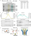
Fig. 6.. In vitro reconstitution establishes SKR-1/2 as SC subunits.
(A) Schematic of the polycistronic plasmid used to coexpress SKR-1 and the SYP proteins (top) and a Coomassie-stained SDS-PAGE gel of the SYP/SKR-1 complexes purified using a 6×His tag on SYP-3 (His) and cation exchange chromatography (SP). Two peak fractions eluted from the SP column are highlighted. (B) Table showing the results from mass spectrometry analysis. (C and D) SEC-MALS traces and Flamingo-stained SDS-PAGE gels of the SYP-3/SYP-4/SKR-1 complex (peak 1; 1034 ± 38 kDa; n = 4) and the complex that contains all six proteins (peak 2; 997 ± 169 kDa; n = 3). The absorbance at 280 nm is shown on the left axis, and the measured molecular weight is shown on the right axis. The void volume is indicated on the top. (E) The model for the dual functions of SKR-1/2 within the SCF ubiquitin ligase complex versus the SC.

Similar articles

Cited by

References

    1. Pyatnitskaya A., Andreani J., Guérois R., De Muyt A., Borde V., The Zip4 protein directly couples meiotic crossover formation to synaptonemal complex assembly. Genes Dev. 36, 53–69 (2022). - PMC - PubMed
    1. Zhang L., Köhler S., Rillo-Bohn R., Dernburg A. F., A compartmentalized signaling network mediates crossover control in meiosis. eLife 7, e30789 (2018). - PMC - PubMed
    1. Libuda D. E., Uzawa S., Meyer B. J., Villeneuve A. M., Meiotic chromosome structures constrain and respond to designation of crossover sites. Nature 502, 703–706 (2013). - PMC - PubMed
    1. Capilla-Pérez L., Durand S., Hurel A., Lian Q., Chambon A., Taochy C., Solier V., Grelon M., Mercier R., The synaptonemal complex imposes crossover interference and heterochiasmy in Arabidopsis. Proc. Natl. Acad. Sci. U.S.A. 118, e2023613118 (2021). - PMC - PubMed
    1. France M. G., Enderle J., Röhrig S., Puchta H., Franklin F. C. H., Higgins J. D., ZYP1 is required for obligate cross-over formation and cross-over interference in Arabidopsis. Proc. Natl. Acad. Sci. U.S.A. 118, e2021671118 (2021). - PMC - PubMed

MeSH terms

Substances