[Preprint]. 2024 Jan 29:2024.01.24.577099.
doi: 10.1101/2024.01.24.577099.

A large-scale cancer-specific protein-DNA interaction network

Affiliations

A large-scale cancer-specific protein-DNA interaction network

Yunwei Lu et al. bioRxiv. .

Update in

  • A large-scale cancer-specific protein-DNA interaction network.
    Lu Y, Berenson A, Lane R, Guelin I, Li Z, Chen Y, Shah S, Yin M, Soto-Ugaldi LF, Fiszbein A, Fuxman Bass JI. Lu Y, et al. Life Sci Alliance. 2024 Jul 16;7(10):e202402641. doi: 10.26508/lsa.202402641. Print 2024 Oct. Life Sci Alliance. 2024. PMID: 39013578 Free PMC article.

Abstract

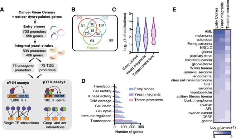
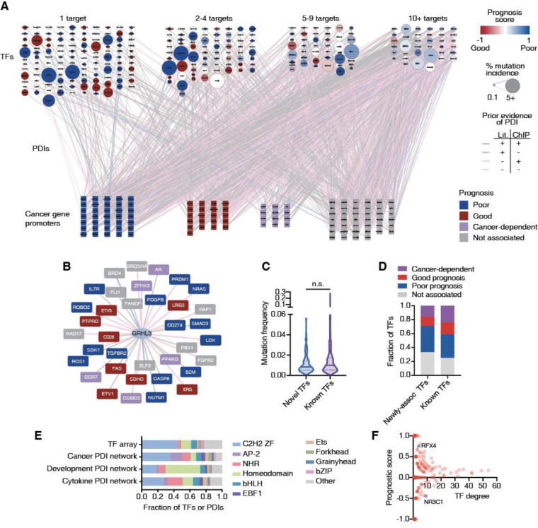
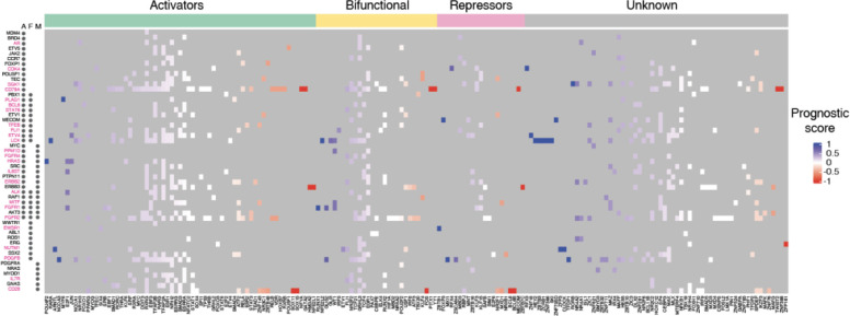
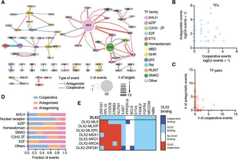
Cancer development and progression are generally associated with dysregulation of gene expression, often resulting from changes in transcription factor (TF) sequence or expression. Identifying key TFs involved in cancer gene regulation provides a framework for potential new therapeutics. This study presents a large-scale cancer gene TF-DNA interaction network as well as an extensive promoter clone resource for future studies. Most highly connected TFs do not show a preference for binding to promoters of genes associated with either good or poor cancer prognosis, suggesting that emerging strategies aimed at shifting gene expression balance between these two prognostic groups may be inherently complex. However, we identified potential for oncogene targeted therapeutics, with half of the tested oncogenes being potentially repressed by influencing specific activator or bifunctional TFs. Finally, we investigate the role of intrinsically disordered regions within the key cancer-related TF estrogen receptor ɑ (ESR1) on DNA binding and transcriptional activity, and found that these regions can have complex trade-offs in TF function. Altogether, our study not only broadens our knowledge of TFs involved in the cancer gene regulatory network but also provides a valuable resource for future studies, laying a foundation for potential therapeutic strategies targeting TFs in cancer.


Conflict of interest statement

Conflict of Interest The authors declare no conflicts of interest.

Figures

Figure 1:
Figure 1:. Generation of clone and yeast resource for cancer gene promoters.
(A) Schematic of the Gateway-compatible cancer gene promoter resource. Cancer genes were selected from the Cancer Gene Census as well as genes dysregulated in cancer. An entry clone resource of 700 promoters (556 genes) was generated as well as a yeast integrant resource corresponding to 508 promoters (426 genes). This yeast resource was tested in eY1H and pY1H assays for TF-DNA interactions. (B) Venn diagram of the number of oncogenes (OG), tumor suppressor genes (TSG) and genes involved in fusions for which yeast integrants were generated. (C) Violin plots correspond to the distribution of the number of publications per gene included in the entry clone resource, the yeast integrant collection, and the yeast integrants tested by eY1H/pY1H. (D) Number of genes associated with different biological functions for genes included in the entry clone resource, the yeast integrant collection, and the yeast integrants tested by eY1H/pY1H. (E) Number of genes associated with different cancer types among those in the set of entry clones, yeast integrants, and tested by eY1H assays.
Figure 2:
Figure 2:. Large-scale cancer TF-DNA interaction network.
(A) Cancer TF-DNA interaction network determined using eY1H and pY1H assays. Circular nodes represent TFs, while squares represent cancer gene promoters. Interactions are represented by edges colored based on whether there is evidence by ChIP-seq (pink), literature (blue), both (purple) or neither (gray). TF nodes are colored based on the prognostic score calculated as (#poor prognosis targets - #good prognosis targets)/(# total number of targets). The borders of TF nodes are colored based on whether the TF is listed (red) in CGC. TF node size indicates the % of non-synonymous mutations across all cancers. Cancer gene promoters are colored based on whether their expression is associated with poor (blue), good (red), or cancer-dependent prognosis (purple). (B) Interaction network involving GRHL3. (C) Violin plot depicting the mutation frequency across cancers for TFs known to bind/regulate our set of cancer genes (purple), and novel TFs (blue). Statistical significance determined by two-tailed Mann-Whitney’s U test. (D) Fraction of TFs whose expression levels are associated with poor or good cancer prognosis for TFs known to bind/regulate our set of cancer genes and novel TFs. (E) Fraction of TFs and TF-DNA interactions corresponding to different TF families in the TF array and the cancer promoter, cytokine promoter, and developmental enhancer networks. (F) Scatter plot of the prognostic score versus degree (number of targets) for each TF.
Figure 3:
Figure 3:. Heatmap of potentially targetable TFs to reduce oncogene expression.
Heatmap of prognostic scores for TFs that bind to oncogene promoters. TFs are classified as potential activators, bifunctional, or repressors based on annotated effector domains in TFRegDB. Oncogenes that contribute to cancer development through amplifications (A), fusions (F), or mutations (M) are indicated next to the gene name. Oncogenes indicated in magenta have at least one TF that is activator/bifunctional with a prognosis score > 0.33.
Figure 4:
Figure 4:. TF-binding cooperativity and antagonism at cancer gene promoters.
(A) Network of cooperative (blue) and antagonistic (red) relationships between TFs at the cancer gene promoters screened. Node size indicates the number of binding events for that TF. Edge width represents the number of cooperative or antagonistic events involving a specific TF-pair. (B) Number of cooperative and antagonistic events observed for individual TFs. (C) Number of cooperative and antagonistic events observed for TF-pairs. (D) Fraction of events where a TFs binds cooperatively, is antagonized by another TF, or antagonizes the binding of another TF for each TF family. (E) Heatmap of interactions involving DLX2, either on its own, or together with other TF partners at 13 cancer gene promoters. Dark blue – cooperative binding; light blue – indicates DLX2 binding not influenced by partner TF; white – no DLX2 binding; and red – DLX2 binding antagonized by TF partner.
Figure 5:
Figure 5:. Role of ESR1 intrinsically disordered regions on DNA binding and transcriptional activity.
(A) Schematic of ESR1 constructs used. IDRs are indicated in green, DNA binding domain in purple, and ligand binding domain in yellow. (B) Examples of eY1H screens for binding of 18 different ESR1 constructs to the promoters of BRCA1, AFF2, and NBL1 in the presence or absence of 100 nM estradiol. (C) eY1H binding activity scored from 0 (no binding) to 5 (very strong binding) for different ESR1 constructs. Connected lines correspond to the same cancer gene promoter. Circle sizes indicate the number bound cancer promoters, whereas color intensity indicates the average eY1H activity across bound promoters. Type-1 promoters (blue) are those where wild type ESR1 binds; type-2 promoters (red) are those that wild type ESR1 does not bind. (D, E) Luciferase assays in HEK293T cells where the promoters of AFF2 (D) or NBL1 (E) are cloned upstream of firefly luciferase. ESR1 constructs are fused to 10 copies of the VP16 activator domain. Experiments were conducted in biological triplicates. *p<0.05 Statistical significance determined by two-tailed Student’s t-test. (F, G) Mammalian one-hybrid assays measuring the transcriptional activity of different ESR1 constructs. ESR1 fusions with the Gal4 DNA binding domain (DB) are recruited to 4 copies of the Gal4 binding site (UAS) cloned upstream of firefly luciferase. Experiments were conducted in biological sextuplicates. *p<0.05 Statistical significance determined by two-tailed Student’s t-test.

Similar articles

References

    1. Djakiew D. (2000) Dysregulated expression of growth factors and their receptors in the development of prostate cancer. Prostate, 42, 150–160. - PubMed
    1. Vervoort S.J., Devlin J.R., Kwiatkowski N., Teng M., Gray N.S. and Johnstone R.W. (2022) Targeting transcription cycles in cancer. Nat Rev Cancer, 22, 5–24. - PubMed
    1. Hanahan D. and Weinberg R.A. (2011) Hallmarks of cancer: the next generation. Cell, 144, 646–674. - PubMed
    1. Carrasco Pro S., Hook H., Bray D., Berenzy D., Moyer D., Yin M., Labadorf A.T., Tewhey R., Siggers T. and Fuxman Bass J.I. (2023) Widespread perturbation of ETS factor binding sites in cancer. Nat Commun, 14, 913. - PMC - PubMed
    1. Sjostedt E., Zhong W., Fagerberg L., Karlsson M., Mitsios N., Adori C., Oksvold P., Edfors F., Limiszewska A., Hikmet F. et al. (2020) An atlas of the protein-coding genes in the human, pig, and mouse brain. Science, 367. - PubMed

Publication types