. 2024 Jan 24;25(3):1410.
doi: 10.3390/ijms25031410.

Mechanism of DNA Intercalation by Chloroquine Provides Insights into Toxicity

Affiliations

Mechanism of DNA Intercalation by Chloroquine Provides Insights into Toxicity

Joha Joshi et al. Int J Mol Sci. .

Abstract

Chloroquine has been used as a potent antimalarial, anticancer drug, and prophylactic. While chloroquine is known to interact with DNA, the details of DNA-ligand interactions have remained unclear. Here we characterize chloroquine-double-stranded DNA binding with four complementary approaches, including optical tweezers, atomic force microscopy, duplex DNA melting measurements, and isothermal titration calorimetry. We show that chloroquine intercalates into double stranded DNA (dsDNA) with a KD ~ 200 µM, and this binding is entropically driven. We propose that chloroquine-induced dsDNA intercalation, which happens in the same concentration range as its observed toxic effects on cells, is responsible for the drug's cytotoxicity.

Keywords: AFM; DNA binding; DNA melting; chloroquine; intercalation; isothermal titration calorimetry; optical tweezers; single molecule.


Conflict of interest statement

The authors declare no conflicts of interest, and the funders had no role in the design of the study; in the collection, analyses, or interpretation of data; in the writing of the manuscript; or in the decision to publish the results.

Figures

Figure 1
Figure 1
Structure of chloroquine, showing aromatic rings that may intercalate into DNA and a basic tail.
Figure 2
Figure 2
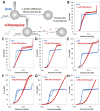
Force extension of λ DNA in the absence and presence of chloroquine (red diamonds). (A) Schematic of λ DNA tethering in optical tweezers experiment. Representative force extension and release data (blue solid circles and open circles, respectively) for λ DNA stretched in the presence of (B) 1 μM, (C) 3 μM, (D) 10 μM, (E) 30 μM, (F) 50 μM, (G) 100 μM, and (H) 200 μM chloroquine (red circles).
Figure 3
Figure 3
Chloroquine titration with DNA determines binding parameters. (A) Chloroquine titration with DNA (from Figure 2) reveals the change in DNA length versus ligand concentration across forces ranging from 15 pN (red) through 70 pN (purple), including the ligand-free length below overstretching (note the break ‘//’ in the x axis). Solid lines are χ2 minimized fits to the binding isotherms, as described in the Methods. (B) Values of the measured chloroquine induced DNA end to end (contour) length (red) compared to models of DNA elasticity (blue), as described in the text. High force (>60 pN) yields a change of Δx = 0.29 ± 0.04 nm. (C) Fitted equilibrium dissociation constant (KD) and (D) binding site size (N) at each force, as described in the text. The equilibrium dissociation may be extrapolated to find a value in the absence of any external force of KD(F = 0) = 170 ± 90 μM (Equation (4), Section 4). The force and extension errors are standard errors from three DNA stretches. Uncertainties in KD, Δx, and N are all deduced from values of χ2 + 1.
Figure 4
Figure 4
ssDNA stretching curves in the presence of 200 μM of chloroquine. (A) Representative stretch and release curves for 8.1 kbp dsDNA in dark blue ((A), 1). After returning to its original position, 20 μL of 5 M NaOH is added to the flow cell to melt the DNA. A representative ssDNA stretch curve is shown in light blue ((A), 2). (B) Representative curve for the first stretch of ssDNA in the absence (blue) and presence of 200 μM chloroquine at 20 nm step size, with total time for extension and release of ~10 s (red closed and open circles, respectively). (C) The same DNA molecule on its nineteenth stretch in the absence (blue) and presence (red) of chloroquine.
Figure 5
Figure 5
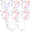
Free energy of DNA overstretching increases with chloroquine binding. The energy of converting dsDNA to ssDNA, or base melting, is shown as the shaded, integrated area between the force extension data for dsDNA (blue without chloroquine, red with chloroquine) and ssDNA (purple), below the critical melting force (Fm, dotted line). This value may be found for each ligand concentration where melting is observed; in the absence of (A) and in the presence of (B) 1 µM, (C) 3 µM, (D) 10 µM, (E) 30 µM, and (F) 50 µM chloroquine. (G) The resulting integrated free energy change per base pair and (H) the change in the critical melting force both increase with ligand concentration. Errors represent the standard error from three DNA stretches, where larger than the symbols used.
Figure 6
Figure 6
AFM measurement of chloroquine-mediated changes to DNA properties. AFM images of 500 bp dsDNA constructs in the absence (A) and presence (B) of 1 mM chloroquine. (C) DNA molecules are traced to acquire the orientation of the molecule over its entire length. The relative angle change (θ) between every two points separated by contour length ranging from 5 to 150 nm is calculated. The average cosine of this value, averaged over all observed molecules, decays as this length is increased (diamonds with standard error bars). Best fits (solid lines) to the DNA only (blue) and DNA with chloroquine (purple and red) are obtained to determine the persistence length. (D) The measured persistence length (yellow) and contour length (green) of DNA–chloroquine complexes. Error bars represent the standard error.
Figure 7
Figure 7
Measuring the effect of chloroquine on DNA stability. (A) Thermal melting profiles in 20 mM NaCl of A20:T20 duplex DNA (4 µM dsDNA) in the absence (black) and presence of 50 μM, 100 μM, 150 μM, and 200 μM (from navy to sky blue, following the grey arrow) of chloroquine diphosphate. (B) First derivative of melting profiles used to determine Tm at each concentration of chloroquine diphosphate. (C) ΔTm values versus added chloroquine, in 20 mM Na+ (blue) and 100 mM Na+ (red). Errors (bars) are standard deviations from three independent replicates in 20 mM Na+ and estimated from individual profiles in 100 mM Na+. Reduced chi-squared fits (dotted lines) determined KD = 90 ± 10 µM (20 mM Na+) and KD = 650 ± 150 µM (100 mM Na+), as described in the text.
Figure 8
Figure 8
Representative ITC measurement of the interaction of chloroquine with A-T duplex DNA. (A) Plot shows the heat of reaction as a function of time. (B) Enthalpy change plotted as a function of the chloroquine/DNA molar ratio during the titration.

Similar articles

References

    1. Hoekenga M.T. The treatment of acute malaria with single oral doses of amodiaquin, chloroquine, hydroxychloroquine and pyrimethamine. Am. J. Trop. Med. Hyg. 1954;3:833–838. doi: 10.4269/ajtmh.1954.3.833. - DOI - PubMed
    1. Fong W., To K.K.W. Repurposing Chloroquine Analogs as an Adjuvant Cancer Therapy. Recent Patents Anti-Cancer Drug Discov. 2021;16:204–221. doi: 10.2174/1574892815666210106111012. - DOI - PubMed
    1. Zhou W., Wang H., Yang Y., Chen Z.S., Zou C., Zhang J. Chloroquine against malaria, cancers and viral diseases. Drug Discov. Today. 2020;25:2012–2022. doi: 10.1016/j.drudis.2020.09.010. - DOI - PMC - PubMed
    1. U.S. Food & Drug Administration Coronavirus (COVID-19) Update: FDA Revokes Emergency Use Authorization for Chloroquine and Hydroxychloroquine [Press Release] [(accessed on 28 December 2023)]; Available online: https://www.fda.gov/news-events/press-announcements/coronavirus-covid-19....
    1. Yang J., Guo Z., Liu X., Liu Q., Wu M., Yao X., Liu Y., Cui C., Li H., Song C., et al. Cytotoxicity Evaluation of Chloroquine and Hydroxychloroquine in Multiple Cell Lines and Tissues by Dynamic Imaging System and Physiologically Based Pharmacokinetic Model. Front. Pharmacol. 2020;11:574720. doi: 10.3389/fphar.2020.574720. - DOI - PMC - PubMed

LinkOut - more resources