. 2024 Jan 25;13(3):232.
doi: 10.3390/cells13030232.

The Opioid Receptor Influences Circadian Rhythms in Human Keratinocytes through the β-Arrestin Pathway

Affiliations

The Opioid Receptor Influences Circadian Rhythms in Human Keratinocytes through the β-Arrestin Pathway

Paul Bigliardi et al. Cells. .

Abstract

The recent emphasis on circadian rhythmicity in critical skin cell functions related to homeostasis, regeneration and aging has shed light on the importance of the PER2 circadian clock gene as a vital antitumor gene. Furthermore, delta-opioid receptors (DOPrs) have been identified as playing a crucial role in skin differentiation, proliferation and migration, which are not only essential for wound healing but also contribute to cancer development. In this study, we propose a significant association between cutaneous opioid receptor (OPr) activity and circadian rhythmicity. To investigate this link, we conducted a 48 h circadian rhythm experiment, during which RNA samples were collected every 5 h. We discovered that the activation of DOPr by its endogenous agonist Met-Enkephalin in N/TERT-1 keratinocytes, synchronized by dexamethasone, resulted in a statistically significant 5.6 h delay in the expression of the core clock gene PER2. Confocal microscopy further confirmed the simultaneous nuclear localization of the DOPr-β-arrestin-1 complex. Additionally, DOPr activation not only enhanced but also induced a phase shift in the rhythmic binding of β-arrestin-1 to the PER2 promoter. Furthermore, we observed that β-arrestin-1 regulates the transcription of its target genes, including PER2, by facilitating histone-4 acetylation. Through the ChIP assay, we determined that Met-Enkephalin enhances β-arrestin-1 binding to acetylated H4 in the PER2 promoter. In summary, our findings suggest that DOPr activation leads to a phase shift in PER2 expression via β-arrestin-1-facilitated chromatin remodeling. Consequently, these results indicate that DOPr, much like its role in wound healing, may also play a part in cancer development by influencing PER2.

Keywords: beta-arrestin; circadian rhythm; keratinocyte; opioid receptor.


Conflict of interest statement

The authors declare no conflicts of interest.

Figures

Figure 1
Figure 1
N/TERT keratinocytes exhibit robust circadian rhythms: upon synchronization with 1 μM dexamethasone, N/TERT-1 keratinocytes were synchronized, and gene expression was measured at indicated times. The cells exhibited peak expression in BMAL1 at 10 h (A) and PER2 at 15–20 h (B).
Figure 1
Figure 1
N/TERT keratinocytes exhibit robust circadian rhythms: upon synchronization with 1 μM dexamethasone, N/TERT-1 keratinocytes were synchronized, and gene expression was measured at indicated times. The cells exhibited peak expression in BMAL1 at 10 h (A) and PER2 at 15–20 h (B).
Figure 2
Figure 2
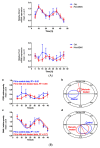
Met-ENK treatment induces internalization of the DOPr and a phase shift in PER2 expression: (A) N/TERT-1 keratinocytes were synchronized, and gene expression profiles were obtained at indicated times. The upper panel and lower panel show expression profiles of BMAL1 and PER2 in control cells and cells treated with Met-ENK, respectively. It is apparent that the control group cells exhibited peak expression in BMAL1 at 10 h and PER2 at 15–20 h, whereas the Met-ENK-treated group cells exhibited peak expression in BMAL1 at 5–10 h and PER2 at 25 h. n = 3 in all panels; results are expressed as mean ± SEM. (B) In all cases, the period is fixed at 24 h. The R2 coefficients indicate goodness of fit or percentage of rhythm in the data. (a) Regression of the cosinor model against the PER2 mRNA expression profiles (pooled data from n = 3 experiments) shows a 5.6 h phase shift in the Met-ENK-treated (red) cells compared to control (blue). (b) The 95% confidence regions (ellipses) obtained from the PER2 regressions are distinct; acrophases φ (clock times indicated by the dashed lines) are significantly different. (c) Regression against the BMAL1 control and Met-ENK treatment data yields no difference in rhythmicity. (d) The BMAL1 95% confidence regions overlap; A and φ are not significantly different.
Figure 3
Figure 3
Met-enkephalin treatment induces a change in clock-controlled gene expression: There was no significant change in Dec2 expression as a result of Met-ENK treatment. However, 25 h post synchronization, the Met-ENK-treated group showed significant changes in DBP and Tef expression. The data here are the means ± SEM of three independent experiments (n = 3). Two-way ANOVA reveals * p < 0.05 and *** p < 0.001.
Figure 4
Figure 4
Activation of DOPr leads to internalization and nuclear co-localization of DOPr and βarr1: (A) DOPr-GFP overexpressing N/TERT-1 keratinocytes exhibit DOPr to be localized to the cell membrane in control (untreated) cells. This localization remains unaffected in cells treated with DOPr antagonist, i.e., NTI, and combined NTI and agonist (Met-ENK). However, upon Met-ENK treatment, the receptor is internalized and appears to undergo relocalization to the perinuclear region in the cytoplasm. Scale bar 20 μm. (B) Confocal visualization of N/TERT-1 keratinocytes overexpressing GFP-DOPr, and CFP-βarr-1 incubated without (Ctrl) or with 100 nM Met-ENK for 5 min before fixation. Upon Met-ENK treatment, both DOPr and βarr1 co-localize in the nucleus. (C) The pictures shown are representative of three independents since all blots appeared the same. Values are expressed as the mean ± SD. ** p < 0.01. (D) N/TERT-1 keratinocytes overexpressing GFP-DOPr and CFP-βarr-1 were incubated without (Ctrl) or with 100 nM Met-ENK for 5 min, and the nuclear extracts were then used for a pulldown using an anti-CFP antibody. The pulldown lysates were then tested for βarr1 and DOPr. (E) shows the graph of the semiquantification of DOPr or βarr1. Each bar represents the signal from one representative experiment, n = 1.
Figure 4
Figure 4
Activation of DOPr leads to internalization and nuclear co-localization of DOPr and βarr1: (A) DOPr-GFP overexpressing N/TERT-1 keratinocytes exhibit DOPr to be localized to the cell membrane in control (untreated) cells. This localization remains unaffected in cells treated with DOPr antagonist, i.e., NTI, and combined NTI and agonist (Met-ENK). However, upon Met-ENK treatment, the receptor is internalized and appears to undergo relocalization to the perinuclear region in the cytoplasm. Scale bar 20 μm. (B) Confocal visualization of N/TERT-1 keratinocytes overexpressing GFP-DOPr, and CFP-βarr-1 incubated without (Ctrl) or with 100 nM Met-ENK for 5 min before fixation. Upon Met-ENK treatment, both DOPr and βarr1 co-localize in the nucleus. (C) The pictures shown are representative of three independents since all blots appeared the same. Values are expressed as the mean ± SD. ** p < 0.01. (D) N/TERT-1 keratinocytes overexpressing GFP-DOPr and CFP-βarr-1 were incubated without (Ctrl) or with 100 nM Met-ENK for 5 min, and the nuclear extracts were then used for a pulldown using an anti-CFP antibody. The pulldown lysates were then tested for βarr1 and DOPr. (E) shows the graph of the semiquantification of DOPr or βarr1. Each bar represents the signal from one representative experiment, n = 1.
Figure 5
Figure 5
DOPr expression is essential for maintaining rhythmicity in PER2 expression: (A) Constitutive knockdown of the DOPr (KD DOPr) was possible using a lentiviral construct. This was validated by Western blot using an anti-Delta Opioid Receptor antibody (ab176324; Abcam), and a band was seen at the 40 kDa mark when used at a 1/1000 dilution with anti-β-actin antibody (A5441; Sigma-Aldrich) as the loading control and (B) quantitative real-time polymerase chain reaction (qPCR), n = 1. (C) To test for rhythmicity in PER2 expression, qPCRs were carried out using non-targeting control (KD Ctrl) and DOPr KD cells. It was observed that the DOPr KD cells did not exhibit rhythmicity in PER2 expression when compared to the KD Ctrl cells which exhibited a trough in PER2 expression at 15h and 35h post synchronization, n = 2. (D) Chromatin immunoprecipitation experiments (n = 2) targeting the PER2 promoter in GFP-tagged DOPr overexpressing N/TERT-1 keratinocytes showed that DOPr binds rhythmically to the PER2 promoter.
Figure 6
Figure 6
Met-enkephalin treatment enhances and induces a phase shift in rhythmical βarr1 binding on the PER2 promoter and enhances βarr1 binding to acetylated H4 in the PER2 promoter: (A) Chromatin immunoprecipitation (ChIP) experiments were carried out with anti-βarr1 antibodies, and PER2 promoter sequences in the input DNA and that recovered from antibody-bound chromatin segments were analyzed by qPCR. The data were normalized to the corresponding input control. All data were then normalized to the corresponding 0 h control. The data shown are the means ± SEM of three independent experiments (n = 3). Two-way ANOVA reveals * p < 0.05, ** p < 0.01, *** p < 0.001. (B) Re-ChIP experiments were carried out on N/TERT-1 keratinocytes which were synchronized for 25 h with or without Met-ENK treatment. In these experiments, antibodies targeting βarr1 were used. The consequent eluates were then used for pulling down CREB and acetylated histone 4 (H4K16ac), and the presence of the PER2 promoter sequences in the input DNA and that recovered from antibody-bound chromatin segments was analyzed by qPCR. The data were normalized to the corresponding input control. All data were then normalized to the corresponding 25 h control. The means ± SEM of three independent experiments (n = 3) are presented. The data were then subjected to a paired two-tailed t-test, and the p values obtained for CREB and H4K16ac were 0.0024 and 0.0171, respectively.
Figure 7
Figure 7
Dexamethasone treatment does not affect DOPr expression in N/TERT-1 keratinocytes. Ligand Metenk-TAMRA binding assay on (A) control N/TERT-1 and (B) dexamethasone-treated cells identifies DOPr positive population. (C) Graphical representation of DOPr positive population percentage. n = 4, results are expressed as mean ± SEM.

Similar articles

References

    1. Salazar A., von Hagen J. Circadian Oscillations in Skin and Their Interconnection with the Cycle of Life. Int. J. Mol. Sci. 2023;24:5635. doi: 10.3390/ijms24065635. - DOI - PMC - PubMed
    1. Junker U., Wirz S. Review Article: Chronobiology: Influence of circadian rhythms on the therapy of severe pain. J. Oncol. Pharm. Pract. 2010;16:81–87. doi: 10.1177/1078155209337665. - DOI - PubMed
    1. Plikus M.V., Van Spyk E.N., Pham K., Geyfman M., Kumar V., Takahashi J.S., Andersen B. The circadian clock in skin: Implications for adult stem cells, tissue regeneration, cancer, aging, and immunity. J. Biol. Rhythms. 2015;30:163–182. doi: 10.1177/0748730414563537. - DOI - PMC - PubMed
    1. Welz P.S. Clock Regulation of Skin Regeneration in Stem Cell Aging. J. Invest. Dermatol. 2021;141:1024–1030. doi: 10.1016/j.jid.2020.10.009. - DOI - PubMed
    1. Bigliardi P.L., Tobin D.J., Gaveriaux-Ruff C., Bigliardi-Qi M. Opioids and the skin—Where do we stand? Exp. Dermatol. 2009;18:424–430. doi: 10.1111/j.1600-0625.2009.00844.x. - DOI - PubMed

LinkOut - more resources