. 2024 Feb 7;15(1):33.
doi: 10.1186/s13287-024-03639-x.

Osteogenic human MSC-derived extracellular vesicles regulate MSC activity and osteogenic differentiation and promote bone regeneration in a rat calvarial defect model

Affiliations

Osteogenic human MSC-derived extracellular vesicles regulate MSC activity and osteogenic differentiation and promote bone regeneration in a rat calvarial defect model

Niyaz Al-Sharabi et al. Stem Cell Res Ther. .

Abstract

Background: There is growing evidence that extracellular vesicles (EVs) play a crucial role in the paracrine mechanisms of transplanted human mesenchymal stem cells (hMSCs). Little is known, however, about the influence of microenvironmental stimuli on the osteogenic effects of EVs. This study aimed to investigate the properties and functions of EVs derived from undifferentiated hMSC (Naïve-EVs) and hMSC during the early stage of osteogenesis (Osteo-EVs). A further aim was to assess the osteoinductive potential of Osteo-EVs for bone regeneration in rat calvarial defects.

Methods: EVs from both groups were isolated using size-exclusion chromatography and characterized by size distribution, morphology, flow cytometry analysis and proteome profiling. The effects of EVs (10 µg/ml) on the proliferation, migration, and osteogenic differentiation of cultured hMSC were evaluated. Osteo-EVs (50 µg) or serum-free medium (SFM, control) were combined with collagen membrane scaffold (MEM) to repair critical-sized calvarial bone defects in male Lewis rats and the efficacy was assessed using µCT, histology and histomorphometry.

Results: Although Osteo- and Naïve-EVs have similar characteristics, proteomic analysis revealed an enrichment of bone-related proteins in Osteo-EVs. Both groups enhance cultured hMSC proliferation and migration, but Osteo-EVs demonstrate greater efficacy in promoting in vitro osteogenic differentiation, as evidenced by increased expression of osteogenesis-related genes, and higher calcium deposition. In rat calvarial defects, MEM with Osteo-EVs led to greater and more consistent bone regeneration than MEM loaded with SFM.

Conclusions: This study discloses differences in the protein profile and functional effects of EVs obtained from naïve hMSC and hMSC during the early stage of osteogenesis, using different methods. The significant protein profile and cellular function of EVs derived from hMSC during the early stage of osteogenesis were further verified by a calvarial bone defect model, emphasizing the importance of using differentiated MSC to produce EVs for bone therapeutics.

Keywords: Bone regeneration; Extracellular vesicles; Mesenchymal stem cells; Naïve-EVs; Osteo-EVs; Rat calvarial defects.


Conflict of interest statement

The authors declare no competing interests.

Figures

Fig. 1
Fig. 1
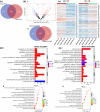
Characterization of Osteo- and Naïve-EVs. a i and b i Histogram graphs depicting the average size of EVs particles (n = 3) in Osteo- and Naïve-EVs, measured by dynamic light scattering (DLS). a ii and b ii Representative transmission electron microscopy (TEM) images of Osteo-and Naïve-EVs (Magnification of the EVs in the main image x10k; scale bar = 2 µm, and in the small box x70K; scale bar = 200 nm). a iii and b iii Representative flow cytometry analysis of transmembrane proteins CD63, CD81, and CD9. b i Venn diagram across all 3 donors in the Osteo-EVs group and inter-donor comparison, and across the common Osteo-EV (882) proteins and both Vesiclepedia Top 100 proteins and Vesiclepedia datasets. c ii The enriched GO terms (BP, CC, and MF) for the common Osteo-EVs (882) proteins using FunRich tool (Version 3.1.3). d i Venn diagram across all 3 donors in the Naïve-EVs group and compared to one another, and across the common Naïve-EV (895) proteins and both Vesiclepedia Top 100 proteins and Vesiclepedia datasets. d ii The enriched GO terms (BP, CC, and MF) for the common Naïve-EVs (895) proteins using FunRich tool (Version 3.1.3)
Fig. 2
Fig. 2
Analysis of DEPs in proteins (745) common to Osteo- and Naïve-EVs groups. a Venn diagram representing the numbers of unique and overlapping proteins among the common protein in each EV group. b i and ii Volcano plot and Venn diagram showing differentially expressed proteins (p < 0.05) when comparing Osteo-EVs to Naïve-EVs. c Heat map showing the distribution of DEPs among the three donors of each EV group. Values represent protein level quantification (log2). d and e The enriched GO terms (BP, and MF) and REAC pathways for the DEPs in each EV group, respectively, are retrieved from the gene ontology resource
Fig. 3
Fig. 3
Effects of Osteo- and Naïve-EVs (10 µg/ml each) in cultured hMSC. a Representative images from the uptake experiments. Cultured hMSC internalised labelled Osteo- and Naïve-EVs: blue, DAPI-labelled nuclei; red, Phalloidin; green, DiOC18-labelled EVs. b Cell proliferation assay indicated that Osteo- and Naïve-EVs stimulate cultured hMSC proliferation after 24 and 72 h compared to the control medium group (BPS and medium). c i Representative images from the wound-healing experiments; scale bar 50 µm. Time course of cell migration wound-healing assay for cultured hMSC as the “recipient” cells with the control medium, Naïve- and Osteo-EVs. Scale bar 100 µm. c ii Quantification of the wound-healing assay from three independent donors. *p < 0.05, **p < 0.01
Fig. 4
Fig. 4
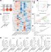
Osteogenic effects of Osteo- and Naïve-EVs in cultured hMSC. a Representative images of ALP after 14 days. b Representative images of ARS after 21 days. Matrix mineralization was greater after Osteo-EVs treatment than after treatment with the Naïve-EVs and control medium. Quantification of the ARS experiment from three independent donors indicated that Osteo-EVs enhanced matrix mineralization of hMSC after 21 days. c Osteogenic gene expression array heatmap among the three donors of each group: control-medium, Naïve- and Osteo-EVs, after 14 days. d Prediction ellipses showed a new observation of donors from the same group falling within the prediction ellipse (confidence interval 95%). e Gene expression level (fold change) between the control medium and EV groups showed upregulation of several genes related to osteogenesis in the EV groups. Genes with red colours are identified in the Osteo-EVs and those with green colours are identified in the Naïve-EVs; green boxes refer to the downregulated genes in the Naïve-EVs f Gene expression level (relative fold change) between the Naïve- and Osteo-EVs showed upregulation of several genes related to osteogenesis in the Osteo-EVs group. g Validation of gene array findings with RT-qPCR: Osteo-EVs triggered higher mRNA expression levels of osteogenesis-related markers SPP1, BSP and BGLAP. *p < 0.05, **p < 0.01, ****p < 0.0001
Fig. 5
Fig. 5
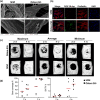
Characterization of Osteo-EVs and MEMs in vitro and in vivo. a Representative SEM images of the MEM loaded with Osteo-EVs, compared with MEM loaded with only SFM. Scale bars, top: 2 µm, bottom: 200 nm with an average size of 61.47 nm (± 17.47 nm). b Representative images from the uptake experiments. Labelled Osteo-EVs were released from the MEM and entered targeted cultured hMSC after 48 h. MEM loaded with SFM served as a control. Blue, DAPI-labelled nuclei; red, Phalloidin; green, DiOC18-dye; scale bar 50 µm. c Representative reconstructed µCT images after 2 (in vivo) and 4 weeks (ex vivo) showing maximum, average, and minimum bone formation in Osteo-EVs and SFM groups. d Quantification of bone coverage (%) and bone volume per tissue volume (BV/TV %) in SFM and Osteo-EVs groups
Fig. 6
Fig. 6
Micro-CT images, histological and histomorphometric analysis of MEM with Osteo-EVs. a Representative histological and μCT images of central slices at 4 weeks. Images showing maximum, average, and minimum bone formation in the Osteo-EVs and SFM groups, scale bar: 1 mm. b Representative histological images of Levi-Lazko dye staining at higher magnification, showing the different tissues analysed in the Osteo-EVs and SFM groups. The upper panel in each experimental group shows a region of interest with outlined sub-regions, which are enlarged in the lower panel (scale bars: upper 200 um, lower panel 50 um). Each sub-region shows a specific tissue type indicated by letters (A-F). (A and D) new bone (B and E), hybrid bone (C and F) and membrane. Numbers on the side panel indicate relative percentages of new bone for the treatment group (red), hybrid bone (cyan), mineralized membrane fibres (pink), residual unmineralized membrane (yellow) and soft tissue area (white). Yellow arrows; mineralized membrane, pink arrows; residual membrane. c Quantification of histomorphometric parameters. Data represent means (n = 5). SFM: serum-free medium; MEM: collagen membrane; Total bone (New Bone + Hybrid Bone Area): TtBAr; nB.Ar: New bone area; hB.Ar: Hybrid bone area; mMb.Ar: Mineralized membrane area; rMb.Ar: Residual Membrane area; Vd.Ar: Soft Tissue Area

Similar articles

Cited by

References

    1. Wang W, Yeung KWK. Bone grafts and biomaterials substitutes for bone defect repair: a review. Bioact Mater. 2017;2(4):224–247. - PMC - PubMed
    1. Gjerde C, Mustafa K, Hellem S, Rojewski M, Gjengedal H, Yassin MA, et al. Cell therapy induced regeneration of severely atrophied mandibular bone in a clinical trial. Stem Cell Res Ther. 2018;9(1):213. - PMC - PubMed
    1. Iso Y, Spees JL, Serrano C, Bakondi B, Pochampally R, Song YH, et al. Multipotent human stromal cells improve cardiac function after myocardial infarction in mice without long-term engraftment. Biochem Biophys Res Commun. 2007;354(3):700–706. - PMC - PubMed
    1. Konala VB, Mamidi MK, Bhonde R, Das AK, Pochampally R, Pal R. The current landscape of the mesenchymal stromal cell secretome: a new paradigm for cell-free regeneration. Cytotherapy. 2016;18(1):13–24. - PMC - PubMed
    1. Al-Sharabi N, Xue Y, Udea M, Mustafa K, Fristad I. Influence of bone marrow stromal cell secreted molecules on pulpal and periodontal healing in replanted immature rat molars. Dent Traumatol Off Publ Int Assoc Dent Traumatol. 2016;32(3):231–239. - PubMed